
yogabook / functional exercises / tibialis posterior training with slantboard
Feedback: We’d love to hear what you think of this description. Please send us your feedback at:
postmeister@yogabuch.de
Last updated: 12 February 2026
Common name: Posterior tibial muscle training on a slant board
Level: A
- Classification
- Contraindication
- Effects
- Preparation
- Follow-up
- derived asanas
- similar asanas
- Diagnostics
- Instructions
- Details
- Variants
Contents
Classification
classical: functional exercise
physiological: strengthening of the tibialis posterior
Contraindication
Effects
- (552) Strengthening the tibialis posterior
Preparation
Follow-up
Derived asanas:
Similar asanas:
Diagnostics (No.)
Variants:
Instructions
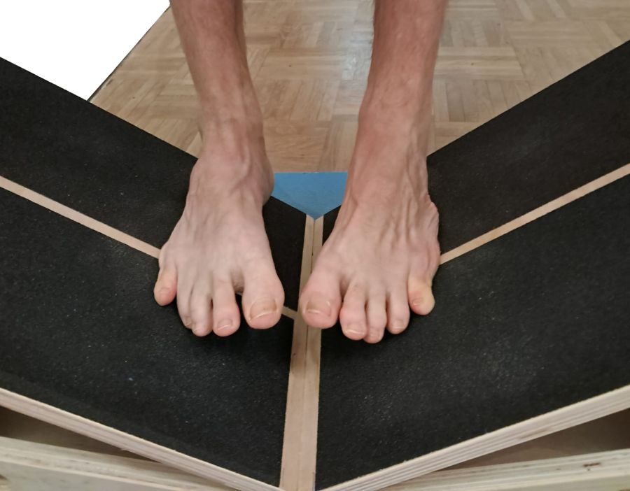
- Place two slantboards side by side so that the sloping surfaces slope towards each other, forming a V shape.
- Stand with both feet on the respective slant board so that the midlines of your feet are roughly parallel and your feet are as close together as possible without touching. The resulting foot position is significantly pronated.
- Shift your weight onto your heels and keep it there.
- Slowly lift your inner feet.
- Avoid lifting your big toes as well. Similarly, avoid using the abductors of the hip joint to push your feet away from each other.
Details
- The tibialis posterior is the primary muscle responsible for supination in this exercise, as the triceps surae—which also supinates—is not engaged here, given that the outer edge of the foot and the heel are to remain on the floor. In the literature, the ratio of the tibialis posterior in the directions of plantar flexion and supination is given as values between 1:1.5 and 1:4.
- Nevertheless, this exercise cannot replace heel raises, as although the range of motion of the tibialis posterior is significantly greater for supination than for plantar flexion, the load arm is also disproportionately smaller compared with heel raises.
- The next supinators in order of strength are the tibialis anterior and the extensor hallucis longus, which should also be avoided as far as possible during the exercise, as the outer forefoot remains firmly on the floor.
- So do not lift your big toes, as the extensor hallucis longus also generates a supinatory moment and can relieve the tibialis posterior of some of the work. If possible, try not to use the tibialis anterior either. However, this requires that the centre of gravity is not too far back, and runs counter to the above requirement that the centre of gravity must not be too far forward, so that the triceps surae does not become active.
- Given these guidelines, this is likely to be a good way of targeting the tibialis posterior quite specifically amongst its synergists.
- In this exercise, the tibialis posterior is therefore strengthened solely through supination of the foot. Unfortunately, the lever arm involved is very short, meaning that in some cases no significant strengthening is felt. In such cases, a different strengthening exercise, such as the posterior tibial muscle strengthening exercise with a band, may be more successful.
- As the supination of the foot is inextricably linked to its adduction, the midlines of the foot cease to be parallel, which is entirely irrelevant for the purposes of the exercise.
- The heels should remain firmly on the slant board so that the triceps surae does not generate any significant supinatorymoments that would place a strain on the tibialis posterior of its ability to work and be strengthened.
- In principle, instead of a slant board (or two), any object that meets the two requirements of being sufficiently stable and non-slip can be used. Ideally, it should allow for a suitable angle of inclination. However, the choice of the angle of the inclined plane is not insignificant. It is in any case limited by the individual’s pronation capacity, which is considerably less in humans than the supination capacity. It is recommended that, on steeper surfaces, one holds on with one hand to avoid a pronation injury caused by uncertainty or carelessness should the inner foot be lowered too quickly. Even though the force involved is unlikely to be as great as in the ankle-twisting injuries that occur in everyday life (which are far less common than supination injuries) involving pronation, this must nevertheless be strictly prevented, after all, pronation injuries can easily lead to far more serious injuries involving tears of the syndesmotic ligament and fractures than supination injuries. In this context, the angle of inclination should be such that the inner foot can just about touch the floor, provided that one hand is used for support. This should place the tipping point at which the supinatory action of the triceps surae changes to a pronatory one (the triceps surae undergoes a functional reversal in this dimension of movement during pronation (exceeding 10°, depending on the source)), so as to ensure that, at the start of the movement, the inner foot is lifted, the triceps surae does not act synergistically, and the potentially possible synergists are thus limited to the tibialis anterior and the extensor hallucis longus, which remain largely unused as the forefoot remains on the ground and the hallux is not raised. If neither of these occurs at all, one can be certain that these synergists are not contributing; after all, a muscle always generates torques in all dimensions of movement that it acts upon, simultaneously. In the case of the extensor hallucis longus, one would observe that the big toe begins to lift off the ground.
- The main purpose of this exercise is to achieve effective and efficient strengthening of the tibialis posterior that goes beyond simply maintaining balance, in order to counteract PTTD and thereby prevent the development of calcaneal valgus and, consequently, drop foot and, later, flat foot. Of course, this strengthening can only be achieved if the tendon of the posterior tibial muscle is so elongated that, despite adequate innervation, it no longer generates any supinatorytorque. This elongated late stage of PTTD does not respond to conservative treatment.
- It is best not to perform this exercise whilst wearing shoes, as much of the effect can be lost due to their elasticity; conversely, this elasticity makes it more difficult to control the movement proprioceptively. Thin barefoot shoes should still allow for a sensible execution of the exercise. If the supporting surface feels uncomfortably hard when the inner foot is raised, or if you experience specific pressure-related discomfort, use as few cushioning aids as possible, such as patches of mat.
- The foot should be positioned roughly so that its longitudinal axis is perpendicular to the gradient of the surface – that is, the direction of the steepest slope; more precisely, the physical base of support, idealised as a line of support from the processes of the calcaneus to the MTP 5, is perpendicular to the gradient. This creates the greatest possible force requirement for supination.
- In the starting position, i.e. with pronounced pronation, if there is subfibular impingement between the fibula and the calcaneus, this may cause the corresponding pain. In this case, the inclined plane can be set slightly flatter; alternatively, the foot would only be lowered to the extent necessary to prevent the pain from occurring. This issue primarily affects people with sagging arch and flat feet, as the calcaneus valgus or hindfoot valgus significantly narrows the space between the fibula and the calcaneus and can compress the tendons of the plantarflexing components of the fibularis group, ( fibularis longus and fibularis brevis as well as any other plantarflexing fibularis variants).
- If you need to maintain your balance, hold on to something.
- If the object provides sufficient friction, the feet will not slide inwards.
- It is generally considered not entirely easy to avoid actively using the abductors of the hip joint. However, this is important for posture, as an abductive force in the hip joint tends to cause the feet to tilt into supination due to friction on their outer edges. In principle, one can try instead to perform adduction in the hip joints – that is, to push the feet towards one another – provided that the friction between the feet and the object allows this to be done safely. This would increase the potential for strengthening, as the adduction generates a pronatorymoment via the friction of the outer edge of the foot against the object, which must be compensated for additionally.
- If the isolated contact between the outer edge of the foot and the object is perceived as uncomfortably hard, a cushioning aid such as a patch can be used under the foot; however, this must also provide good friction.