
yogabook / movement physiology / circulation
Contents
- 1 Circulatory and vascular systems
- 1.1 peripheral circulation
- 1.2 systemic circulation
- 1.3 Coronary
- 1.4 TPR (total peripheral resistance)
- 1.5 Cardiac output (CO)
- 1.6 Maximum oxygen uptake VO2max
- 1.7 metabolic equivalent
- 1.8 Blood vessels: veins and arteries
- 1.9 Capillarisation / Capillarisation
- 1.10 Vascularisation / Vascularisation
- 1.11 Hypervascularisation
- 1.12 Blood
- 1.13 Blood pressure
- 1.14 Pulse pressure / blood pressure amplitude
- 1.15 Wall shear stress (arterial blood)
- 1.16 parasympathetic saturation
- 1.17 Ankle Brachial Index ABI
- 1.18 Aorta
- 1.19 Capillaries
- 1.20 Arterioles
- 1.21 Venules
- 1.22 Lymph
- 1.23 Collaterals, anastomoses
- 1.24 Venous valves
- 1.25 lymphatic vessel
- 1.26 Lymph valves
- 1.27 Lymph nodes
- 1.28 Swollen lymph nodes
- 1.29 Lymphatic system / Lymphatic system
- 1.30 Blood volumes in the circulatory systems
- 1.31 Windkessel function
- 1.32 Interstitium
- 1.33 Arteries
- 1.34 Venen
- 2 heart
- 2.1 Systole
- 2.2 Diastole
- 2.3 systolic
- 2.4 diastolic
- 2.5 systolic blood pressure
- 2.6 diastolic blood pressure
- 2.7 Stroke volume
- 2.8 Cardiac Function / Cardiovascular Function
- 2.9 Heart rate
- 2.10 Bradycardia
- 2.11 Tachycardia
- 2.12 Puls
- 2.13 Pulse deficit
- 2.14 Recovery Heart Rate / HRR
- 2.15 Pulse wave velocity (PWV)
- 2.16 Maximum heart rate / HRmax / maximum heart rate
- 2.17 Exercise heart rate / exercise pulse
- 2.18 Cardiac output
- 2.19 Heart weight
- 2.20 Structure of the heart
- 2.21 Stimulus generation and transmission
- 2.22 Heart Campaign: the stages
- 2.23 Layers of the heart’s walls
- 2.24 EKG
- 2.25 Ischaemia
- 3 Mediastinum
- 4 Windpipe / trachea
- 5 Lung
- 5.1 Abdominal breathing
- 5.2 Chest breathing
- 5.3 Bradypnea
- 5.4 Tachypnea
- 5.5 Tidal volume
- 5.6 Respiratory volumes
- 5.7 inspiratory reserve volume
- 5.8 expiratory reserve volume
- 5.9 Residual volume
- 5.10 Total capacity
- 5.11 Dead space volume
- 5.12 Vital capacity
- 5.13 Reclining position for breathing
- 5.14 Lung function tests
- 5.15 Obstructive
- 5.16 Restrictive
- 5.17 Inspiration
- 5.18 Intercostal muscles
- 5.19 Exspiration
- 5.20 respiratory muscles
- 5.21 inspiratory respiratory muscles
- 5.22 inspiratory accessory muscles
- 5.23 expiratory respiratory muscles
- 5.24 Expiratory accessory muscles
- 5.25 Breathing
- 5.26 portal vein
Circulatory and vascular systems
The blood circulation is a system driven by the heart that circulates blood, enabling it to perform its various functions to sustain the body:
– Transport of O2 and nutrients
– Removal of CO2 and metabolic and waste products
– Sealing of leaks
– Immune functions
– Transport of hormones to regulate the body
– Heat distribution
There are two circulatory systems:
peripheral circulation
This circulates from the heart to the lungs and back, and serves primarily to oxygenate the blood (whilst simultaneously releasing CO2), but also, to a lesser extent, to promote thrombolysis and hormone activation.
systemic circulation
This serves to supply the body with O2 and all other substances and cells that can be transported by the blood. Owing to the size of the systemic circulation and the higher blood pressure required, the associated left ventricle is equipped with stronger musculature. The pressure in the systemic circulation must be sufficiently high to passively force most of the required substances into the relevant regions by diffusion. The lower pressure in the lungs, on the other hand, facilitates gas diffusion (O2/CO2). The nutrients collected from the digestive organs via the portal vein are transported via the liver into the venous system and thus throughout the body. Waste products produced during metabolism are transported to the liver for detoxification or to the kidneys for excretion.
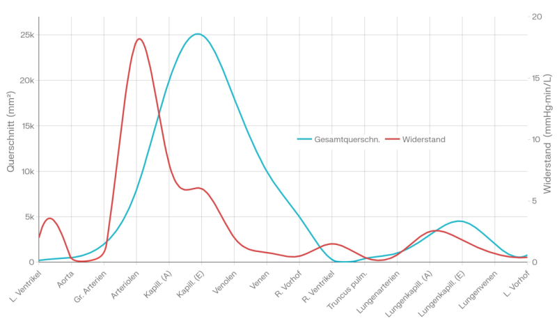
In all circulatory systems, the vessels leading away from the heart are referred to as arteries, whilst all others (those leading towards the heart and those not connected to the heart) are referred to as veins. The cross-sectional area of the arterial vessels decreases from the heart towards the periphery, they eventually give way to smaller arterioles and finally to myriads of capillaries arranged in parallel, which in turn merge into venules and later into veins, the cross-sectional area of which increases towards the heart. The capillaries are just large enough to allow individual erythrocytes to pass through, which drastically improves their ability to release oxygen and take up CO2 compared with larger cross-sections.
Coronary
The coronary arteries supply the heart muscle with blood: the A. coronaria dexter supplies the right ventricle and the right atrium, as well as the posterior wall of the heart, whilst the A. coronaria sinister supplies the left ventricle and the left atrium, as well as a large part of the interventricular septum. The A. coronaria dexter runs almost horizontally towards the back; the A. coronaria sinister is very short and also runs almost horizontally before dividing into two branches: the Ramus circumflexus goes round to the back, and the RIVA (ramus interventricularis anterior) runs downwards and divides the heart into its two halves (right and left). During diastole, the heart itself is supplied with blood (the systolic contraction constricts the coronary arteries, so this is not possible during systole). The apex of the heart lies at the 5th intercostal space (ICR) on the medioclavicular line. When palpating, note that the clavicle lies above the 1st ICR, so that the first palpable point is the 2nd.
Arteriosclerosis of the coronary arteries is referred to as coronary sclerosis and causes coronary heart disease (CHD), with possible consequences including angina pectoris and myocardial infarction.
TPR (total peripheral resistance)
The resistance that the vascular system offers to cardiac output depends most of all on the TPR (total peripheral resistance) of the capillaries and arterioles. The TPR depends on neurological and hormonal factors, the pH value and the NO concentration. The exact composition of TPR is:
– large and medium arteries: 19%
– smaller arteries and arterioles: 47% (regulatable resistance vessels)
– capillaries: 27%
– venules: 4%
– medium and large veins: 3%
The total resistance encountered by the heart is also referred to as afterload.
Cardiac output (CO)
In physiology, the minute is typically used as the unit of time, which is why the term ‘cardiac output’ (CO) is used. This is the volume that the heart ejects in one minute. It must be assumed that, physiologically (in healthy individuals without shunts (see, for example, doccheck.com) or septal defects (see also, for example, doccheck.com)), the output of the right heart is, on average over the short term, exactly the same as that of the left heart, as any deviation from this would imply an accumulation of blood in one of the circulatory systems (pulmonary circulation or systemic circulation) and, beyond a certain level, would no longer be compatible with life. Even a deviation of 1 ml per heartbeat would, at a heart rate of 60 bpm and a stroke volume of 50 ml, correspond to an accumulation of 3 l of blood in one of the circulatory systems. The pulmonary circulation would be completely unable to accommodate such a volume of blood, and in the systemic circulation this would correspond to an extreme pooling of blood in the periphery, as is known to occur, for example, in anaphylactic shock. It would certainly be impossible to survive beyond the second hour at the latest. A distinction is made between the systemic cardiac output—the volume of blood ejected from the left ventricle into the systemic circulation—and the pulmonary cardiac output—the volume of blood ejected from the right ventricle towards the lungs. In a right-to-left shunt, the cardiac output is greater than the pulmonary cardiac output; in a left-to-right shunt, it is smaller.
At rest, the average cardiac output is 4.5–5 litres, which can increase by a factor of approximately 4 during exercise.
If the cardiac output is multiplied by the arteriovenous oxygen difference, this yields the VO2max per minute; as a rule, healthy lungs are not the limiting factor, but rather internal respiration and metabolism are the key determinants of how much of the inhaled oxygen is utilised.
Maximum oxygen uptake VO2max
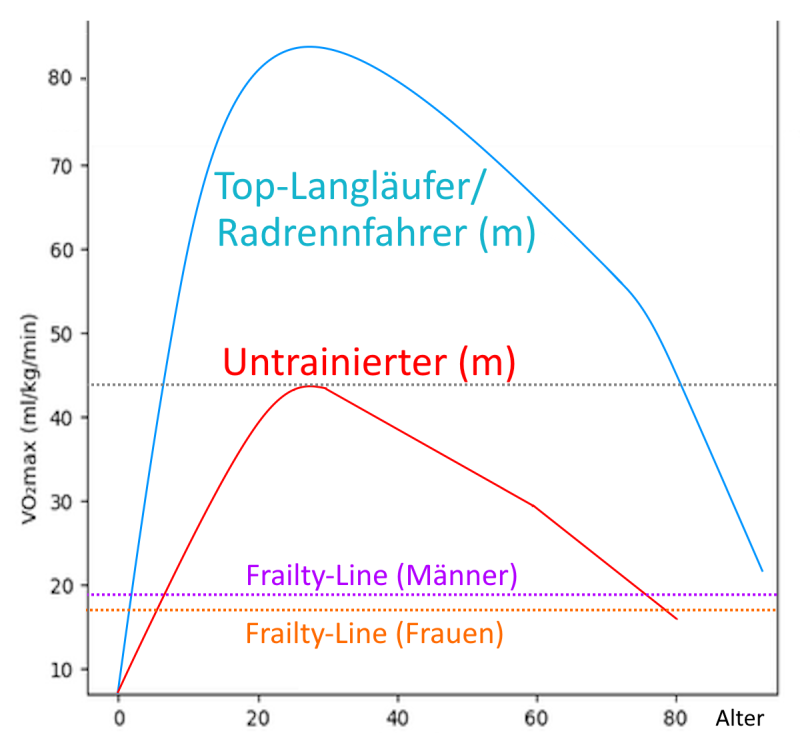
VO₂max refers to a person’s maximum oxygen uptake. In exercise physiology, it is usually not the absolute VO₂max that is used, but rather the VO₂max relative to body weight. This measure depends primarily on the heart’s stroke volume (cardiac output) and only secondarily – except in pathological cases – on the lungs or internal respiration. VO2max indicates an upper limit for a person’s endurance capacity; the extent to which this limit can be reached depends on many other factors, many of which can be improved through endurance training. Among other things, this training aims to bring the anaerobic threshold closer to the performance level corresponding to VO2max. From the age of 40, VO2max decreases by around 10 per cent with every decade of life, meaning that in old age it can fall below the frailty line of 18 (men) or 16 (women). Untrained people typically have values of 35 ml/min/kg (women) or 40 ml/min/kg for (men). Competitive athletes, through training and weight optimisation, can achieve values of up to 80 ml/min/kg. VO₂max is measured quite accurately using specialised equipment in a non-invasive exercise test, although there are several ways to obtain good estimates:
- Fick’s principle: VO₂max = HMV * (arterial oxygen content CaO₂ – venous oxygen content CvO₂). This measurement is invasive and the most accurate.
- Uth–Sørensen–Overgaard–Pedersen estimate: VO2max = 15.3 * HXmax / resting heart rate (RHR). This estimate requires an accurate measurement of the resting heart rate (RHR). There are many factors that can lead to false positives in this measurement.
- Cooper-Test: VO2max = (v * 12m – 505m) / 44,7m = (s-505) / 44,7
- Rockport Walk Test (1-mile walk test): VO₂max = 132.853 − (0.1695 × weight {kg}) − (0.3877 × age) + (6.315 × gender [m:1, w:0]) −(3.2649 × time {min}) − (0.1565 × HR {bpm})
The following tables can be calculated using the formula for the Cooper test,
The following tables show, in deciles, the maximum oxygen uptake (VO₂max) as determined by the Cooper Institute, as it can be achieved by both sexes of different ages on a treadmill or ergometer. The decline with age is due solely to the decrease in maximum heart rate (HRmax).
Running/Treadmill – Men:
| Alter | 10 % | 20 % | 30 % | 40 % | 50 % | 60 % | 70 % | 80 % | 90 % |
|---|---|---|---|---|---|---|---|---|---|
| 20…29 | 34,6 | 37,8 | 41,0 | 42,6 | 44,2 | 47,4 | 49,0 | 52,1 | 55,1 |
| 30…39 | 33,0 | 36,2 | 39,4 | 41,0 | 42,6 | 44,2 | 47,4 | 50,6 | 52,1 |
| 40…49 | 31,4 | 34,6 | 36,2 | 39,4 | 41,0 | 44,2 | 45,8 | 49,0 | 50,6 |
| 50…59 | 29,9 | 31,4 | 34,6 | 36,2 | 37,8 | 39,4 | 41,0 | 44,2 | 49,0 |
| over 60 | 26,7 | 28,3 | 31,4 | 33,0 | 34,6 | 36,2 | 37,8 | 41,0 | 44,2 |
Running/Treadmill – Women:
| Alter | 10 % | 20 % | 30 % | 40 % | 50 % | 60 % | 70 % | 80 % | 90 % |
|---|---|---|---|---|---|---|---|---|---|
| 20…29 | 29,4 | 31,6 | 33,8 | 35,5 | 37,4 | 39,5 | 41,1 | 44,0 | 47,0 |
| 30…39 | 27,4 | 29,9 | 32,3 | 31,8 | 35,2 | 36,7 | 38,8 | 41,0 | 44,7 |
| 40…49 | 25,6 | 28,0 | 29,7 | 31,6 | 33,3 | 35,1 | 36,7 | 38,9 | 42,4 |
| 50…59 | 23,7 | 25,5 | 27,3 | 28,7 | 30,2 | 31,4 | 32,9 | 35,2 | 38,1 |
| over 60 | 21,7 | 23,7 | 24,9 | 26,6 | 27,5 | 29,1 | 30,2 | 32,3 | 34,6 |
Cycling – Men
| Alter | 10 % | 20 % | 30 % | 40 % | 50 % | 60 % | 70 % | 80 % | 90 % |
|---|---|---|---|---|---|---|---|---|---|
| 20…29 | 32,9 | 35,91 | 38,95 | 40,5 | 41,9 | 45,0 | 46,55 | 49,5 | 52,3 |
| 30…39 | 31,3 | 34,4 | 37,4 | 38,9 | 40,5 | 41,9 | 46,0 | 48,1 | 49,5 |
| 40…49 | 29,8 | 32,9 | 34,4 | 37,4 | 40,5 | 41,9 | 43,5 | 46,6 | 48,1 |
| 50…59 | 28,4 | 29,8 | 32,9 | 34,4 | 35,9 | 37,4 | 41,9 | 41,9 | 46,6 |
| over 60 | 25,4 | 26,9 | 29,8 | 31,3 | 32,9 | 34,4 | 35,9 | 41,9 | 41,9 |
Cycling – Women
| Alter | 10 % | 20 % | 30 % | 40 % | 50 % | 60 % | 70 % | 80 % | 90 % |
|---|---|---|---|---|---|---|---|---|---|
| 20…29 | 26,9 | 31,3 | 31,3 | 34,4 | 35,9 | 37,4 | 38,9 | 42,0 | 46,6 |
| 30…39 | 25,4 | 28,4 | 31,3 | 31,3 | 32,9 | 34,4 | 37,4 | 38,9 | 43,5 |
| 40…49 | 23,8 | 26,9 | 28,4 | 31,3 | 31,3 | 32,9 | 34,4 | 37,4 | 40,5 |
| 50…59 | 20,8 | 23,8 | 25,4 | 26,9 | 28,4 | 29,8 | 31,3 | 32,9 | 35,9 |
| over 60 | 19,3 | 20,8 | 22,3 | 23,8 | 25,4 | 26,9 | 29,8 | 31,3 | 32,9 |
VO₂max by age
metabolic equivalent
The metabolic equivalent (MET) describes the ratio of the energy expenditure of an activity to that at rest (1 MET = 3.5 ml O₂/kg/min or 1 kcal/kg/h), i.e. the ratio of energy expenditure during exercise to energy expenditure at rest. MET is therefore a relative measure of intensity and not an absolute value. The value per activity and intensity is standardised. To convert this into calories, the individual’s weight must be taken into account: calories = MET × weight (kg) × time (h). METs are average values and may vary slightly depending on fitness level, technique or terrain. On inclines (e.g. on a treadmill or over rough terrain), the MET value increases due to greater muscular effort, particularly in the legs and trunk. Formulas such as the ACSM equation take this into account: VO₂ = 0.2 × speed + 0.9 × speed × gradient + 3.5 (ml/kg/min), which results in higher METs.
5% incline: +20–30%,
10% incline: +40–50%.
Tree roots or sand (e.g. sand: +40–60%).
Uneven terrain, such as trail running, increases METs by 20–60% due to stabilisation. Downhill runs may be lower, but the performance on uphill sections dominates the average. Here are some examples (based, amongst others, on Ainsworth et al. 2011):
- Sitting quietly: 1
- Exercise bike, 30–50 watts, very light to light exertion: 3.5
- Yoga, Pilates: 2,5-3
- Walking at 4 km/h: 3
- Running, flat, 6 km/h (10 min/km): 6
- Slow swimming: 4–6
- Walking at 6 km/h: 5
- Strenuous weight training: 6
- Badminton: 7
- Basketball, football, tennis, a simple circuit training session with no long breaks: 8
- Running, flat, 8 km/h (7:30 min/km): 8.3
- Exercise bike, 100–160 watts, 8.8
- Backstroke: 9.5
- Running, flat, 10 km/h (6 min/km): 9.8
- Freestyle: 9.8
- moderate breaststroke: 10.3
- Running, flat, 11.4 km/h (5:15 min/km): 11
- Exercise bikes, 160–200 watts: 11
- Handball: 12
- Running, flat, 13.3 km/h (4:30 min/km): 12.8
- Butterfly: 13.8
- Cycling, exercise bike, 200–270 : 14
- Running, flat, 15 km/h (4:00 min/km): 14.5
- Running, flat, 16.5 km/h (3:40 min/km): 16
- Cycling, exercise bike at 32 km/h: 16
Blood vessels: veins and arteries
The veins are also known as blood vessels, whilst the arteries are referred to as arteries, as the blood pulse or pulse caused by the contraction of the heart is clearly perceptible within them. The term ‘blood vein’ reflects the fact that, in living humans, the veins contain a significantly greater volume of blood than the arteries, which in turn have a significantly higher pressure than the veins. The qualification ‘in living humans’ is necessary, as the heart maintains an arteriovenous pressure difference throughout life and the venous blood volume is significantly greater. Post-mortem, blood pressures in the arterial and venous systems equalise at around 6–7 mmHg.
Arteries
Arteries are blood vessels that lead away from the heart. In the systemic circulation, the blood in the arteries is rich in oxygen.
Venen
Veins are blood vessels that do NOT lead away from the heart. These are blood vessels that are not directly connected to the systemic circulation or pulmonary circulation, such as the portal veins, or those that lead towards the heart. In the latter case, they are oxygen-poor in the systemic circulation and oxygen-rich in the pulmonary circulation.
Capillarisation / Capillarisation
Capillarisation refers to the network of tiny capillaries that supply the muscles. The degree of capillarisation is not a fixed, genetically determined factor, but can be increased, for example, through regular endurance exercise or repeated, more intense isometric contractions. Capillarisation is an important aspect of vascularisation. Depending on the author, the term ‘capillarisation’ may also refer to the process of new capillary formation.
Vascularisation / Vascularisation
Vascularisation refers to the network of small blood vessels supplying the muscles. The degree of vascularisation is not a fixed, genetically determined factor, but can be increased, for example, through regular endurance exercise or repeated, more intense isometric contractions; these stimulate angiogenesis in order to better supply the muscle with oxygen in real time. In the context of capillaries, the term capillarisation is also used. The term vascularisation is applied not only to muscles but also to other organs. Depending on the author, vascularisation may also refer to the process of forming new small blood vessels. Hypervascularisation, on the other hand, usually refers to a pathological proliferation of small blood vessels, for example in the context of inflammation, injuries or degenerative processes; this is also referred to as neovascularisation.
Hypervascularisation
Hypervascularisation usually refers to a pathological proliferation of small blood vessels, for example in the context of inflammation, injury or degenerative processes; in other words, it is an increase in vascularisation due to pathological causes, also known as neovascularisation. Hypervascularisation reflects an increased metabolic demand in the tissue; causes may include wound healing, chronic inflammation or, in some cases, malignant tumours.
Blood
Blood, as a cell-containing aqueous solution, is one of the body’s most important fluids. It performs a wide range of functions, some of which are supplemented by cells and substances dissolved in water; this is why this system is also referred to as a dual system. The blood circulates in a largely complete system, driven primarily by the heart. The heart supplies a pulmonary circulation (small circulation) to oxygenate the blood and remove carbon dioxide, and a systemic circulation (essentially the rest of the body, including the supply to the heart itself), which supplies all the body’s tissues via arteries and arterioles. The return flow from the tissues via the venous system is incomplete, as a small portion is transported back to the heart via the lymph. In the process, the vascular system branches out further and further from the heart towards the periphery, like a tree, with the cross-sectional area of the vessels decreasing progressively until it finally drops to approximately the diameter of a red blood cell in the capillaries between the arterial and venous regions of the circulatory system. Similarly, the veins expand progressively from the periphery towards the heart. A human has approximately 70 to 80 ml of blood per kg of body weight, i.e. around 5 to 6 litres. At around 44 per cent, the cells make up the largest proportion (known as the haematocrit) of the blood.
- Erythrocytes (red blood cells): for transporting oxygen to the tissues and removing carbon dioxide from the cells
- Leucocytes (white blood cells): cells of the immune system (immune function)
- Thrombocytes (blood platelets): to stop bleeding from injuries to blood vessels on the body’s surface or inside the body
The erythrocytes, which appear red, constitute by far the largest population, numbering between around 4.5 and 5.5 million cells per µl of blood, and outnumber the next largest population – the thrombocytes – by a factor of more than ten, with the latter numbering between 150,000 and 400,000 per µl of blood, which explains the red colour of the blood. The white blood cells, which are responsible for the blood’s immune function, number between 4,000 and 10,000 and are thus once again more than ten times fewer than the platelets. Although all leukocytes originate from pluripotent stem cells, they differentiate and are subdivided into granulocytes, lymphocytes and monocytes, some of which are further subdivided; for example, granulocytes are divided into eosinophils, basophils and neutrophils. In addition to cells, blood contains various proteins, the largest fraction of which is albumin, followed by globulins, which perform a wide range of functions, including transport, clotting and immune functions. The more than a dozen clotting factors and antibodies also belong to the group of proteins. Other components of blood include glucose, lipids, vitamins, minerals, trace elements and, of course, water as a solvent. Substances excreted in the urine are defined as those dissolved in the blood which the blood transports from the site of their origin to the kidneys so that they can be excreted. The humoral component of the blood – that is, blood without its cells – is referred to as plasma. Due to its content of clotting factors, it is still capable of clotting. If the clotting factors are removed, the resulting fluid is referred to as serum. The body maintains the pH of the blood, if necessary with considerable effort, within a narrow range centred on 7.4; depending on the literature, the limit values are given as 7.35 or 7.45. Deviations in blood pH could have serious consequences for the body’s chemistry and electrochemistry, as well as its metabolic processes.
The functions of blood include:
- Transport: the transport of nutrients, the removal of substances that are no longer required, and the transport of substances produced by one tissue for use by other tissues
- Immune function: carried out by both leukocytes and antibodies
- Haemostasis and coagulation: a dual process involving platelets and coagulation factors
- Heat transfer and regulation
- Respiration: internal (cellular respiration) and external (pulmonary respiration) respiration
Blood pressure
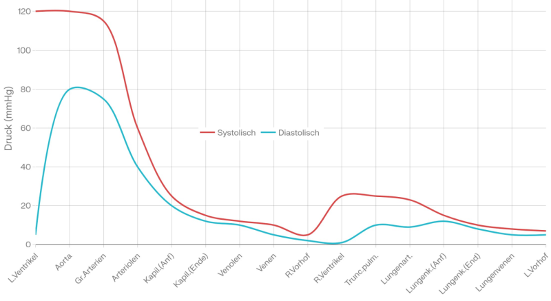
In contrast to the arterial system, there is no pulsation in the venous system and the blood pressure is, on average, significantly lower; above all, however, it is heavily dependent on gravity, being highest at anatomical zero – that is, in the feet and lower legs. At rest, pressure and flow velocity in the arterial system, caused by cardiac contraction, are approximately 120/80 mmHg (millimetres of mercury, measured at heart level without the influence of hydrostatic blood pressure). These values correspond to 160 and 106 mbar or hPa pressure respectively, in addition to ambient pressure. The systolic pressure measured at the origin of the aorta is, due to vascular resistance and the ‘windbag’ function, largely independent of age and is approximately 10–15 mmHg higher than peripheral arterial blood pressure.
The arterial or venous blood pressure measured at heart level must, of course, be increased by the position-dependent hydrostatic blood pressure, approx. 100 mbar/m. In an upright position, venous pressure normally only becomes positive below the heart. In a headstand, the zero point of venous pressure is in the femoral veins. The hydrostatic indifference point – that is, the unambiguous fixed point regardless of any changes in the body’s position – lies approximately 5–10 cm below the diaphragm.
Due to the pressure, the arteries are equipped with a thick layer of contractile muscle, which is particularly pronounced in the region furthest from the heart, to regulate pressure: this allows the vessels to narrow (vasoconstriction) or widen (vasodilation). Towards the periphery, blood pressure falls as the vessels become smaller, resulting in higher vascular resistance: aorta and large arteries: 100 mmHg, arterial branches 40 mmHg, capillaries 25 mmHg, venules 20 mmHg. In the pulmonary circulation, the pressures are significantly lower: pulmonary artery 14 mmHg, pulmonary veins 7 mmHg.
systolic and diastolic blood pressure in both circulatory systems
Total vascular cross-sectional area and vascular resistance in both circulatory systems
Pulse pressure / blood pressure amplitude
Pulse pressure, or blood pressure amplitude, refers to the difference between systole and diastole. Pulse pressure increases roughly linearly with stroke volume (SV) and decreases roughly inversely with vascular elasticity (arterial compliance) and the wind-bag function of the aorta. The duration of diastole also has an influence. Pulse pressure usually refers to resting pulse pressure, as it, like blood pressure itself, varies depending on the load. The following classification applies to elevated pulse pressures:
- 40 – 65: normal
- 66–75: slightly elevated
- 75–89: moderately elevated
- 90 and above: significantly increased
Every 10 mmHg increase in pulse pressure increases cardiovascular risk by around 23 per cent. In addition to reduced vascular compliance (elasticity) caused by
arteriosclerosis, the causes of elevated pulse pressure include:
- Aortic regurgitation
- Hyperthyroidism
- patent ductus arteriosus
- Peripheral shunts
- Aortic dissection
- Anaemia
- increased intracranial pressure
- Pregnancy
A reduced pulse pressure is less common, for example in cases of
- Left-sided heart failure
- Aortic valve stenosis
- cardiogenic shock
The greatest medical risk is the rupture of pre-existing atherosclerotic plaques caused by shear forces; this can affect not only newly formed plaques but also those that have been present for some time. They then rupture from the centre outwards.
Wall shear stress (arterial blood)
The simple blood flow model assumes, in the case of laminar arterial blood flow, a rotationally symmetric parabolic velocity distribution in accordance with Hagen–Poiseuille’s law, in which the maximum flow velocity occurs at the centre of the blood vessel and decreases parabolically towards zero as one moves towards the periphery. According to this model, the cells should float in the fast-flowing blood at the centre. In practice, however, this model proves to be untenable, as in vivo studies show. Instead, cell collisions with the vessel wall occur, which, via the mechanoreceptors stimulated, have positive physiological effects in several ways: A high WSS helps the endothelial cells of the vessel wall to maintain themselves, to align or remain aligned, and to release anticoagulants and vasodilators such as NOx, which also serves to regulate
blood pressure. Furthermore, high WSS counteracts endothelial cell proliferation and has anticoagulant and anti-inflammatory effects. High WSS is therefore atheroprotective. The endothelium reacts adversely, both at the cellular and molecular levels, to chronically low blood flow and to altered blood flow patterns characterised by low WSS; this may manifest as proliferation, an increased
propensity for thrombosis and, at bends and bifurcations, a tendency towards atheroma formation. In combination with other factors, this can lead to
atherosclerosis. WSS is roughly linearly dependent on flow velocity, which reinforces the significance of intense physical activity—where flow velocity is increased due to the muscles’ oxygen demand—as occurs primarily in endurance sports, and to a lesser extent in other activities characterised by high blood flow (higher heart rate). Generally, the peak WSS is higher in men than in women, but falls to a similar level between the second and sixth decades of life (peak: 4.3 Pa to 2.6 Pa and 3.3 Pa to 2.5 Pa respectively; mean: 1.5 Pa to 1.2 Pa and 1.3 Pa to 1.1 Pa respectively in the carotid arteries). In the case of the carotid arteries, the decrease in WSS is accompanied by an increase in cross-sectional area, which is interpreted as an attempt by the vascular system to counteract an age-related reduction in vessel compliance (increased vascular stiffness, fibrosis, increased thickness of the muscularis).
parasympathetic saturation
The influence of the vagus nerve on the heart is mediated by the neurotransmitter acetylcholine. The dose-response relationship of acetylcholine remains linear for a long time, until a concentration is reached at which a further increase in acetylcholine concentration no longer causes any change in the response. This effect is known as parasympathetic saturation. In this state, a further increase in vagal tone does not result in a rise in HRV but may even lead to a decrease. This effect occurs rarely and, as far as is known, only in elite endurance athletes. The following are assumed to be necessary conditions for its occurrence:
Ankle Brachial Index ABI
The ankle-brachial index (ABI) is the ratio, calculated using the ankle-brachial ratio, of the systolic blood pressure at the ankle to that at the wrist.
Physiologically, the two values should be very similar; deviations either upwards or downwards are pathological and often indicate
peripheral arterial disease (PAD).
| over 1.4 | Arteries that may not be compressible (Mönckeberg’s sclerosis) |
| 1,0 – 1,4 | normal |
| 0,91 – 0,99 | borderline |
| 0,8 – 0,9 | relevant pAVK |
| 0,5 – 0,79 | more severe vascular changes |
| less than 0.5 | Clinically confirmed ischaemia (CLTI), high risk of ulceration and necrosis |
Aorta
The aorta is the arterial blood vessel that originates from the left ventricle and whose branches supply the whole body, including the heart, with oxygenated blood from the lungs. The aorta is divided into four sections:
- ascending aorta / aorta ascendens
- Aortic arch / Arcus aortae
- descending aorta / aorta descendens
- Abdominal aorta
The blood supply to the heart itself originates from the ascending aorta.
The aortic arch has three branches:
- A. subclavia sinister: supplies the left arm
- A. carotis communis sinistra
- Truncus brachiocephalicus. The right subclavian artery and the common carotid artery arise from it, thus supplying part of the head and the right arm.
The descending aorta divides into the abdominal aorta and the thoracic aorta. The aorta is elastic and therefore acts as a windbag; that is to say, its elasticity allows it to accommodate a certain volume of blood, which is then immediately released again by the elastic forces. This elasticity moderates the systolic blood pressure peaks resulting from the contraction of the heart and prolongs the diastole, thereby stabilising the blood flow to some extent. Various examination methods are available for examining the aorta; these include invasive and non-invasive techniques, imaging, acoustic and palpation methods, whilst pulse wave analysis and the measurement of PWV also provide insights into the condition of the aorta. Since 2024, the aorta has been classified as a separate organ.
Capillaries
(Blood) capillaries are the smallest blood vessels and are situated between the
arterioles and the venules. They supply the surrounding tissues with oxygen and nutrients and remove
carbon dioxide and metabolic waste products.
Due to the size of the pores in the capillary walls, cells and, for the most part, proteins remain in the blood, whilst O2, fluid and small molecules are forced into the interstitium (the space between cells), where the pressure is lower.
Unlike arterioles and venules, they do not have three layers of wall, but only the intima with a basement membrane. Capillaries are permeable to low-molecular-weight substances (selectively permeable) and are very short, approximately 0.5 mm in length and, at 5–10 µm, even finer than arterioles and venules. An erythrocyte, with a thickness of 2.5 µm and a diameter of 7.5 µm, just fits through the capillaries, so that the diffusion path for the blood gases it transports is as short as possible, thereby ensuring that the exchange takes place as quickly as possible and that the total exchange capacity per unit of time is as high as possible. The capillary density in the tissues depends on their metabolic activity. The structure of the capillaries varies depending on the tissue they supply. Only a few very bradytrophic tissues, such as the cornea, other horny structures, the lens of the eye, hyaline cartilage and epithelia, lack capillaries.
As the blood passes through the capillaries, the blood pressure continues to fall. In the venous limb, fluid containing substances to be removed is then absorbed, aided by the lower blood pressure and the now higher colloid-osmotic pressure (the ability of proteins to bind water). In most cases, the volume of fluid absorbed by the venous limb is less than that released by the arterial limb; the remainder is then transported away via the lymph. If the blood contains too little protein – for example, due to malnutrition, MAS or protein loss via the kidneys – oedema results due to insufficient colloid-osmotic pressure, as too much fluid remains in the tissues. The same applies if the permeability of the membrane is pathologically increased and proteins pass into the tissues. Of course, oedema can also be caused by inadequate venous return, for example due to insufficient venous valves in chronic venous insufficiency.
In three organs (the liver, spleen and bone marrow), the capillaries are expanded into sinusoids with a discontinuous endothelium. The venules can regulate vascular tone via a thin layer of muscle, thereby forming a blood reservoir; the venules have very little muscle tissue. The second drainage of the capillary bed via the lymph transports hydrophobic substances (proteins and lipids from the digestive organs) and cells, flowing very slowly towards the right heart.
The term ‘capillary’ is usually applied to blood capillaries; however, the lymphatic system also begins with capillaries – the lymphatic capillaries – which are embedded between the cells of the tissues and originate there.
Arterioles
Arterioles are the smallest arterial vessels still visible to the naked eye and are therefore the equivalent of venules. Unlike arteries, they are capable of completely constricting via their middle wall layer, the media or muscularis. This makes them the most important factor in TPR and, consequently, in afterload and blood pressure. They are therefore also referred to as resistance vessels. The internal diameter of arterioles is around 100 µm, or 1/100 of a millimetre. Arterioles give way to capillaries.
Venules
Venules are the smallest venous vessels visible to the naked eye and are therefore the equivalent of arterioles. They arise from the capillaries. Many venules join together to form veins. Venules contain hardly any smooth muscle in the media and have no venous valves. They are classified according to their internal diameter into
- post-capillary venules: 10–30 µm
- Collector veins: 30–50 µm, containing individual, branched smooth muscle cells
- muscular venules: 50–100 µm, containing a layer of overlapping, branched smooth muscle cells
Lymph
Lymph is a watery fluid that serves to remove certain substances from the interstitium of tissues which must not be transported in the venous bloodstream because they are, or may be, infectious, or because they are hydrophobic, for example. These include, for example, certain blood proteins, fatty acids, and metabolic or inflammatory products. Substances with a high molar mass, which cannot be transported venously due to the limited size of the pores in the membranes, must also be transported via the lymphatic system. Dietary fats produced in the digestive tract during fat metabolism are transported in the lymph in the form of chylomicrons. Due to the substances it contains, the lymph is pale yellow/milky in colour. The lymphatic vessels contain lymph nodes, which, amongst other things, inactivate pathogens present in the lymph.
The flow rate of lymph is approximately 2–3 cm per hour. It therefore only removes a very small proportion of the fluid produced in the tissues (2–3 l per day compared with 7,000–8,000 l of blood). It returns to the venous bloodstream via the ductus thoracicus (thoracic duct) at the left venous angle (the confluence of the subclavian vein and the internal jugular vein to form the brachiocephalic vein), just before the right heart. At 7.41, its pH is barely higher than that of blood.
As a filtrate of the interstitial fluid, lymph also contains urea, creatinine, glucose, sodium, potassium, phosphate and calcium ions, enzymes such as amylases, catalase, dipeptidases and lipases, as well as fibrinogen and other coagulation factors, which enable the lymph to coagulate in the event of stasis (see Virchow’s triad). Lymph production can be accelerated by lymphagogues (lymph-stimulating agents) such as chicken protein, bile, peptone, salts, urea and sugar. Physiologically, the volume of lymph, together with the blood returning via the veins, is exactly equal to the arterial blood flow. If the flow of lymph is obstructed or restricted, lymphoedema occurs. Due to the much lower flow rate and the much lower volume of return flow per unit of time, lymphoedema develops much more slowly than venous oedema.
The lymph plays an important role in the immune system because it transports pathogens to the lymph nodes, where specialised T and B lymphocytes react to the pathogens, multiply and migrate from there into the blood. Despite its low flow rate and velocity, the lymph must not be overlooked: if lymphatic vessels are rerouted or lymph nodes are removed, this results in lymphoedema. Like veins, lymphatic vessels have valves (lymphatic valves).
Collaterals, anastomoses
Adjacent blood vessels supplying the same target area are referred to as collaterals; almost everywhere in the body, there are connections (anastomoses) between them, which can ensure continued blood supply in the event of a vessel becoming obstructed, for example due to a thrombus. Arteries without anastomoses are called terminal arteries. Occlusions of endarteries therefore lead to ischaemia or infarcts. If the anastomosis of an artery is too small to take over the entire blood supply, it is referred to as a functional endartery.
Venous valves
Whilst the arterial circulation is driven by the heart, the venous return flow works quite differently: the veins contain many valve-like venous valves, which are folds in the lining of the vein that prevent the blood from flowing back into the periphery. With their usually two (only rarely one or three) leaflets, they form a non-return valve for the blood against the intended direction of flow. In this respect, they resemble the bicuspid valve (a valve with two leaflets). The section of the vein situated between the valves is called the sinus valvulae and is more elastic than the area near the valve, which is why it is prone to bulging (varicosis: varicose veins).
Venous valves are found in the veins of the extremities to ensure unidirectional blood flow, both in the deep veins and in the superficial veins, as well as in the venae perforantes that connect them. Blood flow is maintained by the ‘muscle pump’, i.e. the repeated pressure exerted by muscles on the sinus valvulae. Venous valves have been known since 1547 (first described by Giovanni Battista Canano).
Particularly in the legs, the muscles use their alternating contractions to push the blood from one segment to the next, thereby overcoming gravity, so that the blood eventually reaches the right heart; this works particularly well during activities involving the legs. In the trunk, where there are no venous valves, pressure differences caused by breathing contribute to the transport. The venous valves are therefore essential for the return flow.
Because of the venous valves, infusion or injection needles in the extremities are always inserted in the direction of blood flow and not against it. Large vascular trunks such as the vena cava, the portal vein, the umbilical vein, cerebral and pulmonary veins, as well as parenchymal veins, do not have venous valves. In the abdomen, where the distance is significantly shorter than in the leg, return transport occurs mainly through pressure changes caused by abdominal breathing and the resulting fluctuations in intra-abdominal pressure. Venous valves are important not only because of the risk of varicose veins, but above all because of the risk of blood clotting due to the ‘stasis’ factor from Virchow’s triad, i.e. excessively slow blood flow. An analogue of this is found in the lymphatic vessels of the extremities in the form of the valvulae lymphaticae (lymphatic valves).
Venous and arterial blood differ in colour: arterial blood is a lighter shade of red. However, the bluish appearance of the veins is due more to the fact that long-wavelength red light penetrates further into the veins and is absorbed to a greater extent.
lymphatic vessel
The vessels through which the lymph flows from the interstitium of the tissues to the heart (venous angle) and through which substances, particles and pathogens that must not enter the venous bloodstream (and consequently the arterial bloodstream) are transported. The lymphatic vessels contain
lymph nodes; in the case of lymphatic vessels that drain a single organ, these are referred to as sentinel lymph nodes. Some regions of the body have their own lymph nodes. All the lymph flows via the thoracic duct (Ductus thoracicus), the largest lymphatic vessel, to the Virchow lymph node (also incorrectly referred to as the ‘Virchow’s gland’) and from there directly into the venous angle of the right heart and thus into the circulating bloodstream. Similar to the veins of the extremities, the lymphatic vessels have valves that open only towards the centre, which serve to facilitate return flow.
Lymph valves
Lymphatic valves are analogous to venous valves, which are physiologically necessary because both fluids are capable of coagulating. The lymphatic system – apart from the lymph nodes – consists of valve-free lymphatic capillaries, precollectors with isolated valves, and collectors, which possess both valves and their own musculature. The main collecting vessels (lymphatic trunks such as the tracheal trunk and thoracic duct) ultimately transport the lymph to the left venous angle.
Lymph nodes
Typically 5–10 mm in size, but during periods of activity they can reach 20 mm or more; these are clusters of lymphatic tissue (reticulum cells, lymphocytes, antigen-presenting cells). All mammals have lymph nodes, although their number and size can vary considerably. Humans usually have 300–700 lymph nodes. Following an initial non-specific phagocytosis of certain components of the primary lymph, the differentiation of lymphocytes is stimulated within these nodes, so that T cells, plasma cells and memory cells are ultimately released into the bloodstream via the secondary lymph and the right venous angle. All regions of the body have regional lymph nodes that drain a corresponding catchment area; see this map. Swollen lymph nodes indicate an immune response, and therefore usually signify an infection.
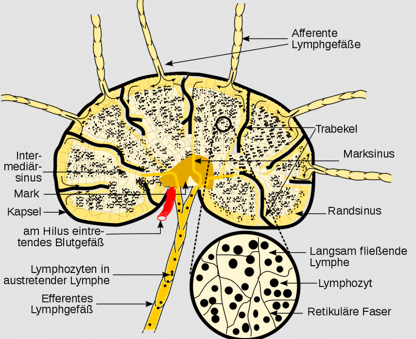
Swollen lymph nodes
Enlargement of lymph nodes. Particularly in cases of inflammation and malignant conditions, the lymphatic tissue in the lymph nodes becomes active, leading to an increase in volume which is subjectively felt as a more or less pronounced tension pain and tenderness on pressure, which is palpable in superficial lymph nodes. Painful, enlarged lymph nodes accompanied by local inflammation are usually caused by an infection. Less commonly, painless, progressive lymph node swelling, on the other hand, often indicates a malignant condition. A further distinction is made based on duration: 1–2 weeks – acute; 2–6 weeks – subacute; over 6 weeks – chronic; and based on location: local or regional (e.g. in the case of sentinel lymph nodes) or, if more than two non-contiguous lymph nodes are affected, generalised. In the case of lymphomas, the cause lies within the lymphatic tissue of the lymph node itself; otherwise, the cause of the swelling lies in the draining area.
Lymphatic system / Lymphatic system
The system consisting of the lymph nodes—which contain lymphatic tissue—and the lymphatic vessels, which provides an (alternative, parallel to the veins) route back from the interstitium to the heart.
Blood volumes in the circulatory systems
Only around 20 per cent of the blood flows through the arteries; post-mortem (following cardiac arrest), due to the drop in pressure and reduced elasticity, this figure falls to around 2 per cent; the heart therefore maintains the arteriovenous pressure difference throughout a person’s lifetime. Without cardiac activity, a uniform pressure of around 6–7 mmHg is established throughout the body when the patient is lying on their back. Changes in blood volume due to blood loss or transfusion are primarily absorbed by the venous low-pressure system and, to a lesser extent, have hardly any effect on the arterial high-pressure system.
Windkessel function
The arteries close to the heart have a Windkessel function, they act as a pulsation reservoir: they are quite elastic and thus stabilise blood flow by largely absorbing spikes in blood pressure. This not only helps to stabilise blood pressure and thus reduce harmful effects on the blood vessels, but also saves energy, as the otherwise intermittent higher flow velocity would otherwise require more energy due to vascular resistance. Arteriosclerosis can cause this function to be partially or completely lost, resulting in higher systolic blood pressure and the associated damage. Arteries further away from the heart are equipped with more muscle tissue so that they can regulate blood pressure by constricting or dilating. The aorta (main artery) has a diameter of approximately 3 cm.
In young adults, the pressure wave of blood reaches approximately 6 m/s; over the course of a lifetime, this doubles due to the diminishing ‘air spring’ function and waning elasticity of the arteries, whilst the volume flow decreases as a result of reduced energy efficiency.
Within the body, blood flow is directed more towards those organs whose activity is currently required; the cardiac output (CO) would not be sufficient to provide maximum blood flow to all organs simultaneously. ‘Shutting down’ organs keeps the TPR – and thus the blood pressure – sufficiently high and reduces the strain on the heart. This means, for example, that digesting food and engaging in high-intensity sport at the same time are mutually exclusive. There are several special features for regulating blood pressure and TPR, as well as for other purposes:
– occlusive arteries, which are normally closed but open in certain situations (e.g. in the penis),
– arteriovenous anastomoses: bypasses around organs,
– constrictive veins, e.g. for the delayed release of nutrients from the gut into the blood.
– Mechanisms for restricting blood flow in the event of injury
– Mechanisms for increasing blood flow in the event of inflammation
Control signals for blood flow are neural, hormonal and local chemical, with the latter being able to override the first two. Information from circulatory sensors is processed in the medulla oblongata: arterial blood pressure, heart rate, filling pressure of the venous system, pH, and the partial pressures of carbon dioxide and oxygen in the blood. The sympathetic nervous system and the parasympathetic nervous system regulate chronotropy (changes in heart rate) and inotropy (change in contractile force) as well as vascular tone.
In the aortic arch and at the bifurcation of the common carotid artery (carotid sinus), there are baroreceptors which relay the adjacent arterial pressure to the medulla oblongata, so that it can adjust the pressure if necessary.
Interstitium
The interstitium is the space between the cells in the body’s tissues. Interstitial fluid is formed from blood that is forced through the pores in the walls of blood vessels (by blood pressure); it is similar to arterial blood and is therefore very similar in composition; the only components missing from the interstitial fluid are blood cells and large molecules such as the proteins albumin and globulin, as the pores in the membranes are too small to allow them to pass through. The serum that is forced into the tissue and altered there by the cells’ metabolic processes is reabsorbed into the bloodstream via the venous branch due to the increased osmotic pressure of the venous blood (colloid-osmotic pressure is elevated: high protein content, low fluid content).
Arteries
Arteries are blood vessels that lead away from the heart. In the systemic circulation, the blood in the arteries is rich in oxygen. Arteries consist of three layers (from the inside out):
1. Intima lies on the inside; it is bare and smooth to minimise resistance
2. Media or Muscularis contains circular and obliquely arranged muscle tissue, which allows the vessel’s cross-sectional area to change: the sympathetic nervous system causes vasoconstriction, whilst the parasympathetic nervous system causes vasodilation; it also contains minute vessels to supply the muscles
3. Adventitia or serosa, consisting of elastic and fibrous connective tissue; together with the vasa vasorum (the vessels of the vessels) and nerves (nervi vasorum), it nourishes and regulates the entire artery.
Hormones help to determine the diameter of the arteries:
adrenaline and noradrenaline cause vasoconstriction, except in the coronary arteries, where they cause vasodilation
Angiotensin II causes vasoconstriction (the most potent vasoconstrictor of all)
Serotonin causes vasodilation in the abdominal region and vasoconstriction in the brain (an excess of it is thought to lead to migraines)
The blood vessels contain alpha receptors, whilst the coronary arteries contain beta receptors (which is why, in cases of hormone-induced hypertension, so-called beta-blockers are prescribed, to prevent the blood pressure-raising – and therefore vasoconstrictive – effect of adrenaline from taking effect there).
beginning behind the aortic valve:
– coronary arteries:
– – right coronary artery,
– – left coronary artery, which divides into the
– – – circumflex branch
– – – RIVA anterior interventricular branch
– ascending aorta → aortic arch, from which the following branches off:
– – the brachiocephalic trunk supplies the right arm, the right side of the brain and the right
half of the head; it divides into:
– – – the right subclavian artery -> the axillary artery -> the brachial artery ->
– – – – right ulnar artery
– – – – right radial artery; the two merge in the palmar arch (of which there are two: the deep palmar arch, which lies deep within the hand, and the superficial palmar arch, which lies more palmar). Branching off from these
– – – – – Aa. digitalis the digital arteries
– – – A. carotis communis dexter passes into
– – – – the right internal carotid artery supplies the right hemisphere of the brain
– – – – the right external carotid artery supplies the right side of the face and skull
– – – the left common carotid artery gives rise to
– – – – The left internal carotid artery supplies the left hemisphere of the brain
– – – – The left external carotid artery supplies the left side of the face and skull
– – The left common carotid artery is analogous to the right; it lies partly within the mediastinum
– – The left subclavian artery
– .. merges into the descending aorta, from which branch the
– – Intercostal arteries (12 on each side)
– .. transitions into the abdominal aorta within the abdominal cavity; the following branches arise from it:
– – Splenic artery to the spleen (caudally)
– – the gastric artery to the stomach (to the left)
– – the hepatic artery to the liver (to the right)
– .. divides at the aortic bifurcation into
– – the right common iliac artery
– – the left common iliac artery; both of these then give rise to
– – – the internal iliac artery, which supplies the small pelvis
– – – the external iliac artery supplies the leg and
– – – .. becomes the femoral artery
– – – .. becomes the popliteal artery at the knee and, after the knee, divides into
– – – – the peroneal artery
– – – – the anterior tibial artery
– – – – posterior tibial artery; this and the anterior tibial artery converge in the forefoot to form the
– – – – arcuate artery, from which the metatarsal arteries then branch off
Venen
Veins are blood vessels that do not carry blood away from the heart. In addition to the vessels that carry blood towards the heart, there are also two vessels whose blood does not flow directly to the heart:
– the portal vein (V. portae) of the abdominal organs, which carries blood from most of the abdominal organs to the liver, and
– the portal vein (V. portae) of the brain between the hypothalamus and the anterior pituitary lobe.
The veins connected to the heart are low in oxygen in the systemic circulation
and rich in oxygen in the pulmonary circulation. Two large veins drain into the heart:
the superior vena cava, into which drain
– the jugular vein from the head
– the subclavian vein, which receives blood from the
– – the axillary vein, which merges into
– – the brachial vein, situated ulnarly, well-suited for blood sampling and infusion, into which flow
– – – the radial vein, deep-seated
– – – the ulnar vein, deep-seated
– – the brachial vein receives:
– – – v. basilica, superficial
– – v. axillaris receives:
– – v. cephalica, lies superficially, radially, well suited for blood sampling and infusion
v. cava inf., into which it drains
– 2 renal veins (kidney)
– 3 hepatic veins (liver)
– internal iliac vein
– external iliac vein, arises from
– femoral vein, arises from
– popliteal vein, arising from
– small saphenous vein, situated laterally, into which two lower leg veins drain
– the great saphenous vein drains into the femoral vein, and two further lower leg veins drain into it
The spleen, pancreas, stomach, large intestine and small intestine have no direct venous connection to the inferior vena cava, but are connected venously via the portal vein to the liver, which in turn is connected to the inferior vena cava via three veins.
heart
The heart, cardia (Greek) cor (Latin) is a muscular hollow organ which, through rhythmic contractions, pumps blood through the body, thereby ensuring that all organs are supplied with blood. It is situated in the mediastinum, protrudes slightly to the right beyond the spine (which is why pain may also be felt on the right-hand side) and is about the size of a fist, weighing approximately 200–350 g: 280–340 grams in men, 230–280 g in women. Over the course of a person’s life, the heart’s mass increases slowly; however, the blood vessels supplying it do not grow at the same rate, which is why a weight of 500 g is generally already considered pathological: critical heart weight (hypertrophy), as the blood vessels can no longer adequately supply a myocardium that has grown to such a size (for further details, see heart weight). Exercise cannot cause hypertrophy exceeding 500–600 g. Each year, younger people produce approximately 1% new heart cells, whilst older people produce only 0.5%.
The heart lies at a slight angle (the right side tilted downwards, the lower side forwards) on the diaphragm, to which it is fused at the bottom; in slender people, it is positioned more upright. In front of the heart lie the thymus and the sternum. The heart is situated in the region of the 2nd to 5th ribs. At the top, the heart extends approximately 2 cm beyond the edge of the sternum.
The heart functions like a two-way pump with valves that ensure the blood flows only in a specific direction. It is rotated to the left, so that the larger part of the left side of the heart, which pumps into the systemic circulation, lies at the back. This is why posterior wall heart attacks (left-sided heart attacks) are also considered more critical.
Systole
Systole normally refers to the contraction phase of the ventricular myocardium, during which the heart ejects blood into the large vessels. In the human heart, systole lasts between approximately 400 ms at rest and 270 ms under maximum exertion. The ejection phase hardly shortens at all as the heart rate increases; this is achieved primarily by a shortening of the diastole. Systole consists of a shorter contraction phase of the ventricular myocardium and a longer ejection phase whilst the myocardium remains contracted. At the start of the ejection phase, the pressure in the ventricles – which has increased due to the tension in the ventricular myocardium – opens the semilunar valves leading to the pulmonary arteries and the aorta (positive pressure difference between the ventricles and the vessels); at the end of the ejection phase, these close because the pressure difference between the ventricles and the large vessels has once again become negative. The semilunar valves remain closed throughout the entire systole to prevent backflow of blood from the ventricles towards the atria. On auscultation, the first heart sound marks the start of the ejection phase, whilst the second heart sound marks the closure of the semilunar valves and thus the end of systole. On the ECG, systole corresponds roughly to the QT interval. By analogy with the ventricular systole described above, one can also speak of an atrial systole, which corresponds to ventricular diastole.
Diastole
Diastole is the filling phase of the heart, that is, the phase during which the ventricular myocardium is passive and the atrial myocardium, with the semilunar valves open, propels the blood accumulated during the preceding systole into the ventricles. During this phase, the semilunar valves are closed to prevent backflow from the large vessels into the ventricles. By analogy with the ventricular diastole described above, one can also speak of an atrial diastole, which corresponds to ventricular systole.
systolic
relating to the ejection phase of the heart. The term is used to describe the heart’s actions or sounds, but also the arterial blood pressure present during this phase. Thus, systolic blood pressure refers to the maximum value recorded during systole and, consequently, throughout the entire cardiac cycle.
diastolic
Relating to the filling phase of the heart. The term is used to describe the heart’s actions or sounds, but also the arterial blood pressure present during this phase. Thus, diastolic blood pressure refers to the value that is at its lowest during diastole and, consequently, throughout the entire cardiac cycle.
systolic blood pressure
the value of blood pressure reached at its peak during systole – and thus also over the entire cardiac cycle – measured in the ventricle or in the aorta immediately behind the semilunar valve. As this value cannot usually be measured non-invasively, the maximum blood pressure in an arm held at heart level is used as an approximation instead.
diastolic blood pressure
the value of the blood pressure which is at its lowest during diastole and thus also throughout the entire cardiac cycle, measured in the aorta directly behind the aortic valve. As this value cannot usually be measured non-invasively, the minimum blood pressure in an arm held at heart level is used as an approximation instead.
Stroke volume
Stroke volume refers to the volume of blood ejected by the heart during a single ventricular contraction. Normally, this refers to the volume ejected into the aorta, i.e. into the systemic circulation; however, the stroke volume of the right ventricle must be identical, as otherwise significant amounts of blood would accumulate in one of the two circulatory systems, which would be incompatible with life even in the short term. At rest, stroke volume is usually in the range of 60–80 ml; with a strong heartbeat, it can reach up to 90 ml, depending on fitness level. Regular endurance training significantly increases stroke volume; well-trained individuals with an RHR of around 40 or in the low 40s bpm have a stroke volume of 80–105 ml at rest and up to about twice that under exertion, approximately 140–180 ml, which corresponds to a cardiac output of just over 7 l at rest and 20–30 l/min under maximum exertion. In elite athletes, stroke volume is roughly twice that of untrained individuals, at around 180–210 ml, and in some cases even higher, so that world-class athletes achieve cardiac output of 30–40 l/min, and in isolated cases even up to 50 l/min. Endurance training not only increases the mass of the heart muscle and its contractile force, but also the ventricular volume. As the heart rate is regulated mainly in accordance with the body’s current oxygen demand, these adaptations lead to a resting heart rate RHR that is, in some cases, significantly lower. Regular endurance training sessions are necessary to increase stroke volume; longer intervals just below the individual’s anaerobic threshold are also helpful. Strength training is far less effective in this regard than endurance training. Stroke volume is usually determined by echocardiography, MRI or using specialised spiroergometry protocols.
Cardiac Function / Cardiovascular Function
An effect that occurs particularly at higher temperatures, whereby the exercise heart rate begins to rise slowly after a certain time, whilst the level of exertion remains constant. It is generally assumed that this is due to a reduced stroke volume resulting from sweat loss. The poorer the subject’s hydration at the start of the exercise, the sooner this effect occurs. The greater the amount of muscle mass used, the more pronounced the effect. The body is cooled via the skin, which must therefore be supplied with a greater blood flow. This results in lower stroke volumes in both circulatory systems. In order to compensate for the decreasing stroke volume whilst maintaining the same performance demand and keeping the cardiac output constant, the heart rate must therefore increase. As a result, the perceived exertion, as measured by the Rate of Perceived Effort on the Borg scale, increases. Another factor that contributes to cardiac drift is muscular fatigue, including from the previous day’s training, as this leads to the recruitment of more type 2 fibres, which are less energy-efficient than type 1 fibres. In addition to the above explanation, there is also debate as to whether an increase in sympathetic tone, via an associated shortening of the diastole, causes a decrease in stroke volume.
Heart rate
Heart rate is the frequency at which the heart beats. A minute is usually used as the time reference, so the heart rate is then expressed in bpm (beats per minute). 60–80 beats per minute (bpm) at rest are considered normal; less than 60/min is termed bradycardia, more than 100 is termed tachycardia, more than 250 is termed flutter, and more than 350 is termed fibrillation. In the case of ventricular fibrillation (usually affecting both ventricles), no haemodynamically meaningful action is possible; in the case of atrial flutter or fibrillation (usually affecting both atria), the AV node can filter out meaningful impulses. In endurance athletes, a physiological bradycardia develops over the years.
Physiologically, every heartbeat generates a pulse wave in the periphery, so that the pulse measured peripherally corresponds to the heart rate. If the measured pulse is lower, a pulse deficit is present.
By way of comparison: the heart rate (HR) of the blue whale is 6/min, whilst that of the Etruscan shrew is 1000/min; generally speaking, larger animals have a lower heart rate than smaller ones, with one exception: the giraffe, at 170/min. The giraffe’s blood pressure is also very high at 300/250, as there is a 2-metre difference in height between its heart and brain; the blood pressures of horses (114/90) and cats (125/75) are very similar to those of humans, whilst other animals often have significantly different pressures: rooster (191/154), hen (162/133), spiny dogfish (32/16), frog (27), sparrow (180/140). The hearts of mammals all have the same structure and account for approximately 0.6 per cent of body mass; the stroke volume increases linearly with body mass. A blue whale weighing 100 t has a heart weighing 600 kg with a stroke volume of 350 l, which is roughly equivalent to the body volume of four adults weighing 85 kg each.
Bradycardia
A reduced heart rate at rest. Bradycardia is present in a small number of medical conditions; it is also a physiological occurrence in endurance athletes. Strictly speaking, a heart rate below 60 beats per minute is bradycardia; however, it is not uncommon for the resting heart rate of well-trained endurance athletes to be well below 50 beats per minute, and in isolated cases even below 40. The Spanish professional cyclist Miguel Indurain had the lowest resting heart rate ever recorded in a healthy person, at 28 bpm. See also tachycardia.
Tachycardia
Tachycardia refers to a heart rate at rest that is significantly higher than the age-related physiological rate, i.e. above 100 beats per minute in adults. In addition to disorders of the heart itself and drug use, psychogenic factors may also play a role in causing it. See also bradycardia.
Puls
The term ‘pulse’ refers to the pulse wave in the peripheral bloodstream that can be detected, for example, by palpation or technical sensors; Examples include the radial pulse on the radial-palmar side of the forearm in front of the wrist or the carotid pulse in the carotid artery ventral-medial to the sternocleidomastoid muscle. If the pulse is lower than the heart rate over a given time interval, this indicates a pulse deficit.
Pulse deficit
A pulse deficit indicates the amount by which a peripheral pulse (e.g. the radial pulse) is reduced compared with the heartbeats (heart rate), i.e. the difference between the heart rate and the pulse. Physiologically, there is no pulse deficit. A pulse deficit may occur in the following cases:
- severe hypotension, e.g. in cases of shock
- severe hypertension
- arterial circulatory disorders, e.g. in the context of peripheral arterial disease (PAD)
- arterial thrombosis
- Arrhythmias in which haemodynamically ineffective cardiac contractions occur, e.g. extrasystoles, absolute arrhythmia or atrial fibrillation
- Aortic dissection
- Carotid sinus pressure bandage
A pulse deficit can be classified as either complete or incomplete;
it is considered complete when a ventricular contraction does not eject any blood.
Typically, a pulse deficit decreases when the head is lowered to the level of the heart, such as when lying down, and increases again when the head is raised. Furthermore, the pulse deficit often decreases as blood pressure rises. Even minimal physical activity significantly reduces the pulse deficit; light exertion, such as climbing stairs, often causes it to disappear altogether. A pulse deficit may be a sign of heart failure. In 50 per cent of patients diagnosed with a pulse deficit, there are no symptoms; in the other half, palpitations, reduced exercise tolerance or even presyncope are experienced.
As extrasystoles can also trigger a pulse deficit, their causes – some of which are harmless – must also be regarded as indirect causes. Supraventricular extrasystoles (SVES, atrial extrasystoles), which originate in the atria (atrial SVES) or in the AV node (nodal SVES), are usually asymptomatic because they have less of an impact on haemodynamics than ventricular extrasystoles (VES). Only occasionally do they cause a fluttering sensation, a racing heart or palpitations. In rare cases, they may also lead to dizziness, anxiety, nervousness or fainting. They are often discovered incidentally on an ECG and do not require treatment in otherwise healthy people. Ventricular extrasystoles (VES) may always appear the same on an ECG (monomorphic) and are not always pathological, or they may often appear different (polymorphic) and indicate damage to the heart muscle. If a regular heartbeat is followed by just one extrasystole, this is termed bigeminy; in the case of trigeminy, there are two extrasystoles; and if there are more than two, the term ‘salvo’ is used. If the premature beat occurs too close in time (within the refractory period) to a regular heartbeat, the latter cannot be carried out effectively from a haemodynamic perspective, resulting in a compensatory pause. This is not pathological, but may be perceived as a skipped beat.
One of the harmless causes of extrasystoles, which also occur in healthy individuals, is exercise. They can occur during physical exertion because the relative lack of oxygen promotes fluctuations in the membrane potential of the heart muscle cells, thereby affecting the electrochemistry of the heart. Due to the adrenergic state, and depending on its severity, dromotropic effects (conduction) and bathmotropic effects (excitability) are enhanced; the latter in particular promotes extrasystoles. In people who are otherwise in good cardiac health, this is not a cause for concern. Extrasystoles can also occur after exercise, because the increasingly active vagus nerve, combined with a simultaneous decline in sympathetic activity, exerts a growing influence on the heart. In addition to exercise, stress can also contribute to extrasystoles in various ways due to the body’s heightened arousal; this applies to both distress and eustress. This is particularly pronounced in cases of cardiac neurosis.
Pregnancy, too, can increase the likelihood of
extrasystoles, particularly at the beginning and towards the end, which makes it advisable to monitor thyroid levels, electrolytes and heart function via ECG. Stimulants such as nicotine and caffeine, as well as alcohol (both consumption and withdrawal), can also lead to extrasystoles
Among the electrolytes, imbalances in potassium, magnesium (deficiency) and calcium are particularly likely to contribute to extrasystoles.
Recovery Heart Rate / HRR
The recovery heart rate is generally defined as the drop in heart rate (or, for simplicity’s sake, a peripheral pulse) after a certain period following exercise; the post-exercise heart rate is the heart rate remaining at that point, i.e. the training heart rate minus the recovery heart rate. Common values are given for 1 (HRR1), 2 (HRR2), 3, 5 and 10 minutes. It has been shown that HHR1 correlates with VO₂ peak and Pmax, whilst HHR5 is an even better predictor of maximum performance capacity. HHR5 also correlates more strongly with weekly training time than HHR1 or HHR3. As a general rule, the higher the level of fitness in terms of endurance performance, the higher the recovery heart rate. For example, a 1-minute recovery heart rate of just 20 is considered a sign of low fitness, whilst a reading of 30 or above indicates good fitness. However, other parameters also influence the recovery heart rate, such as the duration, intensity and type of exercise, current health limitations, and the type of recovery. The recovery heart rate indicates the rapid regulatory phase of the body’s ability to recover.
The initial rapid decline is primarily due to the resurgence of vagal tone and the waning of sympathetic nervous system activation. The subsequent slower decline depends on several factors, such as the continued reduction in the activation of the
sympathetic nervous system, a further decrease in catecholamines, energy supply and temperature. The faster initial decline in trained individuals is primarily due to the more pronounced increase in vagal tone, which is higher even at rest.
Over time, the heart rate drops exponentially following exercise. In athletes who undertake a high volume of endurance training at a moderate intensity, the effect of parasympathetic overtraining may occur, in which the resting heart rate is excessively high, even though this does not correspond to oxygen demand. This is often regarded as a protective response by the body. Another factor that can also lead to a significant drop in heart rate that is not commensurate with fitness level is fatigue.
According to a Dutch study, the 10-second recovery heart rate correlates more closely with the risk of death than that measured at longer time intervals; this applies not only to the consequences of CHD as a cause of death, but in general. Measurements were also taken at 20s, 30s, 40s and 50s. In a study involving 126,356 participants, Serge Harb of the Cleveland Clinic used treadmill data on recovery heart rate, exercise capacity, heart rate response (chronotropic reserve) and metabolic rate (metabolic equivalent, MET), a value called A-BEST (Age Based on Exercise Stress Testing), which is intended to reflect a person’s physiological age. In this context, MET and chronotropic reserve are inversely correlated with the risk of death. Particularly poor recovery heart rates were associated with higher risks of mortality. A-BEST is particularly well suited as a predictor of mortality or life expectancy.
Pulse wave velocity (PWV)
Measuring pulse wave velocity is a non-invasive way of determining the stiffness of the arteries, and as such, the measurement helps to assess cardiovascular risk. It is measured at two points (ideally far apart) along a continuous section of a blood vessel, usually in a limb, e.g. the upper arm (brachial artery) and in front of the wrist (radial artery). The most significant condition that increases pulse wave velocity is
arteriosclerosis (atherosclerosis); increases in pulse wave velocity are also observed in the following cases:
- Obesity
- Arterial hypertension
- Chronic kidney failure
- Diabetes mellitus
- Hypercholesterolaemia
- Hyperuricaemia (gout)
- Coronary heart disease (CHD)
- heavy nicotine abuse
- increased salt intake
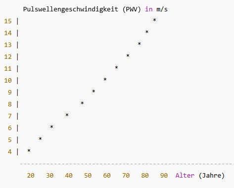
Pulse wave velocity must not be confused with the lower flow velocity. In a young, healthy aorta, the pulse wave velocity in this region is approximately 4–6 m/s due to its elasticity (wind-bag function) and larger lumen; in the periphery, it is then around 8–12 m/s, for example in the radial artery. In an aorta affected by arteriosclerosis, the pulse wave velocity is increased; furthermore, as biological age increases, the elasticity of the aorta decreases, consequently raising the pulse wave velocity. A value of 11 m/s or above is considered to indicate recognised, manifest end-organ damage. People with severe atherosclerosis may already reach values in excess of 16 m/s even in their younger and middle years. The increase with age is largely linear; some sources even assume a non-linear increase in pulse wave velocity with age:
There are several routes on which the PWS is measured:
- Carotid–femoral PWV (cf-PWV, the gold standard, commonly used in studies)
- Carotid–radial PWV (cr-PWV, less clinically significant, but often used in studies of vascular function
- Brachial–ankle PWV (baPWV; very common in Asia (Japan, Korea) because devices such as Omron and Fukuda Denshi were designed for this purpose; shows good correlation with cf-PWV, but yields systematically higher values)
- Aortic PWV by MRI (MRI-PWV; MRI provides the most direct measurement of aortic pulse wave velocity and is used more frequently in modern studies, e.g. MESA, the Multi-ethnic Study of Atherosclerosis)
The results for the various routes generally differ as follows:
- The further out towards the periphery, the stiffer the blood vessels → higher PWV.
- Aorta = elastic → lower PWV than in the legs/arms.
- baPWV → usually 1–2 m/s higher than cf-PWV.
- MRI-PWV measures specific segments, which is why values are often lower at a young age and rise more steeply.
An increased pulse wave velocity leads, as a result of increased pulse wave reflection, to a rise in systolic blood pressure and a fall in diastolic blood pressure, i.e. an increase in amplitude, leading further to an increased workload on the left ventricle (afterload) and reduced perfusion of the coronary arteries during diastole.
Bramwell-Hill described the PWV as the square root of the compliance D, which they expressed as (∆V/V)/∆P, or, in other words, as
the square root of AdP/(rhodA), where
- dP: Difference between systolic and diastolic pressure
- dA: Change in the cross-sectional area of the blood vessel during one cardiac cycle
- rho: density of blood, usually assumed to be approximately 1051 kg/m³
- A: average cross-section of the blood vessel
Factors affecting the PWV
| Influencing factor | Influence m/s |
|---|---|
| The hydration factor is incorporated into the calculation via rho: inadequate hydration increases the cellular proportion in the blood (haematocrit), resulting primarily from MCV * number of erythrocytes per ml. Dehydration is assumed to cause an increase of up to 1 m/s. Whilst dehydration has an increasing effect, hyperhydration (usually caused by excessive fluid intake that cannot be excreted by the kidneys in a timely manner) has only a slight reducing effect. | |
| Blood pressure is not a direct parameter in the equation, but it is one of the most important variables: in general, elevated systolic blood pressure can cause the PWV to rise; in the short term, spikes in blood pressure – caused, for example, by stress, caffeine or pain – can increase the measured PWV by several m/s. This is because the stiffness of the arteries increases as blood pressure rises. The PWV increases by approximately 0.5 to 1 m/s for every 10 mmHg rise in systolic blood pressure. | + 0,5 .. + 1,0 |
| In practice, the heart rate also contributes to the PWV at a rate of approximately 0.1–0.3 m/s per 10 bpm. For this reason, it is often standardised to 75 bpm. | + 0,1 .. + 0,3 |
| The effect of coffee and nicotine consumption is reported to increase this by 0.5–1 m/s; nicotine tends to have a stronger effect. | + 0 ,5 .. + 1,0 |
| Sympathetic nervous system/Parasympathetic nervous system: In general, all sympathicotonic influences have a vasoconstrictive effect, whilst all vagotonic influences have a slightly vasodilatory effect. | |
| Alcohol lowers PWV by causing vasodilation. | |
| Large meals have a vasodilatory effect and lower the PWV. | |
| Cold slightly increases the PWV through vasoconstriction, whilst heat reduces it slightly through vasodilation. The reported effects of heat and cold range from 0.5 to 1.5 m/s. | +/- 0,5 .. +/- 1,5 |
| Circadian: PWV is measured at a slightly higher level in the morning, with the lowest value occurring at night. The circadian variation is less than 0.5 m/s. | < + 0.5 |
| In practice, body position also affects the results of PWV measurements; values measured whilst standing are 1–2 m/s higher than those measured whilst lying down. Previous physical activity also increases the PWV. In the long term, however, regular, intensive physical activity, such as sport, lowers the PWV. Trained athletes generally have a lower PWV. | + 1 .. + 2 |
| Inspiration does not, as one might expect, cause a slight increase in PWV, as the heart rate rises slightly. This effect is outweighed by the fall in intrathoracic pressure, so that the PWV rises slightly. Accordingly, the PWV falls slightly during exhalation. A lower PWV is measured when lying down than when standing or walking. | |
| Rest: It is recommended that you rest in a sitting or lying position for 5–10 minutes before taking the measurement; failing to do so may cause the reading to be 5%–15% higher than it should be. | + 5% .. 15% |
When taking measurements using high-quality equipment, a large number of readings are usually recorded. The Atcor Medical Sphygmocor, for example, which uses an ECG to determine heart activity and an applanation tonometer to measure changes in pressure in a peripheral artery such as the radial artery, also provides a pulse wave analysis with the following parameters:
- ED: Ejection duration (ms)
- SP, DP: systolic pressure, diastolic pressure, both aortic and peripheral (mmHg)
- T1, T2: Time to the first and second peaks, respectively, in the blood pressure curve (ms)
- TR: time to reflection (ms)
- AG: Augmentation (mmHg) Interpretation: low: 0–5 mmHg, moderate: 5–12 mmHg, elevated: >12 mmHg (more common in cases of increased arterial stiffness)
- P1, P2: Pressure at T1, T2 (mmHg)
- PP: Pulse pressure, RRsyt. – RRdia. (mmHg)
- Augmentation index ( = AG/PP),
Interpretation:- Young adults: 0–20 %
- Middle-aged adults (40–60 years): 15–35 %
- 60 years and over: 20–40 %
Higher values are assessed as follows: - 40–45% and above: already unusually high
- 60% and above: highly abnormal
- 80% and above: pathologically high (measurement error or severely increased arterial stiffness)
High values are usually due to long-standing chronic conditions such as atherosclerosis, long-term hypertension, type 2 diabetes mellitus, chronic renal insufficiency and smoking.
A low heart rate (below 55 bpm) can also cause the AIx to rise significantly. Furthermore, the AIx increases as the pulse pressure decreases.
Physiologically, and as is regularly observed in healthy young people, the second peak in the pulse waveform (the curve of intra-arterial pressure) – caused by the pulse wave reflection first described by Otto Frank as far back as 1904 – is lower than the first peak, which is generated directly by ventricular contraction (negative augmentation P2 – P1) and is clearly separated from it. If the pulse wave is reflected too quickly due to an excessively high PWV, it may encounter the pulse wave as early as the systole, thereby increasing left ventricular afterload and consequently contributing to hypertrophic cardiomyopathy, rather than being delayed until the diastole to allow for better myocardial perfusion. In addition to augmentation, the augmentation index – which expresses augmentation as a ratio of the pulse pressure – is an important parameter. When vascular elasticity is significantly reduced, the second peak moves closer to the first and may exceed it (positive augmentation). The augmentation index increases with age and blood pressure and decreases with height and heart rate. It follows that shorter people have, on average, a slightly higher cardiovascular risk than taller people. Various studies have shown a 10–39% increase in mortality for every 1 m/s increase in pulse wave velocity. On average, women have a slightly higher index than men, due to their generally shorter stature.
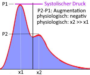
https://www.degag.eu/wp-content/uploads/2013/10/Konsensus_Gefaesssteifigkeit_DMW_Suppl_1_20101.pdf
Maximum heart rate / HRmax / maximum heart rate
There are various formulas for estimating the maximum heart rate based on age and, in some cases, additional factors:
- Spanaus (for athletes): 226 – 1.0 × age (women) or 223 – 0.9 × age (men)
- Tanaka (for athletes): 208 – 0.7 × age
- Hossack: 206 – 0.597 × age (women) or 227 – 1.067 × age (men)
- Edwards: 210 – 0.5 × age (in years) – 0.11 × body weight (women) or 214 – 0.5 × age – 0.11 × body weight (men)
Here you can calculate your maximum heart rate (HRmax) and view the training zones according to Karvonen, which are based on HRmax and resting heart rate.
The formulas become less accurate as athletes get younger and more accurate as they get older, although very significant deviations are possible in individual cases. For example, there are known cases of fit 60-year-old athletes who can still reach 200 bpm and tolerate this rate.
On the other hand, it is also known that, in older people, problems such as cardiac arrhythmia or heart failure can suddenly arise after years of training at the limits of cardiac capacity.
Unlike the calculation of training zones, the resting heart rate – where known – is not relevant for determining the maximum heart rate. If the maximum heart rate is determined experimentally, the amount of muscle mass involved plays a major role.
In a study of cyclists and runners, three-quarters of the 13 different prediction models underestimated the maximum heart rate by up to 5 beats, whilst some overestimated it by up to 5 beats.
In general, maximum heart rate does not appear to depend greatly on fitness level; rather, well-trained people tend to have a
lower HRmax. In the study, the runners’ HRmax was 1 per cent higher and their VO2max 3 per cent higher than that of the cyclists. Generally speaking, according to another study, men have a HRmax that is just under 2% higher, a heart rate reserve that is around 7% higher and heart rate recovery that is half a per cent better; however, the decline in HRmax with ageing appears to be slightly less pronounced in women.
Exercise heart rate / exercise pulse
The exercise heart rate – or, to be precise, the exercise heart rate – is the increase in heart rate caused by exercise compared with the resting heart rate.
Cardiac output
Cardiac output is the product of heart rate and stroke volume and is therefore the most important parameter for assessing the body’s blood supply. In physiology, the minute is typically used as the unit of time, so the term cardiac output (CO) is used. This is the volume that the heart ejects in one minute. It must be assumed that, physiologically (in healthy individuals without shunts; see, for example, doccheck.com) and, septal defects (see also, for example, doccheck.com), the output of the right heart corresponds exactly to that of the left heart on a short-term average, as any deviation from this would result in an accumulation of blood in one circulatory system (pulmonary circulation or systemic circulation) and, beyond a certain level, would no longer be compatible with life. Even a deviation of 1 ml per heartbeat would, at a heart rate of 60 bpm and a stroke volume of 50 ml, correspond to an accumulation of 3 litres of blood in one of the circulatory systems. The pulmonary circulation could not possibly accommodate such a volume of blood, and in the systemic circulation this would correspond to an extreme pooling of blood in the periphery, as is known to occur, for example, in anaphylactic shock. It would certainly be impossible to survive beyond the second hour at the latest. A distinction is made between the cardiac output ejected from the left ventricle into the systemic circulation and the pulmonary output ejected from the right ventricle towards the lungs. In a right-to-left shunt, the cardiac output is greater than the pulmonary output; in a left-to-right shunt, it is smaller. At rest, the average cardiac output is 4.5–5 l, which can increase by a factor of approximately 4 during exercise. Cardiac output is regulated primarily by the body’s oxygen demand.
If the cardiac output is multiplied by the arteriovenous oxygen difference, this gives the VO2max per minute a2> per minute; as a rule, healthy lungs are not the limiting factor, but rather internal respiration and metabolism are the key determinants of how much of the inhaled oxygen is utilised.
Adjustment in response to performance requirements
When the physical demands exceed those of rest, a number of key mechanisms alter the cardiac output. Neurologically, these are primarily brought about by a reduction in vagal tone and activation of sympathetic tone; endocrinologically, they result from the release and action of catecholamines (adrenaline, noradrenaline) at the β₁ receptors.
- Chronotropy (increase in heart rate): sympathetic activation causes an increase in action potentials in the cardioexcitatory centres of the brainstem. The acceleration of the pacemaker potential in the sinus node means that the threshold potential is reached earlier, leading to an increased heart rate. This is an important and the fastest regulator of the HMV. Even a reduction in vagal tone causes an increased heart rate, and an increase in sympathetic tone has an even greater effect.
- Inotropy (increase in contractile force): is primarily caused by catecholamines. The heart beats more forcefully, leading to an increased stroke volume. The Frank-Starling mechanism (increased preload and thus increased stretch stimulus during diastole increases contractile force) also increases contractile force. At the level of the myocardium, this is achieved through increased bridge formation. An increase in heart rate (chronotropy) and contractile force (inotropy) occur in parallel up to approximately 60% of VO₂max. At around 60%, maximum contractile force is reached, which explains the great value of endurance sports in this region. Inotropy is only slightly slower than chronotropy, but, as described, it has a dual effect.
- Dromotropy (increased conduction velocity): the sympathetic tone-mediated increase in the speed of electrical impulse conduction through the cardiac conduction system. This primarily affects the AV node, which is evident on the ECG as a shorter PR interval. This enables higher heart rates and reliable impulse conduction, and thus better synchronisation of the heart’s contractions.
- Bathmotropy: a reduction in the stimulation threshold of cardiac muscle cells
- Lusitropy: causes a more rapid and complete relaxation of the ventricles through the sympathetic-catecholamine-mediated acceleration of calcium removal from the cytoplasm. This allows the systole to be shorter, leaving more time for the diastole whilst the heart rate remains the same. Functionally, lusitropy thus complements inotropy.
At rest, vagotonus has a negative chronotropic and negative dromotropic effect, as well as a slightly negative inotropic effect. Chronotropism begins to take effect as early as around 20–30% of VO₂max, rising more or less linearly with oxygen uptake up to approximately 60%, and then at a slightly less steep rate thereafter. Chronotropism therefore begins to take effect even at moderate levels of exertion. The chronotropic response can be enhanced through regular aerobic training, ideally supplemented by interval training, e.g. 4×4 training. Inotropy sets in somewhat later, at around 30–50% VO2max, increases significantly up to 60–70% and then gradually levels off, reaching a plateau at around 90%. Inotropy is therefore particularly important for moderate and higher levels of exertion, up to high levels. Dromotropy becomes noticeable from around 40–50 VO₂max and increases up to approximately 80 per cent. After that, it remains at a high plateau. Medications, heart disease, autonomic dysfunction and sinus node syndrome can cause chronotropic incompetence, i.e. the failure to achieve an adequate heart rate (defined as 85% of the expected HRmax). This represents a risk factor for morbidity and mortality and results in reduced exercise capacity and premature fatigue during exercise.
Heart weight
The physiological weight of the heart depends on fitness level and gender. As an adaptation to physical demands, various processes take place, such as an increase in the heart’s muscle mass (hypertrophy), as well as improvements in vascularisation and adaptations in the connective tissue and innervation. This is referred to as physiological cardiac hypertrophy. In contrast, there are pathological forms of cardiac hypertrophy, such as those occurring with the use of anabolic steroids, in which the heart’s muscle tissue hypertrophies more rapidly than other necessary adaptations can take place. Another pathological condition is hypertrophic cardiomyopathy, which occurs in cases of arterial hypertension due to increased left ventricular afterload, or in cases of valvular regurgitation and stenosis. Pulmonary hypertension consequently leads to increased afterload on the right side of the heart, potentially resulting in heart failure.
Drugs can also lead to pathological hypertrophy, as can conditions such as diabetes mellitus, hypothyroidism, hyperthyroidism, chronic renal failure, rheumatoid arthritis (RA), lupus erythematosus (SLE), viral myocarditis, amyloidosis, sarcoidosis, haemochromatosis and long-standing pheochromocytoma.
Anaolic abuse also leads to weights in excess of 500 g within a relatively short period and without the necessary adaptations; however, the heart’s pumping function (stroke volume) may be impaired. In other pathological cases, a heart weight of 500 g or more is referred to as a critical heart weight, which is usually accompanied by pathological changes going beyond hypertrophy. In cases of valvular dysfunction, weights of 600–800 g may be reached, for which the arterial supply is not designed to cope.
The normal weight of the heart ranges from 300–350 g (men) and 250–300 g (women); in trained endurance athletes, it can reach up to 500 g, and in elite athletes, 600 g (men) or 450–500 g (women). In athletes, enlarged ventricles are associated with, at most, slightly thicker muscle tissue. Strength athletes fall below the weights stated for endurance athletes, tending instead towards 450–500 g (men). In these athletes, the myocardium is slightly more hypertrophied than in endurance athletes, but the ventricular volumes are less enlarged.
This adaptation in athletes is the result of regular, intense and long-term exercise, which is also reflected in a reduced RHR, as the oxygen demand at rest can be met with far fewer heartbeats. Occasional or very brief periods of physical activity do not yet lead to such an adaptation.
Adaptation to the demands of exercise has a significant beneficial effect on the vascular system and blood pressure, due to the markedly increased wall shear stress experienced during exercise – which leads to increased release of NO – and the lower RHR, which also places less strain on the elasticity of the vascular system.
If athletes stop training, a gradual reduction in cardiac mass begins after a few weeks, down to a level not associated with regular exercise. This reduction continues for months. After 3 months, the ongoing reduction should already be clearly visible or measurable.
Structure of the heart
The heart is divided by a partition (septum) into the left and right hearts, each comprising an atrium and a ventricle: the atria collect the blood and have significantly weaker muscles than the ventricles, which pump it into the circulatory systems under pressure. The left heart pumps blood into the systemic circulation and, due to the significantly higher pressure there than in the pulmonary circulation—which is supplied by the right heart—has significantly stronger muscles. The heart therefore has 2 atria and 2 ventricles:
– Atrium dextrum: right atrium
– Ventriculus dexter: right ventricle
in between: Valva tricuspidale (tricuspid valve), a three-leaflet valve
From the ventricle to the lungs: Pulmonary valve (Valva pulmonale)
– Left atrium: left atrium
– Left ventricle: left ventricle
in between: Valva bicuspidale (bicuspid valve or mitral valve), a two-leaflet semilunar valve.
From the ventricle to the aorta: Valva aortae (aortic valve).
The four valves lie roughly in a single plane, the valvular plane; they are held in place by the cardiac skeleton, a connective tissue structure which not only serves as an attachment point for the muscles and valves, but also electrically separates the atria and ventricles. During ventricular contraction, the valve plane is pulled downwards towards the apex of the heart, thereby increasing the atrial volume, which draws blood from the vena cavae – where the pressure is low – into the atria. As the valve plane, which is now rising again whilst the semilunar valves are open, covers the blood volume in the atria during the ventricular relaxation phase, atrial contraction is not strictly necessary for the haemodynamic function of the heart; in other words, a person can remain viable even in the event of, for example, atrial fibrillation.
Naturally, the filling and stroke volumes of the two halves of the heart are equal; otherwise, blood would pool in the pulmonary or systemic circulation.
Stimulus generation and transmission
The contraction of the heart is triggered electrically by an impulse generated within the heart itself. The cardiac conduction system:
1. The crescent-shaped sinus node is situated at the junction of the superior vena cava; it acts as the pacemaker and is, physiologically speaking, the sole generator of electrical impulses, firing at a rate of 60–80 beats per minute.
2. The AV (atrioventricular) node is situated in the right atrium, in the angle between the septum and the tricuspid valve. It has a natural frequency of 40–60 beats per minute. It acts as a backup system in the event of sinus node failure and, where necessary, moderates the impulses from the sinus node.
3. The bundle of His is the only muscular connection between the atria and the ventricles
4. The left and right Tawara branches in the left and right ventricles. They carry the impulse down to the apex of the heart and innervate the ventricles
5. The fine Purkinje fibres connect to the fibres of the working myocardium
The sinus node has no connection to 2–5; the AV node receives the impulse from the sinus node as an electrical signal from the atrial muscle. The impulse travels: sinoatrial node → atrial muscle → AV node → His bundle → Tawara branches → Purkinje fibres → apex of the heart → back up to the His bundle. It stops before reaching the AV node.
Heart Campaign: the stages
Oxygen-rich venous blood and oxygen-poor arterial blood are found only in the pulmonary circulation between the heart and the lungs; in the systemic circulation, the venous blood is oxygen-poor and the arterial blood is oxygen-rich. The oxygen-poor blood from the lower limbs and the abdominal and thoracic cavities flows via the V. cava inferior (inferior vena cava), whilst the oxygen-poor blood from the upper limbs flows via the V. cava superior (superior vena cava) into the right atrium. During diastole (the filling phase: the semilunar valves are open and the mitral valves are closed), the blood flows on into the right ventricle. During systole (the contraction phase of the heart muscle, with the semilunar valves open and the mitral valves closed), the blood is pumped into the truncus pulmonalis (pulmonary trunk) and flows to the lungs. As it passes through the lungs, it is enriched with oxygen by diffusion across the semi-permeable membranes of the alveoli and flows back through the left pulmonary vein and right pulmonary vein into the left atrium. The mitral valve then opens and the blood flows into the left ventricle during diastole, from where it is pumped into the aortic arch during systole. Systole is further subdivided into the contraction phase (the phase in which the heart muscle has already contracted, but the pressure generated is not yet sufficient to open the semilunar valves and expel blood) and the ejection phase. Similarly, diastole is further divided into the relaxation phase (the heart muscle ceases its contraction and expands again) and the filling phase (blood flows into the atria).
Layers of the heart’s walls
The heart consists of (from the inside out):
Endocardium
The endocardium, also known as the inner lining of the heart, lines the heart from the inside and also forms the valves; it is very smooth.
Function:
1. To smooth out any unevenness and roughness and keep the surface mirror-smooth, so that flow resistance is kept to a minimum
2. To enable the valves to function properly
Myocardium
The heart muscle, also known as the working myocardium. The myocardium of the left heart is 2–3 times as thick as that of the right heart and also has 2–3 times the contractile force: whilst the pressure in the atria is approximately 0–5 mmHg, the pressure in the right ventricle is 40–45 mmHg and in the left ventricle 120 mmHg. All four atria and ventricles are roughly the same size; of the approximately 150 ml they can hold, around 70–80 ml (50–100) is pumped with each heartbeat. At a pulse rate of 80, this amounts to approximately 5 l/min, or the entire blood volume once per minute.
By doubling the stroke volume and multiplying the rate by 2.5, the maximum increase is a factor of 5 during physical exertion.
Cardiac output can be increased under the influence of sympathetic nerve fibres, which release noradrenaline in the myocardium and along the conduction system; in addition, noradrenaline can reach the heart via the bloodstream together with adrenaline. These have chronotropic, inotropic and dromotropic effects (the conduction time through the AV node is shortened). The antagonist of the sympathetic system, the parasympathetic vagus nerve, acts via acetylcholine to produce negative chronotropic, negative inotropic, negative dromotropic and negative bathmotropic effects (it increases the excitation threshold) . Through the Frank-Starling mechanism, the heart adjusts the ejection force in response to filling: a greater volume results in greater stretching, causing the heart muscle to pump more. If there is a persistent excess of blood volume, a diuretic hormone—atrial natriuretic peptide (ANP)—is released in a stretch-dependent manner.
Functions of the myocardium:
1. To propel the blood forwards through the circulatory system by means of contraction.
2. Conduction: to transmit the electrical impulses that trigger the contractions of the four chambers from their point of origin, the sinus node.
Epicardium
The outer wall of the heart lies on the outside of the myocardium and is firmly attached to it
Function: it helps to shape and maintain the heart’s structure, and its mirror-smooth surface minimises friction
Between the epicardium and the pericardium lies serous pericardial fluid, which reduces friction between the two.
Pericardium
Pericardium: strictly speaking, the epicardium transitions into the pericardium at the points where the major vessels emerge, as if the heart were a fist being placed inside an air-filled balloon, so that the balloon’s wall folds over the fist.
EKG
P: Atrial excitation by the sinus node, approx. 100 ms
Q: Onset of ventricular excitation, conduction from the apex to the base of the heart, 40 ms, < ¼ of the R wave; if widened or deepened: a sign of a previous heart attack. As the papillary muscles conduct more rapidly, they contract slightly before the ventricles
P-Q: Conduction time from the sinus node to the AV node
R: Depolarisation of the myocardium. Widened or notched: disturbance of electrical conduction; shortened: indication of a previous infarction
QRS: approx. 110 ms, sometimes up to 120 ms
Ischaemia
Ischaemia is a relative or absolute deficiency in the blood supply to a tissue. Temporary episodes of ischaemia are known as ‘transient’. The time taken for damage to occur is determined by the relationship between the reduction or interruption in blood supply and the tissue’s oxygen demand. In the case of transient ischaemia, the term ‘ischaemic time’ refers to the period during which the blood supply is reduced or interrupted. Well-known forms of ischaemia include those associated with myocardial infarction and its precursor, angina pectoris, but they also occur elsewhere, such as the pain typical of peripheral arterial disease (PAD) in the form of intermittent claudication (window-shopping disease), which makes even moderate walking distances impossible due to pain. Silent apoplexy is also a form of ischaemia, although it does not cause any pain (silent ischaemia). Risk factors for ischaemia include nicotine consumption, lipid metabolism disorders, diabetes mellitus, obesity, arterial hypertension. Pathophysiologically, ischaemia is characterised by a significantly elevated extravascular pressure, an insufficient arterial time volume (for example, due to leakage or stenosis) or impaired microcirculation.
Mediastinum
The space between the lungs is known as the mediastinum. Dorsally, it is bounded by the vertebral column and its ligamentum longitudinale anterius, and ventrally by the sternum; both provide bony protection. Higher up in the mediastinum are: the thymus gland, the large vessels near the heart (the aortic arch and its branches, the pulmonary trunk, the superior vena cava), the trachea, the oesophagus, lymph nodes (mediastinal lymph nodes, tracheobronchial lymph nodes), the thoracic duct (running behind the left clavicle to the left venous angle), the phrenic nerve, the vagus nerve and the recurrent laryngeal nerve; and further down: the oesophagus, aorta, inferior vena cava, as well as the vagus nerve and the recurrent laryngeal nerve. vagus and recurrent laryngeal nerve, as well as further down: oesophagus, aorta, inferior vena cava, azygos vein, hemiazygos vein, thoracic duct and vagus nerve.
Windpipe / trachea
The trachea (windpipe) is shorter than the oesophagus, in front of which it lies; it is approximately 11–14 cm long. It begins at the level of the cricoid cartilage behind the thyroid gland and consists of 16–20 horseshoe-shaped cartilaginous rings; towards the back, it is covered by connective tissue. The trachea lies in front of the oesophagus, which is why there is only tissue behind it. It divides – the point of division is known as the tracheal bifurcation – into the right and left main bronchi (which still contain cartilaginous rings). The right side runs at a steeper angle and is therefore more prone to obstruction when swallowing. The right main bronchus has three main branches, the left two, corresponding to the number of lung lobes. The bronchi continue to branch out; distally, the number of alveoli increases rapidly. The alveoli are surrounded by veins and arteries.
Lung
The lung, Pulmo or Pneumo, consists of a left and a right lobe, with 3 (right) and 2 (left) lobes (Lobus) respectively. Based on the branches of the bronchi, 10 segments can be distinguished on the right and 9 on the left. The left lung is slightly smaller because the heart takes up some space.
The lung consists of the branching tree of bronchi, each of which ultimately ends in countless alveoli (air sacs). Through the interplay of inhalation and exhalation, fresh, oxygen-rich air is constantly delivered to the blood-air barrier, the approximately 2.2 µm-thick, permeable layer of tissue between the air-filled interior of the lung and the capillaries. Inhalation increases the volume of the chest cavity in two ways:
Abdominal breathing
The dome-shaped muscular diaphragm moves downwards, increasing the space available for the lungs, which are fused to it: by 1.5 cm during a gentle inhalation, and by 10 cm during a deep inhalation. Excessive contraction of the diaphragm can pull the ribs downwards, thereby restricting chest breathing.
Chest breathing
The rib arches, which rise forwards and upwards during thoracic respiration, increase the volume of the lungs, which are attached to the inside of the chest cavity: the visceral pleura (pleura pulmonalis), which lies directly against the lung, is attached in a sliding manner by means of a serous fluid to the parietal (pleura parietalis) pleura, which lines the inside of the thoracic cavity. The greatest expansion of the thoracic cavity occurs, particularly in the cranial region, in the sagittal direction, and further caudally also to some extent in the frontal plane.
If, in the event of a pneumothorax, air enters the (solely) fluid-filled pleural space between the parietal and visceral layers, the two layers become separated, and the lung no longer follows the excursion of the chest. If, in the context of pleurisy, the two pleural layers become fused or adhered to one another, their mobility is restricted and inhalation is painfully limited.
During shallow inhalation, almost exclusively the scalene muscles lift the chest from above; during moderate inhalation, the intercostal muscles and, during vigorous inhalation, all the remaining inspiratory muscles plus the accessory inspiratory muscles. Due to the gravity of the costal arches, exhalation occurs even without muscular effort. Even against gravity, the retraction force (intrinsic tension of the lung tissue) is sufficient for this. Furthermore, the tension in the abdominal muscles counteracts abdominal inhalation, and the gravity of the ribcage (in an upright position) causes thoracic exhalation.
When we inhale, the air present in the airways above the lungs is drawn in as well; this space – comprising the nasal and oral cavities, the pharynx, the trachea and the bronchi – is known as the dead space and has a volume of approximately 150 ml. Here, the inhaled air is warmed, moistened and (particularly in the nose) filtered to remove dust. The mouth is very poor at filtering out dust. The ciliated epithelium of the trachea (windpipe) and bronchi can transport particles back out of the respiratory tract.
Bradypnea
Bradypnoea refers to a reduced respiratory rate at rest, defined in adults as fewer than 8 breaths per minute. Under physiological conditions, the respiratory rate depends on the tissues’ oxygen requirements; it is therefore increased during physical activity. At rest, it is usually between 12 and 16 breaths per minute; in newborns, it is 30 to 50, and in premature babies it can be as high as 80. During meditation and relaxation techniques, it can fall significantly below 12, as it can in some pathological cases. A breathing rate that is too fast is called tachypnoea.
Tachypnea
Tachypnoea refers to an increased respiratory rate at rest; in adults, this is defined as more than 20 breaths per minute. Under physiological conditions, the respiratory rate depends on the tissues’ oxygen requirements; it is therefore higher during physical activity. At rest, it is usually between 12 and 16 breaths per minute; in newborns, it is 30 to 50, and in premature babies it can be as high as 80. It can rise significantly during exercise, but a certain degree of tachypnoea is also frequently observed in cases of fever and various illnesses. A respiratory rate that is too slow is known as bradypnoea.
Tidal volume
Tidal volume is the average volume of air inhaled and exhaled at rest, i.e. without any strenuous activity. The average for the general population is around 500 ml. The dead space volume of approximately 150 ml does not contribute to gas exchange.
Respiratory volumes
When at rest, a man weighing 70 kg inhales and exhales a tidal volume of approximately 0.5 l per breath. When inhaling to maximum capacity by engaging all inspiratory respiratory muscles and inspiratory accessory muscles, he can inhale an additional inspiratory reserve volume of approximately 2.5 l. At the end of an unforced exhalation, by engaging all expiratory muscles and expiratory accessory muscles, he is still able to exhale a further expiratory reserve volume of 1.5 litres of air. This results in a vital capacity of approximately 4.5 litres, comprising the sum of the tidal volume, inspiratory reserve volume and expiratory reserve volume. However, after maximum exhalation, a residual volume of approximately 1.5 l of air remains in the lungs and bronchi, which cannot be exhaled, resulting in a total capacity of around 6 l.
Various respiratory volumes are defined:
tidal volume: the volume of air normally moved, ranging from approximately 500 ml at rest to 2.5 l during strenuous exertion
end-expiratory lung volume (lung volume following normal exhalation) = functional residual capacity
This is subdivided into
– expiratory reserve volume: Volume that can still be exhaled with effort, on average 1–2 l
– residual volume: The volume that cannot be exhaled even with the greatest effort; this amounts to 1–1.5 l and is largely expelled when the pleural space opens on both sides
– inspiratory reserve volume: The volume that can be inhaled in addition to a normal inhalation, averaging 2–4 litres
– Vital capacity: The sum of tidal volume, inspiratory reserve volume and expiratory reserve volume
– Total lung capacity: Vital capacity plus residual volume, approx. 5–10 l; slightly lower in women than in men, and highly dependent on fitness level
inspiratory reserve volume
the volume that can be inhaled in addition to a calm, effortless inhalation.
expiratory reserve volume
the volume of air that can be exhaled in addition to that exhaled during a normal, unforced exhalation when performing a forced exhalation.
Residual volume
The volume in the lungs and bronchi that cannot be exhaled after forced exhalation.
Total capacity
The sum of all respiratory volumes, including the inspiratory and expiratory reserve volumes and the residual volume.
Dead space volume
Dead space is the volume of the nasal cavity, oral cavity, pharynx, trachea and bronchi in the respiratory system that is involved in the passage of air. It averages around 150 ml and must be subtracted from the respiratorily effective tidal volume of around 500 ml, meaning that only around 350 ml contributes to gas exchange.
Vital capacity
Vital capacity is the sum of tidal volume, inspiratory and expiratory reserve volumes, and is therefore the maximum amount of air a person can breathe in and out.
Reclining position for breathing
The resting respiratory position is the point of equilibrium between the retraction force of the lungs and the elastically expanding forces of the chest. When transitioning from sitting/standing/walking to a supine position, the resting position of the respiratory system shifts slightly towards expiration due to the greater effect of gravity on the chest and the pressure exerted by the abdominal organs against the diaphragm. A reduction in the elastic recoil force of the lungs leads to a resting respiratory position shifted towards inspiration; in this case (as, for example, in emphysema), the expiratory reserve volume increases and the inspiratory reserve volume decreases. With a very flexible thorax, the inspiratory reserve volume increases whilst the expiratory reserve volume decreases. A low resting position of the lungs – that is, a low expiratory reserve volume with a high inspiratory reserve volume – is desirable, as this means less oxygen-depleted air remains in the lungs and inhaling the same volume of air results in a higher oxygen content in the air remaining in the lungs after inspiration. The ratio of (tidal volume minus dead space volume) to (residual volume plus expiratory reserve volume) is called the ventilation coefficient and averages 1/8. It should be as high as possible and is correspondingly lower in conditions such as emphysema.
The respiratory rate averages 15/min (12–20); approximately 500 ml of air is therefore exchanged in 4 seconds, with the expiratory phase being 3:2 times longer than the inspiratory phase; there is often an end-expiratory pause. On average, 7–8 litres of air are inhaled per minute, including ¼ litre of oxygen. Depending on fitness level, a maximum of approximately 120 l of air is inhaled per minute.
The oxygen transferred to the cells during internal respiration is used there to ‘burn’ nutrients: carbohydrates, fats and proteins. Ideally, these are broken down without leaving any residue to form CO2 and H2O; in the case of proteins, urea is also produced, which must be excreted by the kidneys. Like the heart rate, respiration is also regulated by the medulla oblongata. If this regulation were to fail, during physical exertion or other situations requiring increased energy, the oxygen content in the blood would fall (hypoxia) and the CO2 concentration would rise (hypercapnia), which would, not least, have an adverse effect on blood pH. For this reason, the partial pressures of O2 and CO2 are measured in the aortic arch and in the carotid glomus, whilst the pH and pCO2 are measured by the medulla oblongata itself. Stretch receptors in the alveoli inhibit further breathing (Hering–Breuer reflex). Further impulses from various regions of the body influence respiration. As with the heart, both respiratory rate and tidal volume can be increased; increasing the tidal volume is preferable, as this reduces the impact of dead space.
Lung function tests
Forward Excitation Velocity (one-second expiratory capacity Forward Excitation Velocity, Tiffenau Test): Following a maximum inhalation, a measurement is taken of the maximum amount of air the subject can exhale in one second (relative capacity in seconds). Depending on their age, healthy individuals achieve 70–80 per cent of their vital capacity. Obstructive lung diseases, reduced elastic recoil in the lungs and weakened expiratory (and accessory) muscles reduce this value. The average one-second capacity, FEV1, declines slowly from puberty onwards and is higher in taller people than in shorter ones. In men, the FEV1 is greater than in women of the same height.
Furthermore, the lung volume of people living at higher altitudes is generally greater.
Forced Expiratory Volume in one second: the ratio of FEV1 to FVC (Tiffenau index)
PEF (peak expiratory flow): the maximum respiratory flow rate during exhalation in l/s
PIF (peak inspiratory flow): the maximum respiratory flow rate during inhalation, in l/s
FEF25 (forced expiratory flow at 25% expiration): respiratory flow rate at 25% expiration
Fifty (forced expiratory flow at 50% expiration): respiratory flow rate at 50% expiration
FEF75 (forced expiratory flow at 75% expiration): the respiratory flow rate at 75% expiration
FEF2575 (mean forced expiratory flow between 25% and 75% expiration): the average respiratory flow rate between 25% and 75% expiration
Spirometry: Measurement of tidal volume, vital capacity, inspiratory reserve volume and expiratory reserve volume:
VC (vital capacity), also FCV (forced vital capacity): The difference in volume between maximum inhalation and maximum exhalation. This parameter can also be broken down into:
IVC (inspiratory vital capacity): Determination of VC from the exhaled state
EVC (expiratory vital capacity): Determination of VC from the inhaled state
These two may differ slightly.
VC should be at least 3 l and is primarily limited by restrictive lung diseases. In favourable cases, it amounts to 5 l, which naturally depends on height and build.
The European Coal and Steel Community uses the formula:
height³ / correction factor * (1.03 – (age – 25) / 100) * 0.75, where the correction factor is 1 for men and 1.1 for women.
A reduction of 20 per cent is considered pathological. Other sources give different formulae:
0.052 * height – 0.022 * age – 3.6 for men and
0.041 * height – 0.018 * age – 2.69 for women, or alternatively
(27.63 – 0.112 * age) * height for men and
(21.78 – 0.101 * age) * height for women.
There are several contraindications to determining the FVC via testing:
haemoptysis, pneumothorax, cardiovascular events such as myocardial infarction, angina pectoris, thoracic, cerebral and abdominal aneurysms, cataracts, recent surgery, dizziness, nausea, acute illness, current or recently recovered viral infection, and undiagnosed hypertension.
It follows that:
IC (inspiratory capacity): VT + IRV
In addition, the following is also considered:
VT (AZV): the tidal volume during normal breathing, i.e. the difference between effortless exhalation and normal inhalation. The mid-range is referred to as the respiratory mid-range.
Not measured by spirometry
IRV (inspiratory reserve volume), the volume that can still be inhaled after normal inhalation
ERV: (Expiratory reserve volume), the volume that can still be exhaled after a normal inhalation
At an early stage, the bronchial tree still contains a significant proportion of cartilage to prevent it from collapsing. In the alveoli, a surfactant (surface-active agent) is produced and released onto the surface, which reduces surface tension. Macrophages phagocytose dust and debris from haemorrhages. Due to the structure of the tissue, in the event of congestion in the lungs caused by heart failure, fluid can leak into the interior of the lungs (the air-filled space), resulting in pulmonary oedema. Before birth, the lungs are not essential for life; after birth, they must expand in a flash: the water in the lungs is displaced by air with the help of surfactant, and breathing becomes possible. The newborn’s first cry is proof that the lungs are filled with air.
The term ‘respiration’ is usually understood to refer only to external (pulmonary) respiration; however, respiration also includes internal (cellular) respiration. Respiration consists of:
– Ventilation
– Gas exchange in the lungs
– Gas transport in the blood
– Gas exchange in the tissues
– Utilisation of oxygen within the cell
– Adaptation of respiration to meet the body’s needs
Obstructive
Respiratory restrictions are termed ‘obstructive’ when they result from a narrowing of the cross-sectional area of the airways. The main obstructive lung diseases are COPD, bronchial asthma and chronic obstructive bronchitis. These are usually caused by secretions, foreign bodies or tumours in the airways. In addition to imaging and endoscopic procedures, an obstruction can be detected using the Tiffeneau test as part of a functional diagnostic assessment.
Restrictive
Respiratory limitations are termed ‘restrictive’ when they result from a reduced ability of the lungs to expand. In such cases,
vital capacity and total capacity are reduced. Causes include, for example, pulmonary fibrosis, pleural adhesions or restricted thoracic mobility, as seen in scoliosis and, in particular, hyperkyphosis of the thoracic spine, for instance in the context of severe forms of osteoporosis, Scheuermann’s disease or Bechterew’s disease. Restrictive lung function can be detected by measuring vital capacity.
Inspiration
Inhalation. This takes place at a slow pace whilst maintaining an upright posture, with the assistance of the inspiratory respiratory muscles, beginning with the scalenes, which may be sufficient for gentle inhalation; for slightly deeper inhalation, the intercostal muscles are also engaged; during forced inhalation, the inspiratory accessory muscles are additionally involved. Inhalation (inspiration) consists of two parts: thoracic and abdominal inspiration. During thoracic inhalation, the ribs are raised cranially -ventrally, which enlarges the thorax and draws air into the lungs: without incoming air, a negative pressure relative to the surroundings would develop in the lungs; the inflowing air compensates for this. Abdominal breathing occurs through contraction of the diaphragm, which contracts caudally and thereby also increases lung volume, as the lungs are caudally fused with the diaphragm. In pulmonary function testing, the inspiratory vital capacity, i.e. the maximum volume of air that can be inhaled after forced exhalation or, conversely, the maximum volume of air that can be exhaled after forced inhalation, is measured to screen for restrictive lung diseases.
The following diagram shows the exhaled and forced-inhalation positions of the ribs from cranial, with the axes of movement resulting from the joints at the vertebrae.
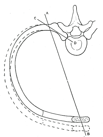
In the lateral view, the lifting movement is clearly visible, as is the change in inclination in the sagittal plane:
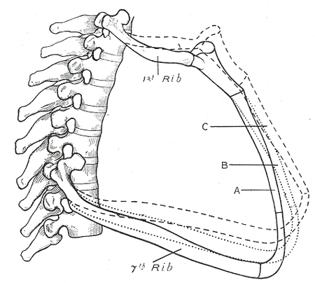
Intercostal muscles
Intercostal muscles, or muscles between the ribs, are muscles situated between two adjacent ribs that assist with breathing:
- Mm. intercostales externi (external intercostal muscles), superficial, inspiratory
- Mm. intercostales interni (internal intercostal muscles), deep, expiratory
- Inner intercostal muscles, branches of the internal intercostal muscles, expiratory
- Subcostal muscles, derived from the internal intercostal muscles, expiratory
- The musculus transversus thoracis influences the elasticity of the ribcage through tension in the costal cartilages
Exspiration
Exhalation. Like inhalation, this has two possible components: chest breathing and abdominal breathing. For abdominal exhalation, the diaphragm relaxes so that it can return to its resting position, which extends further cranially. Chest exhalation occurs naturally at a slow pace whilst maintaining an upright posture (upper body roughly vertical, head raised), in accordance with gravity. When slightly accelerated, it is aided by the expiratory respiratory muscles; when forced, the expiratory accessory muscles are also engaged. In pulmonary function testing, the Tiffeneau test is used to investigate obstructive airway restrictions that impede exhalation.
respiratory muscles
Muscles whose contraction leads to the expansion or constriction of the chest or abdominal cavity and which, as a result, contribute to inhalation or exhalation. Normally, exhalation occurs automatically as a result of gravity, muscle tension and the retraction forces of the lungs as an elastic organ; force therefore only needs to be exerted for inhalation. In addition to the inspiratory muscles (inhalation) and the expiratory muscles the inspiratory accessory muscles and expiratory accessory muscles. One of the most important respiratory muscles, and the one whose contraction causes abdominal inhalation, is the diaphragm. Another important factor, which is not included in the above calculation but is nevertheless significant, is the dead space volume of approximately 150 ml.
inspiratory respiratory muscles
Muscles used during normal inhalation:
- Diaphragm (Diaphragm)
- Mm. intercostales externi (external intercostal muscles)
- M. scaleni
- M. intercartilaginei (the part of the internal intercostal muscles situated between the costal cartilages)
inspiratory accessory muscles
Muscles that can be engaged during a forceful inhalation:
- Muscle of the costal lifters
- M. serratus anterior (anterior serratus muscle)
- M. serratus posterior superior (posterior superior serratus muscle)
- The lesser breast muscle
- Pectoralis major (only when the arm is supported)
- M. sternocleidomastoideus
- M. erector spinae
expiratory respiratory muscles
Muscles used during normal exhalation:
- Mm. intercostales interni and intimi (internal intercostal muscles)
- Muscle of the subcostal
- Transverse thoracic muscle
Expiratory accessory muscles
Muscles that can be engaged during a forceful exhalation:
M. obliquus externus abdominis
M. obliquus internus abdominis
M. transversus abdominis
M. transversus thoracis
M. latissimus dorsi (‚cough muscle‘)
M. serratus posterior inferior (posterior inferior saw muscle)
M. quadratus lumborum
M. rectus abdominis
Breathing
Respiration refers to the exchange of oxygen and carbon dioxide, which enables energy production within the cells. This involves the uptake of oxygen from the external environment via the lungs, whilst simultaneously releasing carbon dioxide into the external environment – a process known as external respiration, and internal respiration, in which the cells take up oxygen from the blood (bound to haemoglobin in the red blood cells) and release the carbon dioxide produced during metabolic processes to the red blood cells.
External respiration consists of two components that can be controlled voluntarily: chest breathing and abdominal breathing.
However, even without voluntary intervention, respiration continues and is autonomously regulated by the respiratory and circulatory control centre in the medulla oblongata in accordance with the body’s needs. Depending on the need, different muscles are engaged with the appropriate intensity.
The abdominal muscles primarily assist rapid exhalation at rates of approximately 40 l/min and above. A distinction is made between costosternal (chest breathing) and costodiaphragmatic (abdominal breathing). In the former, the thorax is primarily expanded forwards; in the latter, the lungs are stretched downwards – mainly by the diaphragm – whilst the abdominal cavity is compressed, causing the abdominal wall to bulge forwards. When the upper body is in an upright position (sitting, standing, walking), costodiaphragmatic breathing predominates, as the diaphragm can contract downwards easily and freely. When lying on the back, there is increased counter-pressure from the abdominal organs against the diaphragm, and thoracic breathing becomes more prominent. In the prone position, chest inspiration is impeded by the gravity-induced pressure on the chest and abdominal breathing is hampered by the gravitational pressure on the abdominal cavity, so that both types of breathing are restricted.
portal vein
The term ‘portal vein’ usually refers to the V. portae hepatis (and only rarely to the V. portae hypophysiales), which collects blood from all the abdominal organs, with the exception of the kidneys, and carries it to the liver. The individual organs are: the stomach, small intestine, large intestine, parts of the rectum, pancreas and spleen. It is 8–15 mm wide, has a flow velocity of up to 23 cm/s and a pressure between 5 and 20 mmHg, which is difficult to measure. If blood becomes congested in the portal vein or the liver, other venous systems must drain the blood and become overloaded, leading to the formation of portocaval anastomoses:
– oesophageal varices (varicose veins in the oesophagus)
– caput medusae (overload of deep and superficial abdominal and thoracic veins)
– haemorrhoids resulting from varices in the rectal region
In addition to the portal vein, there are also the pituitary portal veins (venae portales hypophysiales), which carry various regulatory hormones from the hypothalamus (the highest regulatory centre for all autonomic and endocrine processes) to the anterior pituitary (which produces various effector and regulatory hormones).