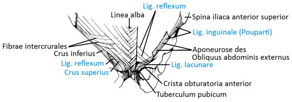
Upper arm/shoulder ventral
Upper arm/shoulder dorsal
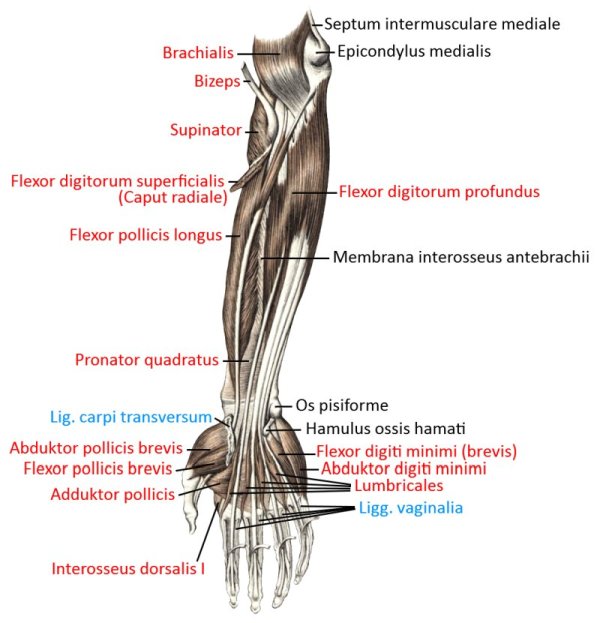
Forearm, hand palmar
Forearm, hand dorsal
Trunk
Back muscles, allochthonous
Back muscles, autochthonous
Abdominal muscles
Thigh, hip ventral
Thigh, hip dorsal
Lower leg ventral, foot dorsal
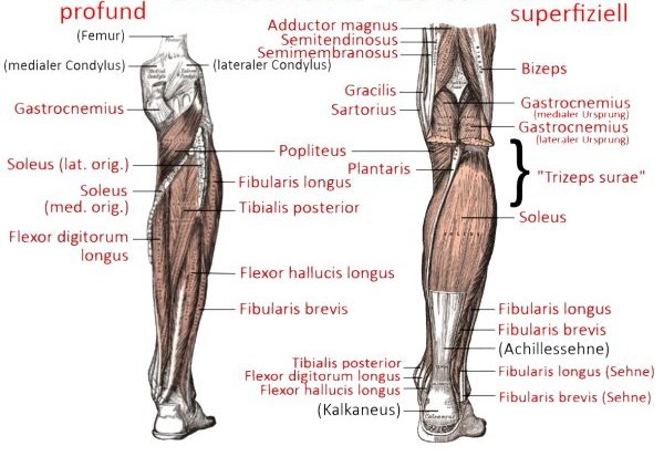
Lower leg dorsal, foot plantar
Foot dorsal
Plantar foot
Contents
- 1 Upper arm/shoulder ventral
- 1.1 Overview (image links to linkmap)
- 1.2 superficial view (image links to linkmap)
- 1.3 profound view (image links to linkmap)
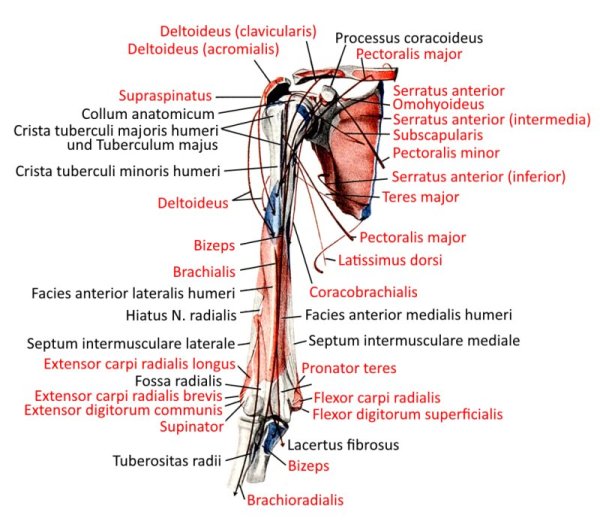
- 1.4 Insertions (image links to linkmap)
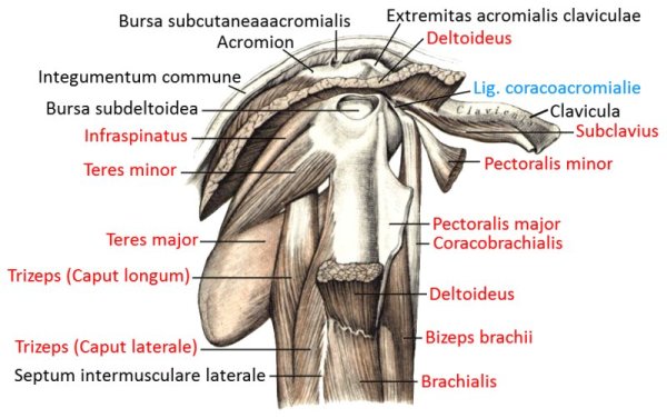
- 1.5 Shoulder joint with arm muscles (image links to linkmap)
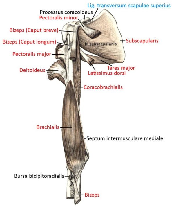
- 1.6 Shoulder joint with scapula, profound (image links to linkmap)
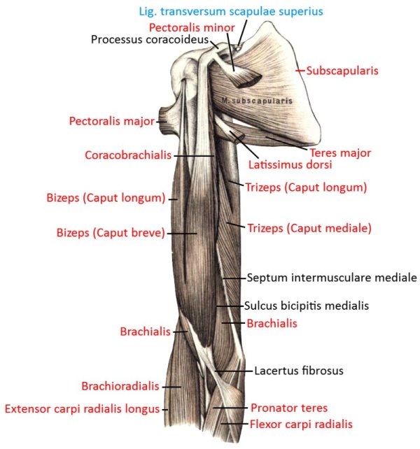
- 1.7 Shoulder joint with scapula, superficial (image links to linkmap)
- 1.8 Medioventral shoulder joint (image links to linkmap)
- 1.9 Shoulder from the side (image links to linkmap)
- 2 Upper arm/shoulder dorsal (image links to linkmap)
- 3 Back, allochthonous musculature (image links to linkmap)
- 4 Back, autochthonous musculature (image links to linkmap)
- 4.1 Autochthonous musculature (image links to linkmap)
- 4.2 Scheme (image links to linkmap)
- 4.3 lateral tract (image links to linkmap)
- 4.4 transversospinal tract (image links to linkmap)
- 4.5 Scheme
- 4.6 Cervical spine and thoracic spine from dorsolateral (image links to linkmap)
- 4.7 Short intervertebral muscles (image links to linkmap)
- 4.8 Abdominal muscles (image links to linkmap)
- 4.9 Profound abdominal muscles (image links to linkmap)
- 4.10 Superficial abdominal muscles (image links to linkmap)
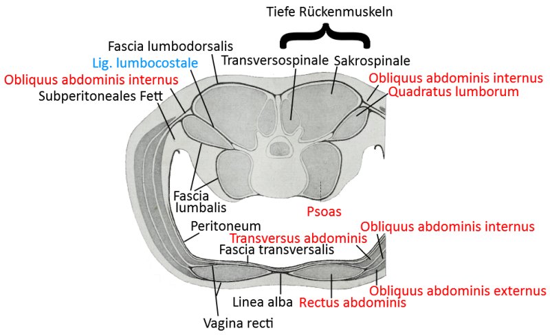
- 4.11 Abdominal muscles, cross-section of the trunk through the lumbar spine region
- 5 Thigh, hip ventral
- 6 Overview (image links to linkmap)
- 6.1 View from ventromedial (image links to linkmap)
- 6.2 another view from ventromedial (image links to linkmap)
- 6.3 Ventral view (image links to linkmap)
- 6.4 Ventral profound view (image links to linkmap)
- 6.5 Schematic view (image links to linkmap)
- 6.6 profound view (image links to linkmap)
- 6.7 medioventrale, very profound view (image links to linkmap)
- 6.8 medioventral profound view (image links to linkmap)
- 6.9 Medial superficial view (image links to linkmap)
- 6.10 superficial view (image links to linkmap)
- 6.11 Exploded view (image links to linkmap)
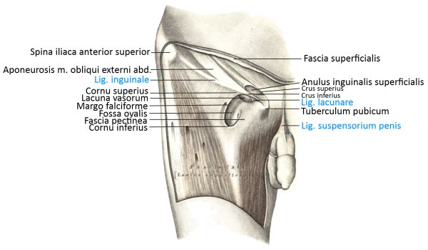
- 6.12 Lateral view
- 6.13 Lateral view of the fasciae
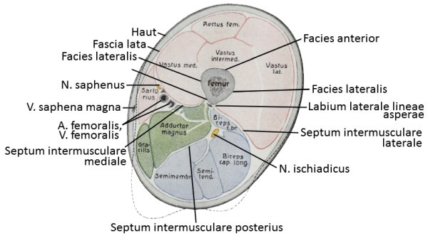
- 6.14 Transversal cut
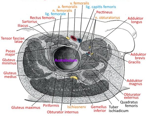
- 6.15 Hip joint, incision (image links to linkmap)
- 7 Thigh, hip dorsal
- 8 Lower leg, foot ventral (image links to linkmap)
- 8.1 Lower leg from ventral (image links to linkmap)
- 8.2 Lower leg superficial from ventral (image links to linkmap)
- 8.3 Knee and lower leg ventrolateral (image links to linkmap)
- 8.4 Knee and lower leg ventromedial (image links to linkmap)
- 8.5 Lower leg, foot ventral, profound (image links to linkmap)
- 8.6 Lower leg, foot ventrolateral superficial (image links to linkmap)
- 8.7 transverse cross-section (image links to linkmap)
- 9 Lower leg dorsal, foot plantar
- 9.1 Overview (image links to linkmap)
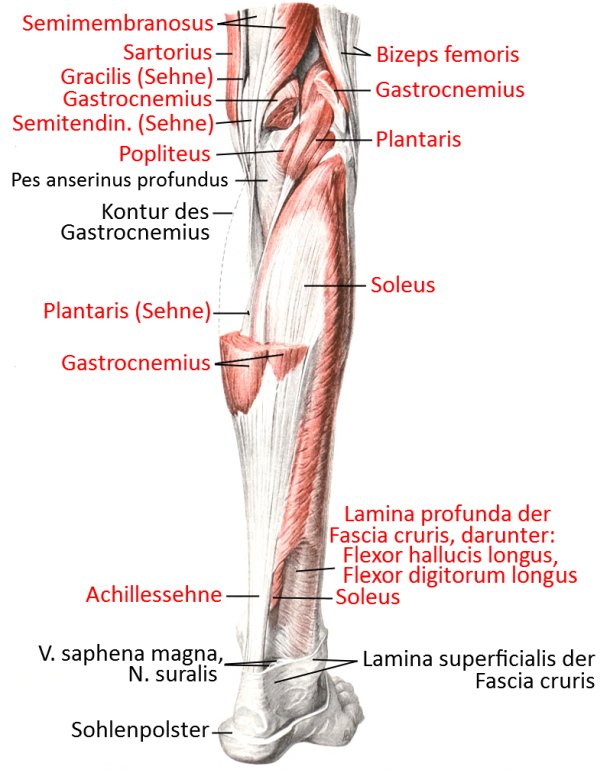
- 9.2 Knee dorsal (popliteal region) with lower leg semi-profound (image links to linkmap)
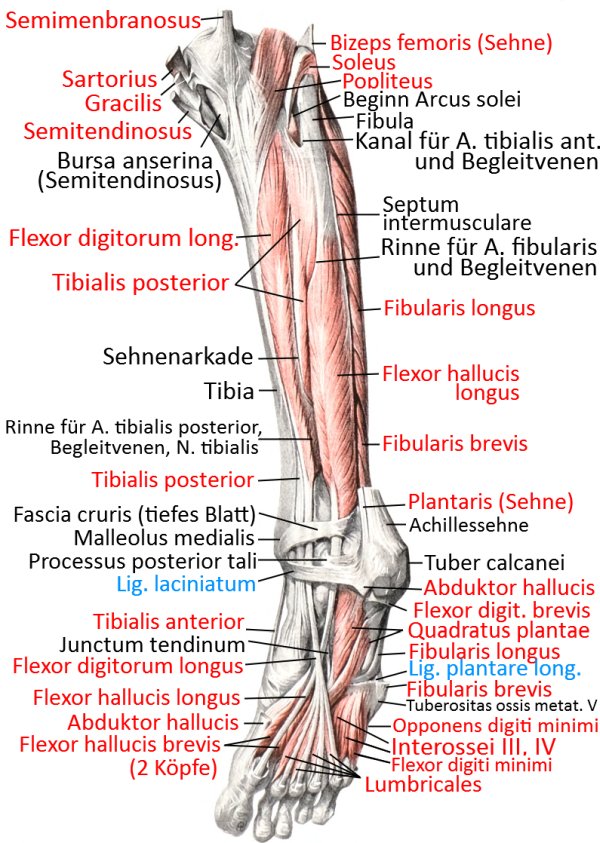
- 9.3 Lower leg, dorsal with plantar base, profound (image links to linkmap)
- 9.4 Lower leg to ankle dorsal (image links to linkmap)
- 9.5 Popliteal region profound (image links to linkmap)
- 9.6 Poplietal region superficial (image links to linkmap)
- 9.7 dorsal, very profound view (image links to linkmap)
- 9.8 dorsal profound view (image links to linkmap)
- 9.9 Dorsal superficial view (image links to linkmap)
- 10 Foot dorsal, deep (image links to linkmap)
- 11 Plantar foot
- 11.1 Overview (image links to linkmap)
- 11.2 Foot medial superficial (image links to linkmap)
- 11.3 Foot medioplantar, extract (image links to linkmap)
- 11.4 Fuß medioplantar deep (image links to linkmap)
- 11.5 Foot medioplantar quite profound (image links to linkmap)
- 11.6 Foot medioplantar superficial (image links to linkmap)
- 11.7 Plantar and sagittal toes (image links to linkmap)
- 11.8 Sole of foot
- 12 Forearm/palmar hand
- 12.1 Overview (image links to linkmap)
- 12.2 Wrist, hand palmar (image links to linkmap)
- 12.3 Muscles (image links to linkmap)
- 12.4 Cross section at the metacarpal level (image links to linkmap)
- 12.5 Pronators and supinators, palmar view (image links to linkmap)
- 12.6 Insertions, palmar view (image links to linkmap)
- 12.7 Profound palmar view (image links to linkmap)
- 12.8 superficial palmar view (image links to linkmap)
- 12.9 superficial view (image links to linkmap)
- 12.10 profound view (image links to linkmap)
- 12.11 very profound view (image links to linkmap)
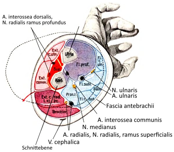
- 12.12 Forearm cross-section
- 13 Forearm, dorsal hand
- 14 Rumpf
- 14.1 Lateral superficial view (image links to linkmap)
- 14.2 Lateral superficial view (2) (image links to linkmap)
- 14.3 Lateral profound view (image links to linkmap)
- 14.4 Lateral very profound view (image links to linkmap)
- 14.5 Lateral mid-profile view
- 14.6 Dorsal view (image links to linkmap)
- 14.7 Dorsal profound view (image links to linkmap)
- 14.8 Cervical spine and thoracic spine from dorsolateral (image links to linkmap)
- 14.9 Back, parts, profound (image links to linkmap)
- 14.10 Neck, dorsal
- 14.11 ventral profound view (image links to linkmap)
- 14.12 Ventral view, semi-profound (image links to linkmap)
- 14.13 ventral superficial view (image links to linkmap)
- 14.14 Abdominal and thoracic cavity from ventral (image links to linkmap)
- 14.15 Posterior abdominal wall from ventral (image links to linkmap)
- 14.16 Chest wall from the inside, dorsal view (image links to linkmap)
- 14.17 Insertions of the chest and neck muscles from the side (image links to linkmap)
- 14.18 Insertions of the chest and neck muscles from the ventral side (image links to linkmap)
- 15 ventral neck muscles (image links to linkmap)
Upper arm/shoulder ventral
Overview (image links to linkmap)
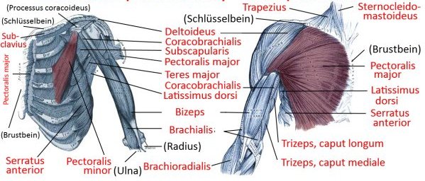
superficial view (image links to linkmap)
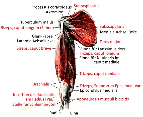
profound view (image links to linkmap)
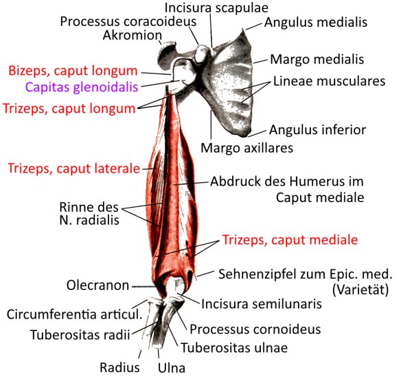
Insertions (image links to linkmap)
Shoulder joint with arm muscles (image links to linkmap)
Shoulder joint with scapula, profound (image links to linkmap)
Shoulder joint with scapula, superficial (image links to linkmap)
Medioventral shoulder joint (image links to linkmap)
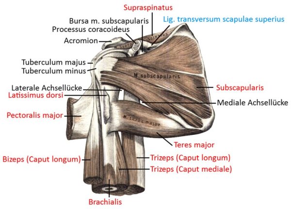
Shoulder from the side (image links to linkmap)
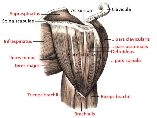
Upper arm/shoulder dorsal (image links to linkmap)
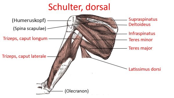
Insertions (image links to linkmap)
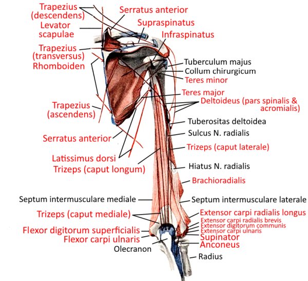
Dorsal upper arm (image links to linkmap)
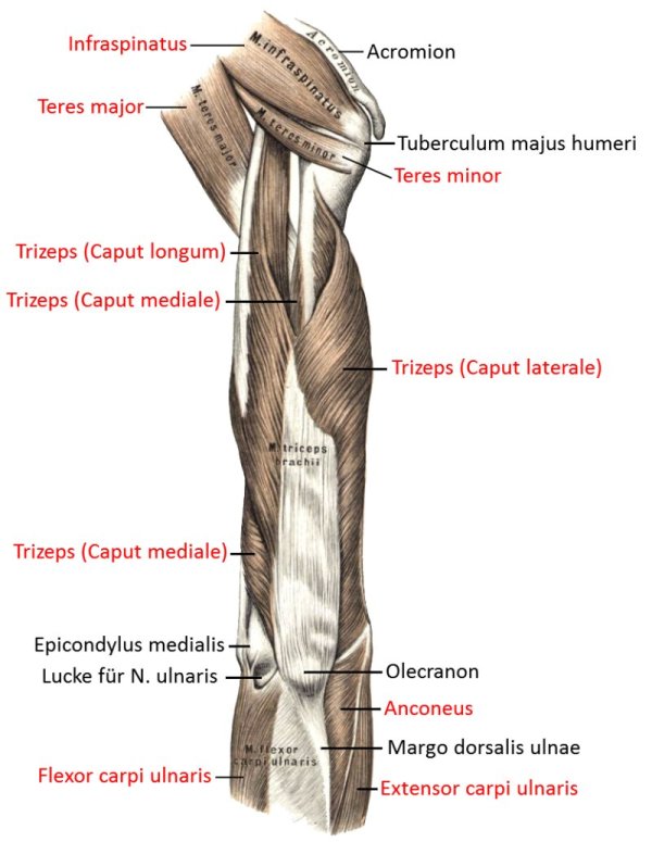
Shoulder joint with scapula (image links to linkmap)
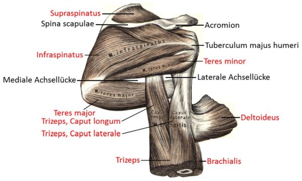
Shoulder joint with scapula, exploded (image links to linkmap)
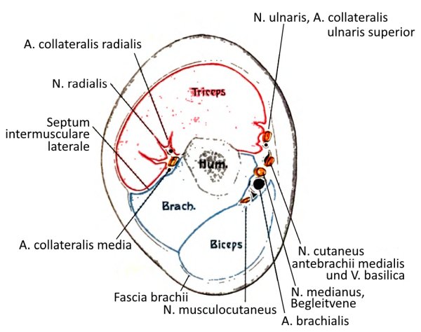
Upper arm cross-section
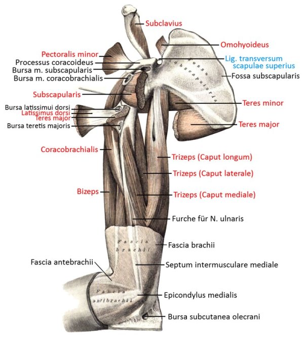
Back, allochthonous musculature (image links to linkmap)
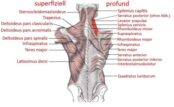
Back, autochthonous musculature (image links to linkmap)
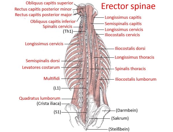
Autochthonous musculature (image links to linkmap)
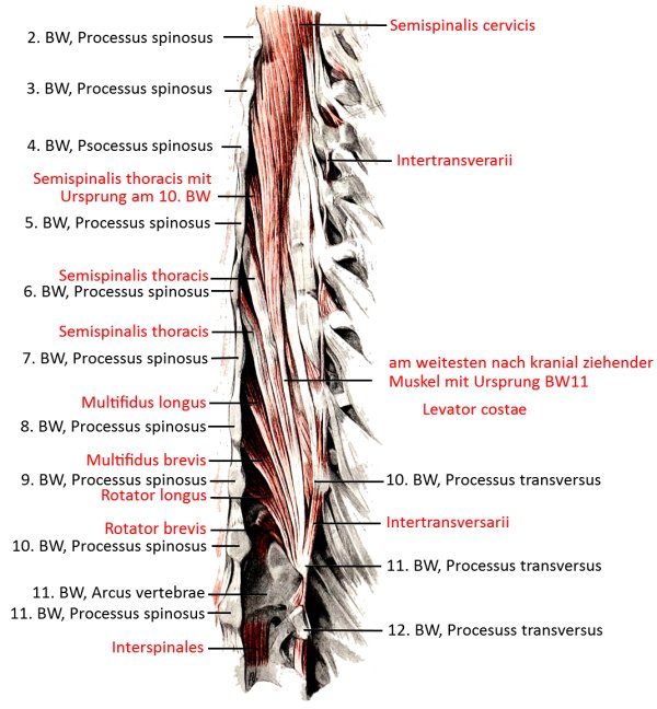
Scheme (image links to linkmap)
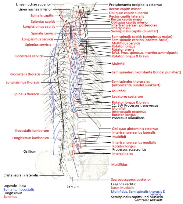
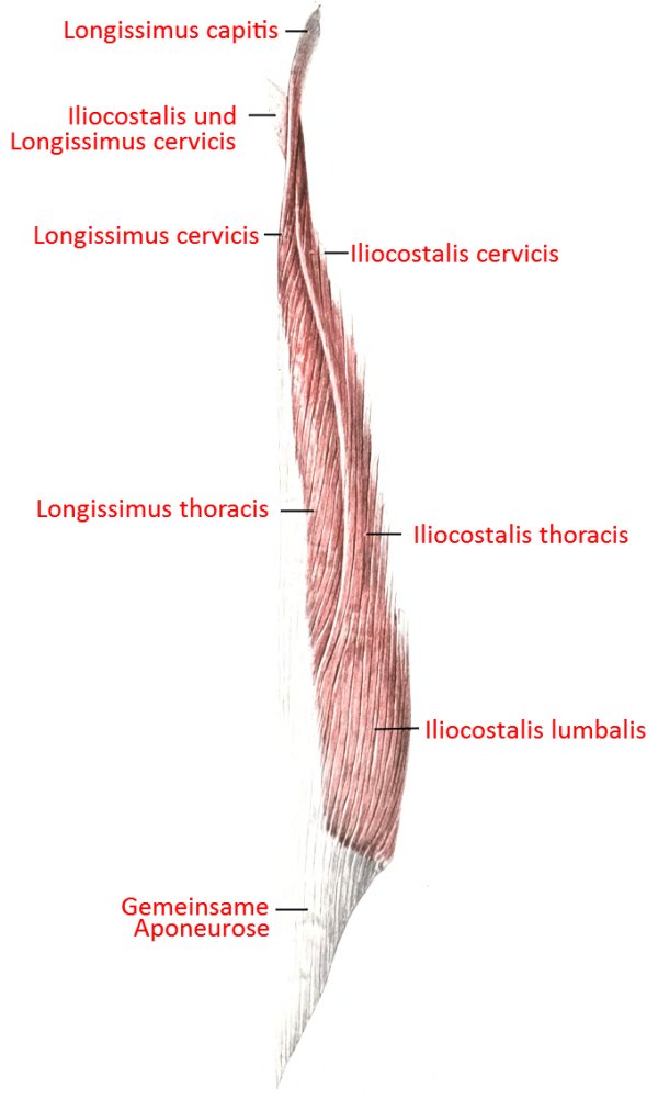
lateral tract (image links to linkmap)
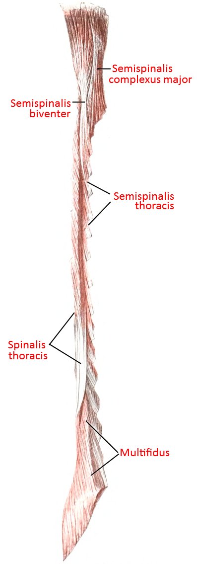
transversospinal tract (image links to linkmap)
Scheme

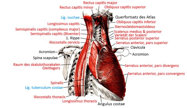
Cervical spine and thoracic spine from dorsolateral (image links to linkmap)
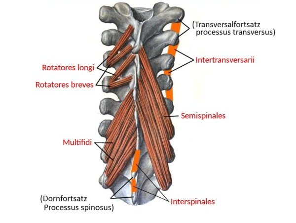
Short intervertebral muscles (image links to linkmap)
Abdominal muscles (image links to linkmap)
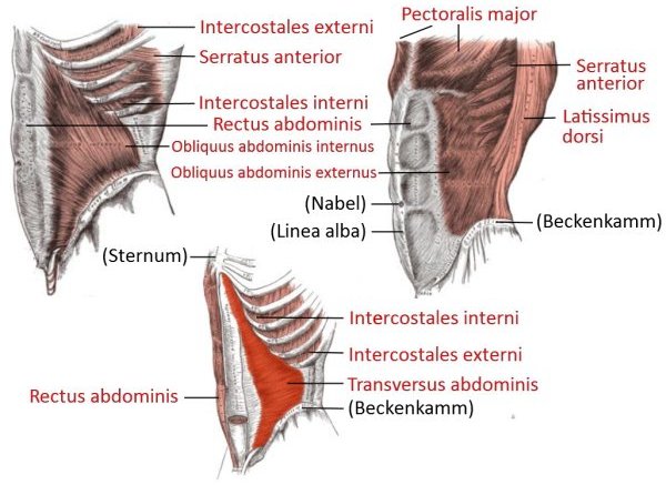
Profound abdominal muscles (image links to linkmap)
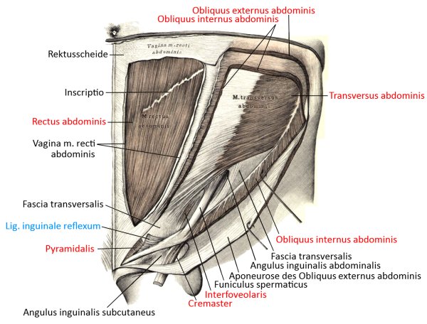
Superficial abdominal muscles (image links to linkmap)

Abdominal muscles, cross-section of the trunk through the lumbar spine region
Thigh, hip ventral
Overview (image links to linkmap)
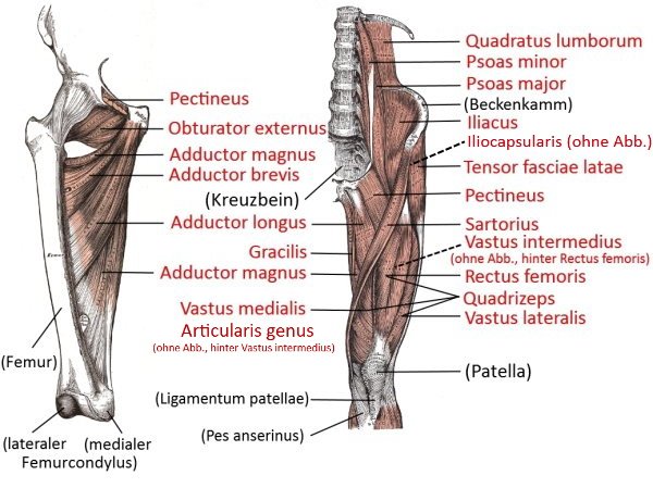
View from ventromedial (image links to linkmap)
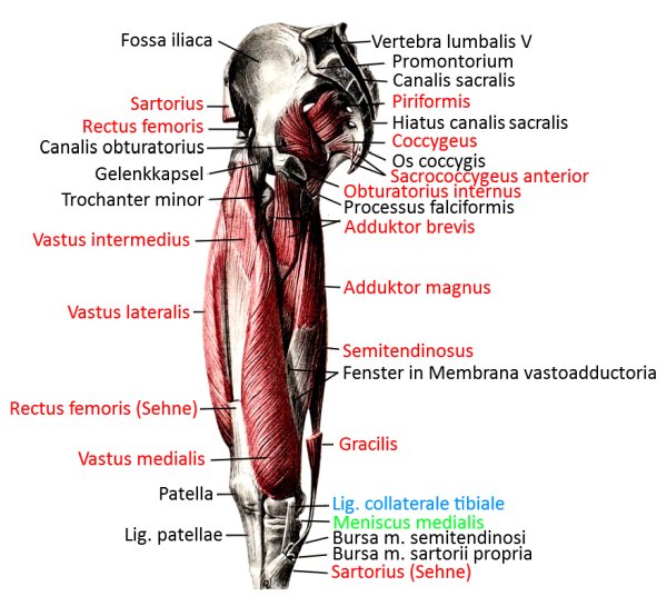
another view from ventromedial (image links to linkmap)
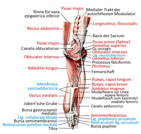
Ventral view (image links to linkmap)
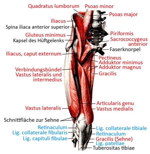
Ventral profound view (image links to linkmap)
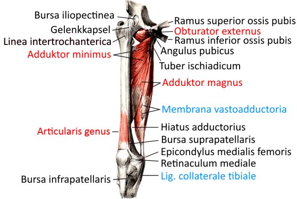
Schematic view (image links to linkmap)
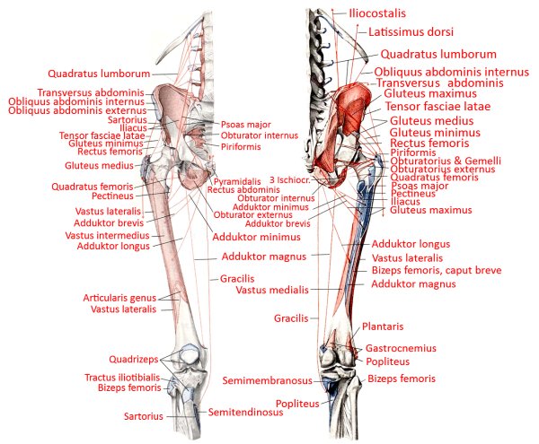
profound view (image links to linkmap)
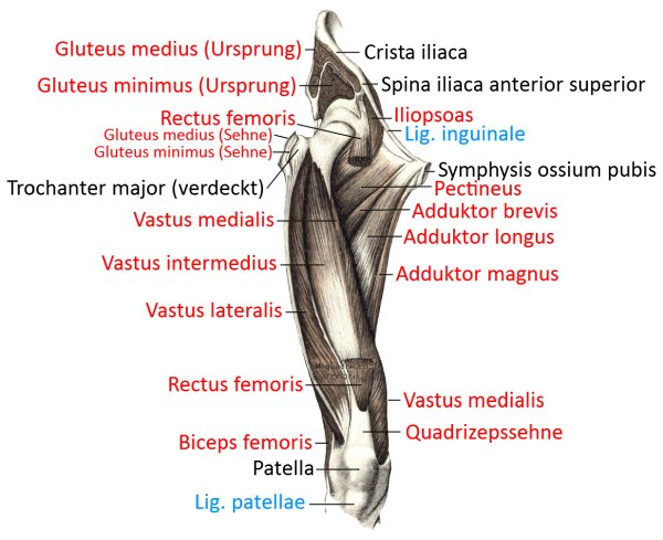
medioventrale, very profound view (image links to linkmap)
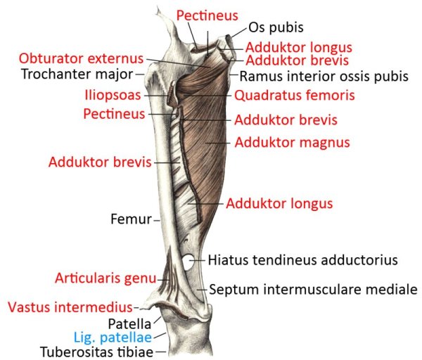
medioventral profound view (image links to linkmap)
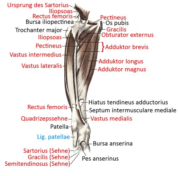
Medial superficial view (image links to linkmap)
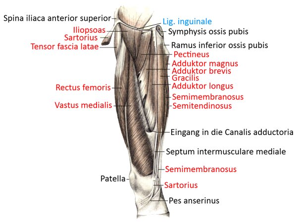
superficial view (image links to linkmap)
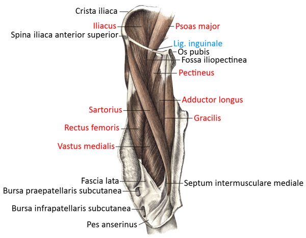
Exploded view (image links to linkmap)
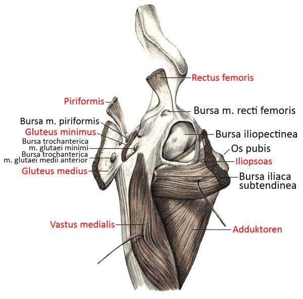
Lateral view
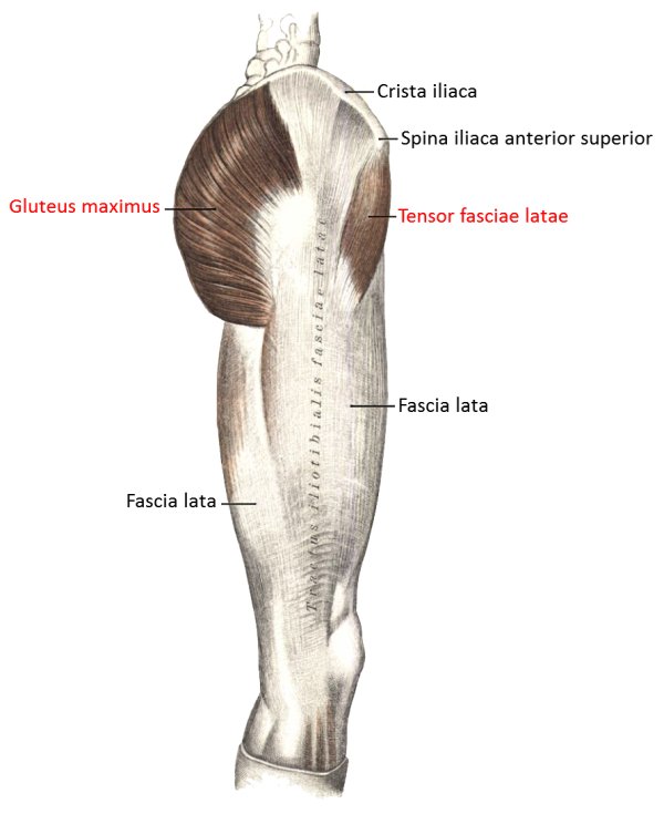
Lateral view of the fasciae
Transversal cut
Hip joint, incision (image links to linkmap)
Thigh, hip dorsal
Overview (image links to linkmap)
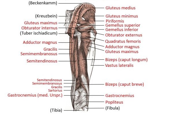
Scheme (image links to linkmap)

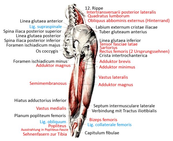
Hip, thigh from dorsolateral (image links to linkmap)
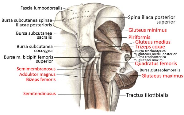
Profound view of the hip region (image links to linkmap)
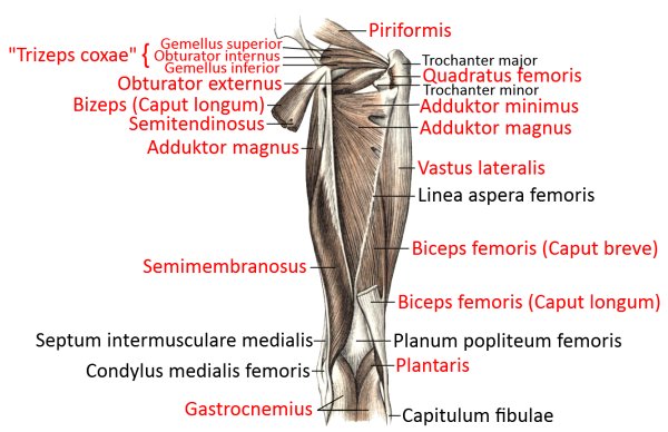
profound view (image links to linkmap)
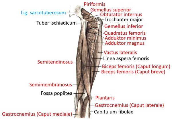
superficial view (image links to linkmap)
Lower leg, foot ventral (image links to linkmap)
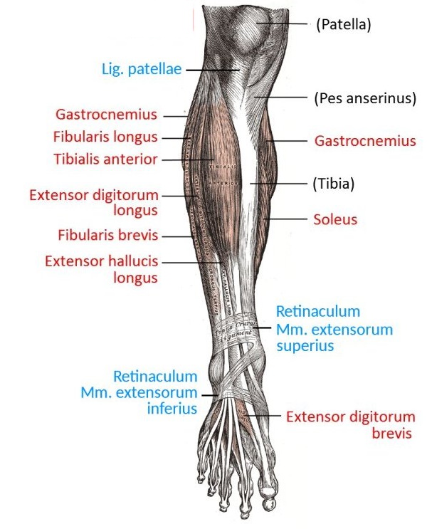
Lower leg from ventral (image links to linkmap)
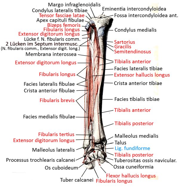
Lower leg superficial from ventral (image links to linkmap)
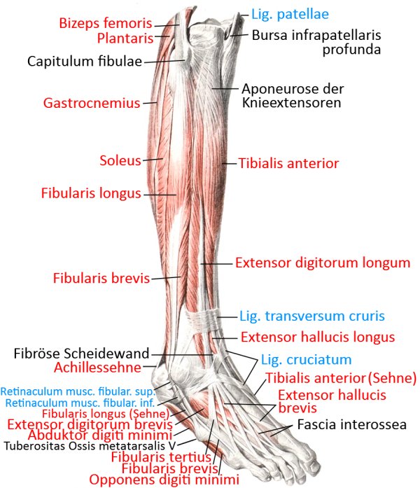
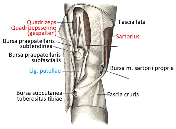
Knee and lower leg ventrolateral (image links to linkmap)
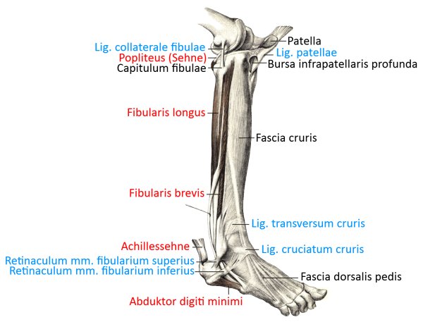
Knee and lower leg ventromedial (image links to linkmap)
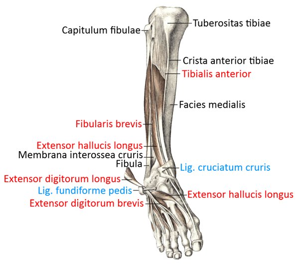
Lower leg, foot ventral, profound (image links to linkmap)
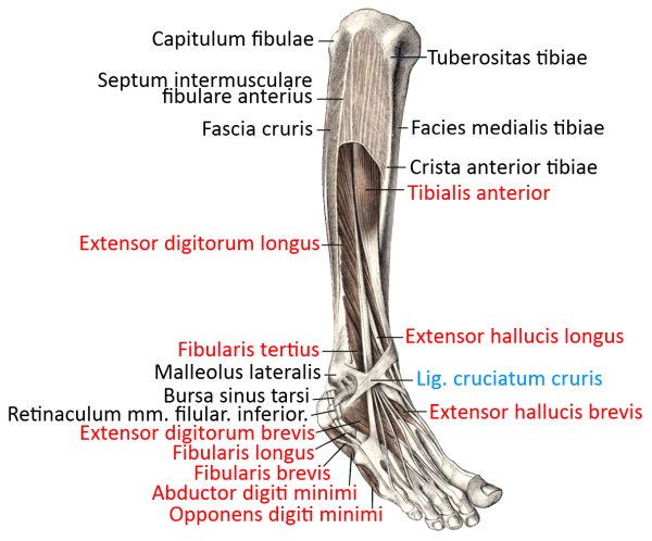
Lower leg, foot ventrolateral superficial (image links to linkmap)
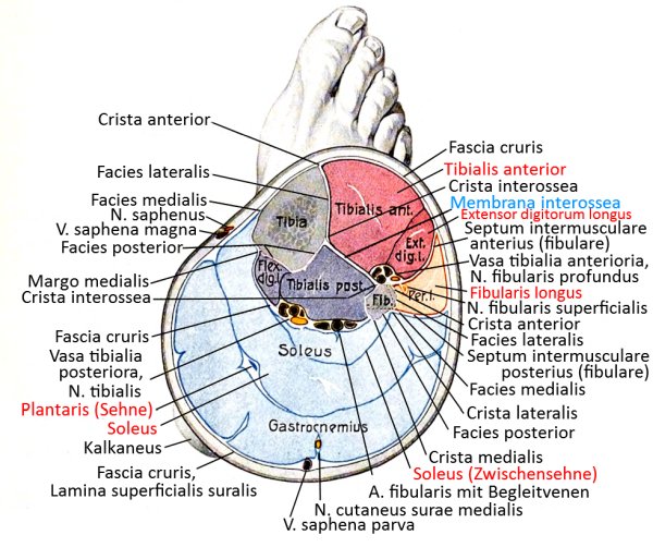
transverse cross-section (image links to linkmap)
Lower leg dorsal, foot plantar
Overview (image links to linkmap)
Knee dorsal (popliteal region) with lower leg semi-profound (image links to linkmap)
Lower leg, dorsal with plantar base, profound (image links to linkmap)
Lower leg to ankle dorsal (image links to linkmap)
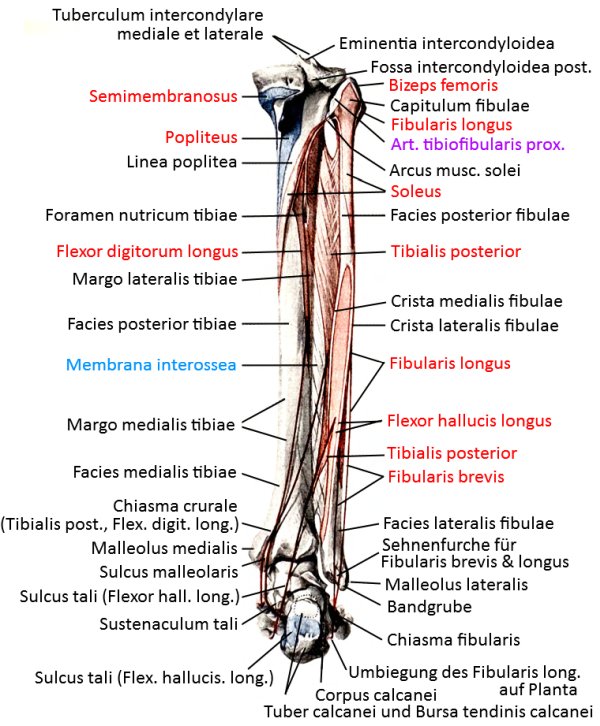
Popliteal region profound (image links to linkmap)
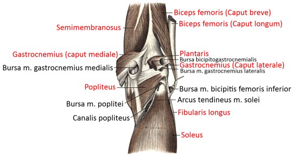
Poplietal region superficial (image links to linkmap)
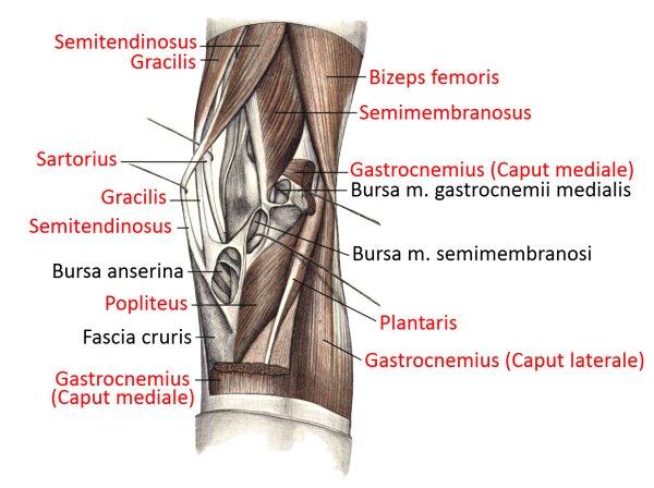
dorsal, very profound view (image links to linkmap)
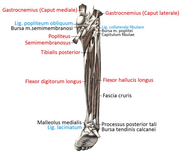
dorsal profound view (image links to linkmap)
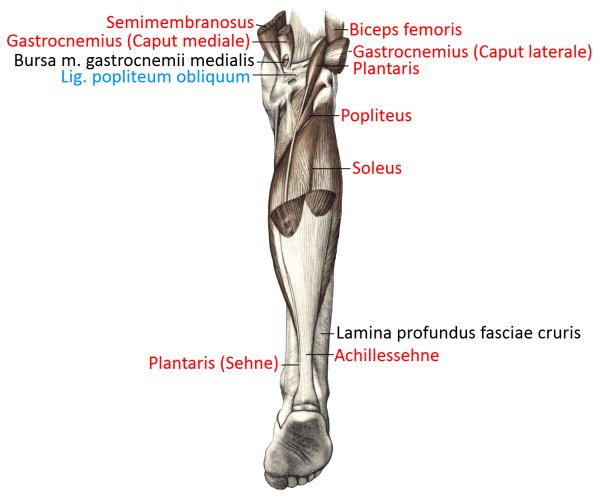
Dorsal superficial view (image links to linkmap)
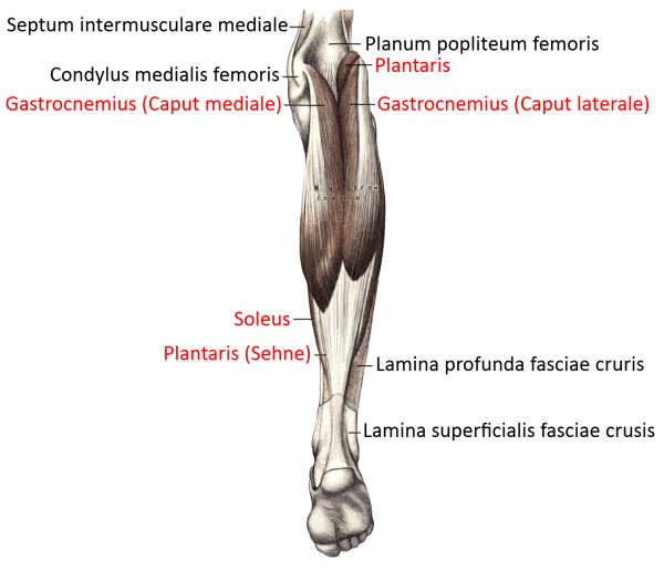
Foot dorsal, deep (image links to linkmap)
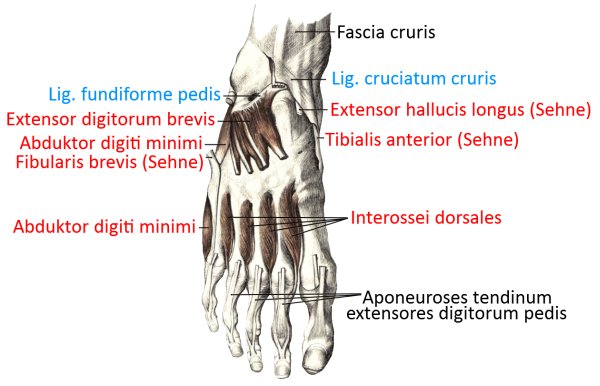
Dorsolateral foot (image links to linkmap)
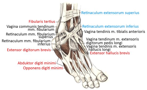
Plantar foot
Overview (image links to linkmap)
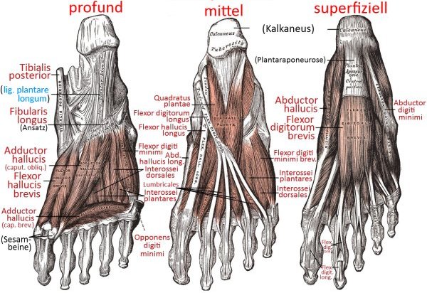
Foot medial superficial (image links to linkmap)
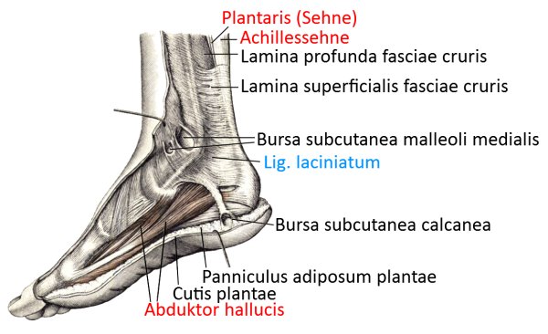
Foot medioplantar, extract (image links to linkmap)

Fuß medioplantar deep (image links to linkmap)
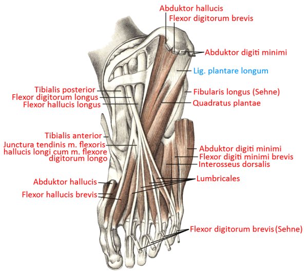
Foot medioplantar quite profound (image links to linkmap)
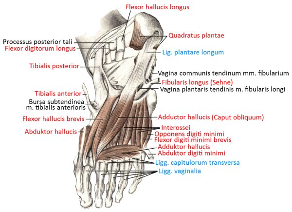
Foot medioplantar superficial (image links to linkmap)
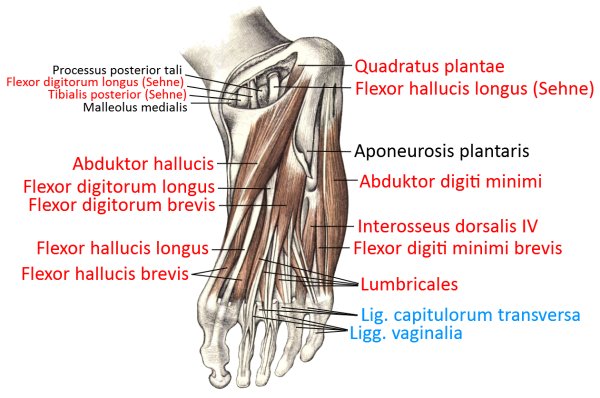
Plantar and sagittal toes (image links to linkmap)
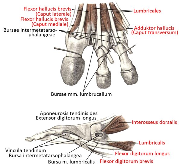
Sole of foot
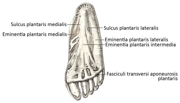
Forearm/palmar hand
Overview (image links to linkmap)
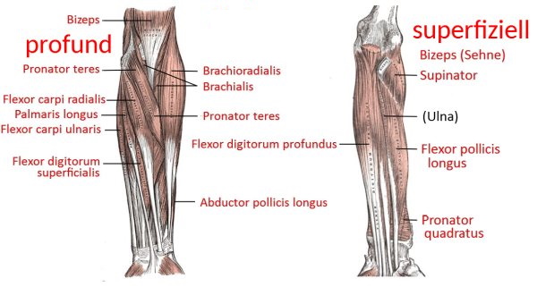
Wrist, hand palmar (image links to linkmap)
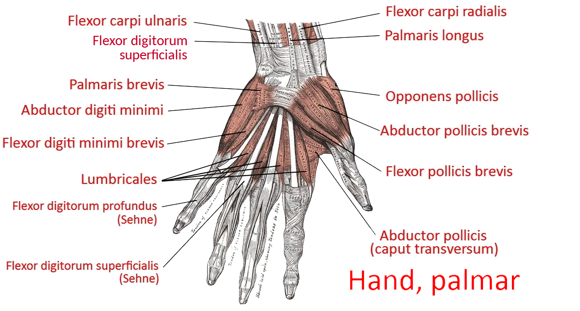
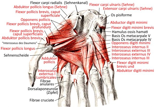
Muscles (image links to linkmap)
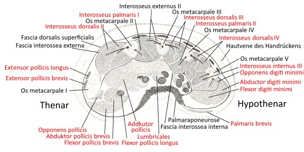
Cross section at the metacarpal level (image links to linkmap)
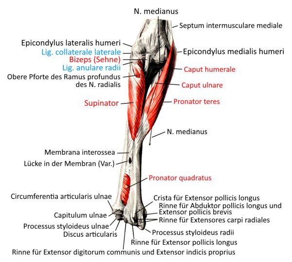
Pronators and supinators, palmar view (image links to linkmap)
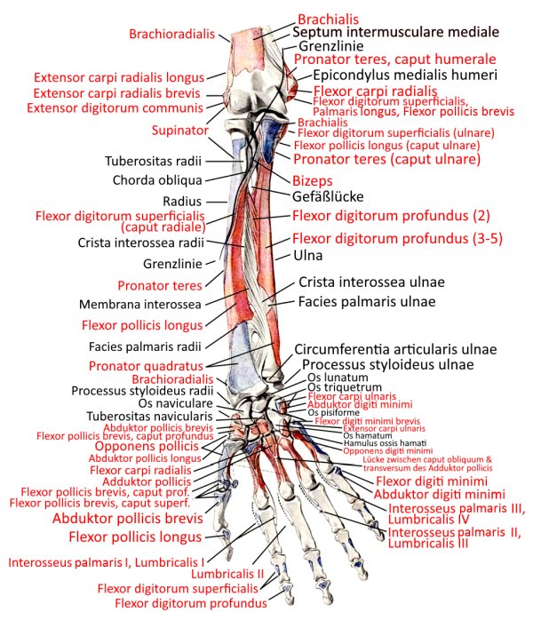
Insertions, palmar view (image links to linkmap)
Profound palmar view (image links to linkmap)
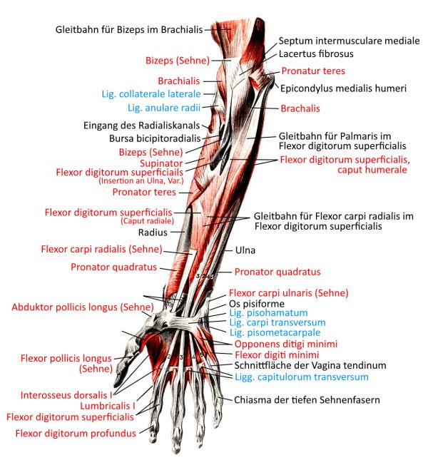
superficial palmar view (image links to linkmap)
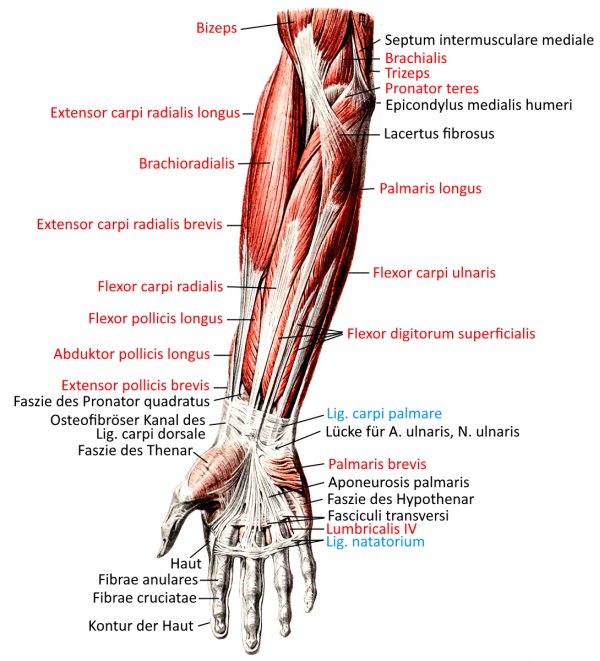
superficial view (image links to linkmap)
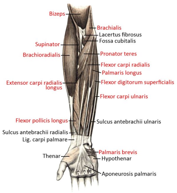
profound view (image links to linkmap)
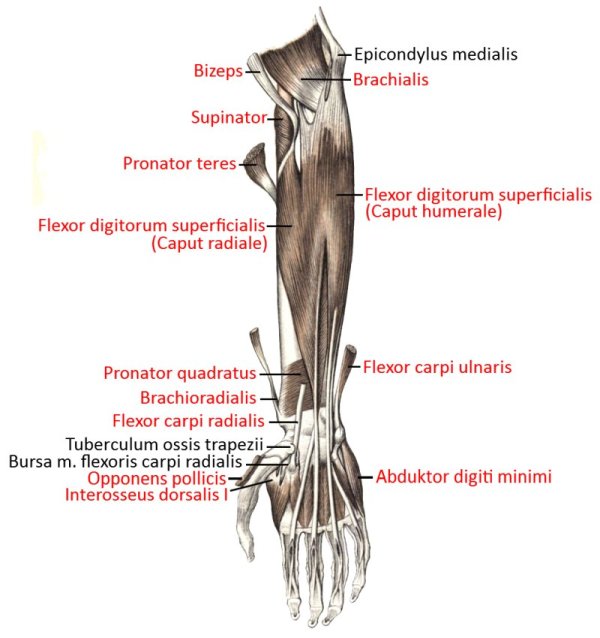
very profound view (image links to linkmap)
Forearm cross-section
Forearm, dorsal hand
Overview (image links to linkmap)
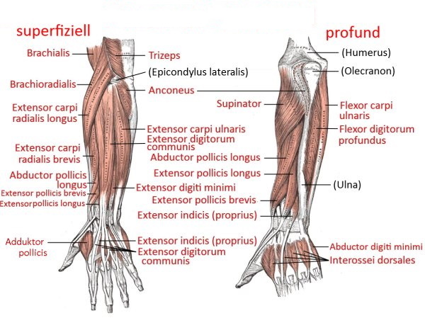
profound view (image links to linkmap)
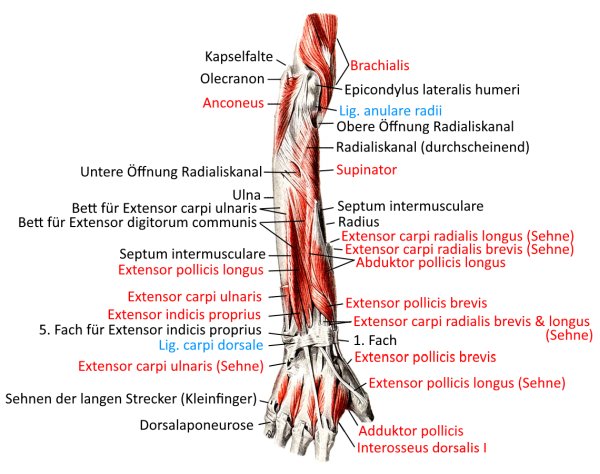
superficial view (image links to linkmap)
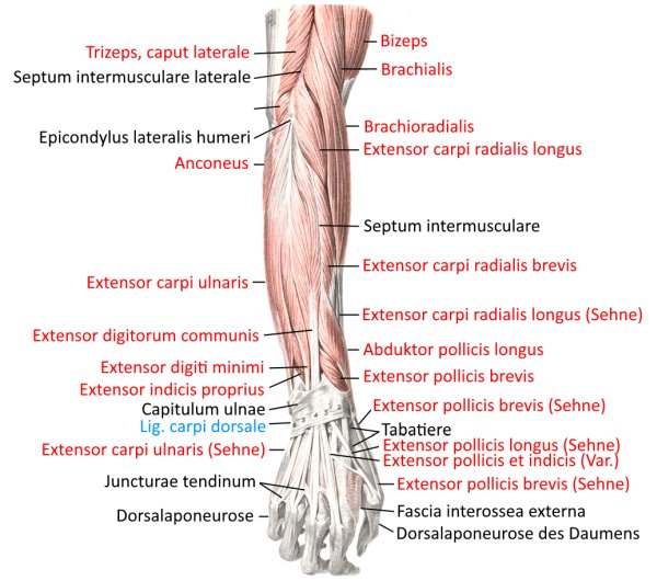
Insertions (image links to linkmap)
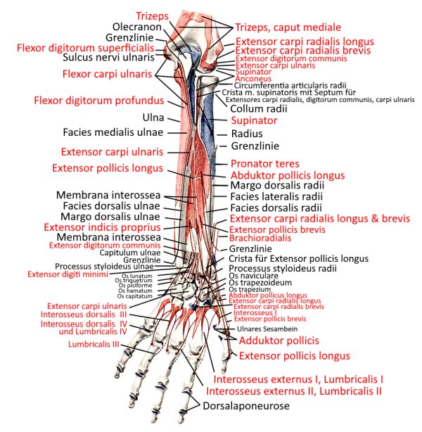
Forearm dorsal from lateral (image links to linkmap)
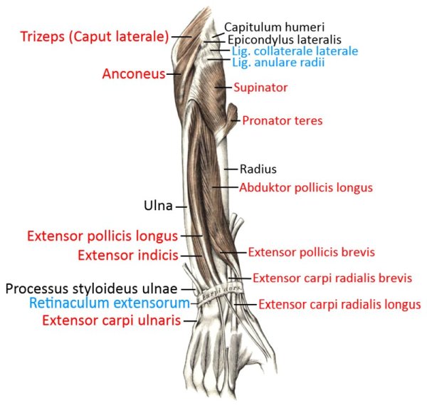
Forearm dorsolateral (image links to linkmap)
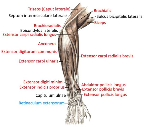
Rumpf
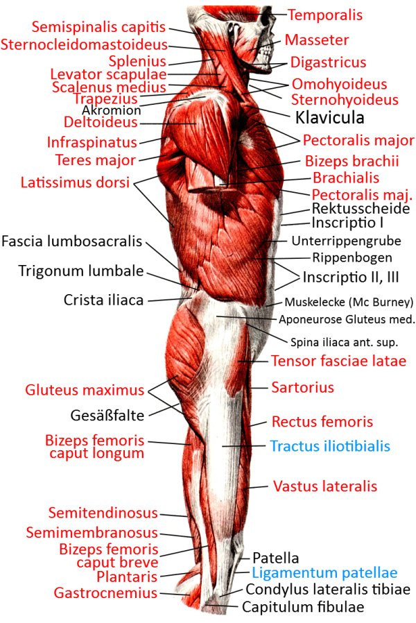
Lateral superficial view (image links to linkmap)
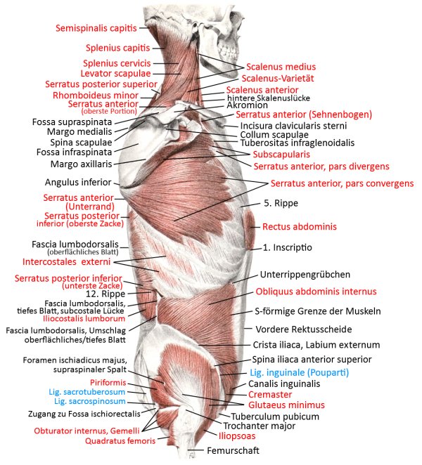
Lateral superficial view (2) (image links to linkmap)
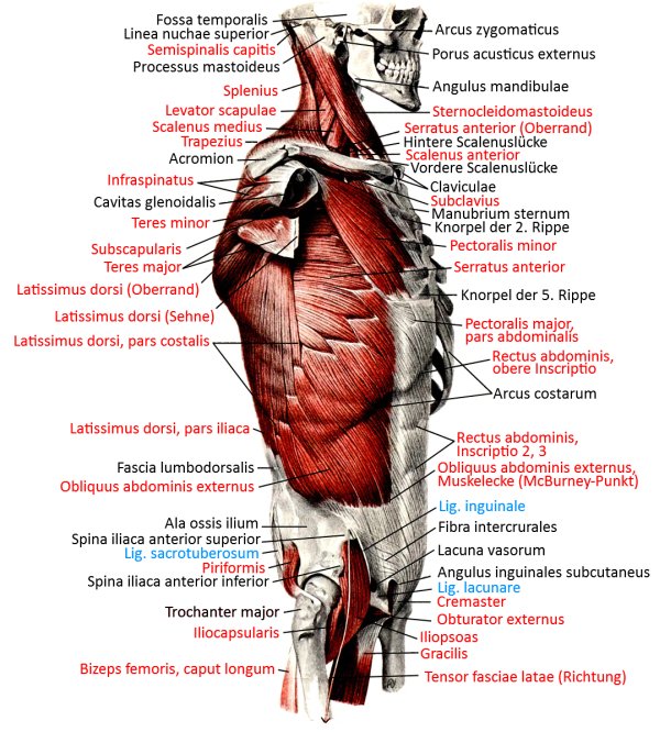
Lateral profound view (image links to linkmap)
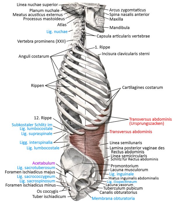
Lateral very profound view (image links to linkmap)
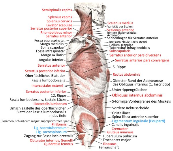
Lateral mid-profile view
Dorsal view (image links to linkmap)
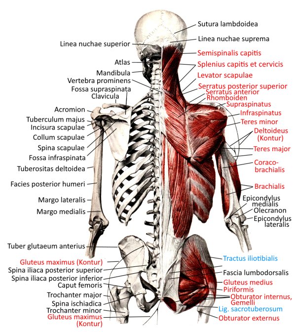
Dorsal profound view (image links to linkmap)
Cervical spine and thoracic spine from dorsolateral (image links to linkmap)

Back, parts, profound (image links to linkmap)
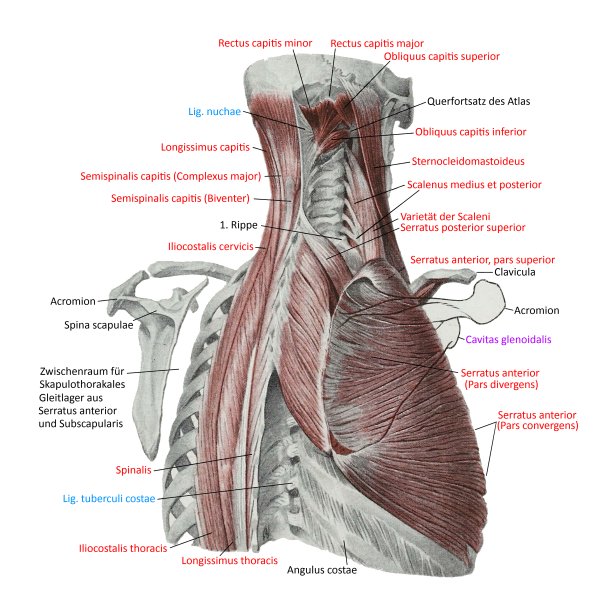
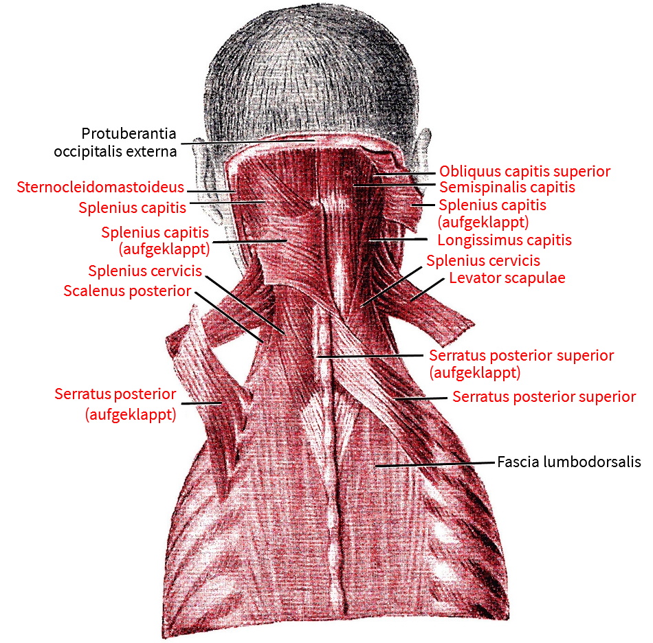
Neck, dorsal
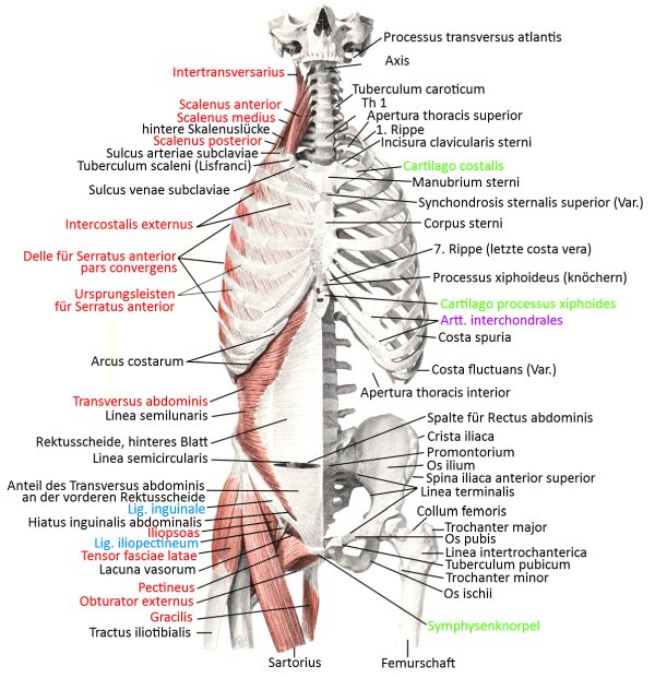
ventral profound view (image links to linkmap)
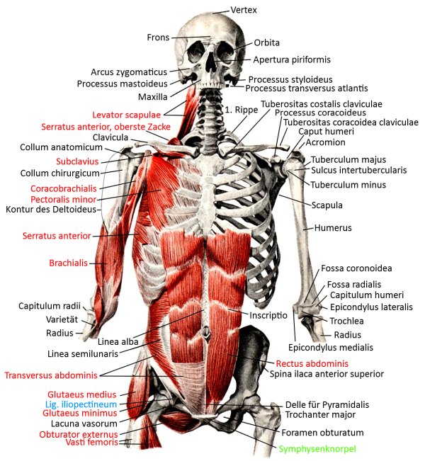
Ventral view, semi-profound (image links to linkmap)
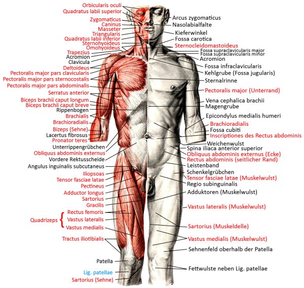
ventral superficial view (image links to linkmap)
Abdominal and thoracic cavity from ventral (image links to linkmap)
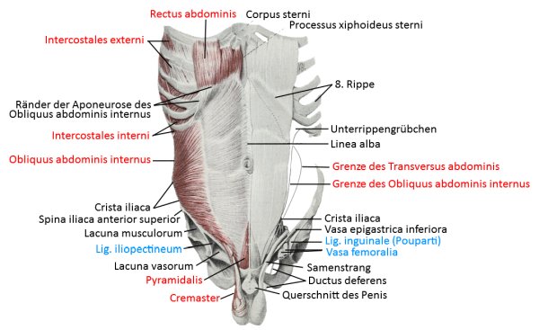
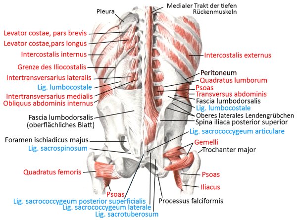
Posterior abdominal wall from ventral (image links to linkmap)
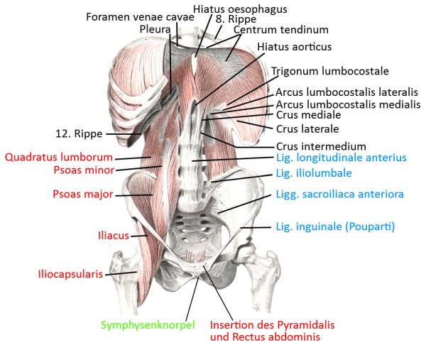
Chest wall from the inside, dorsal view (image links to linkmap)
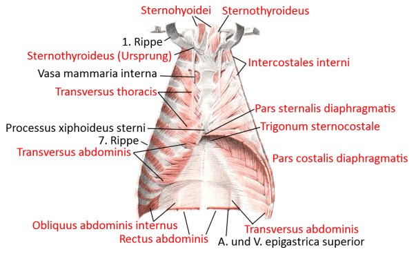
Insertions of the chest and neck muscles from the side (image links to linkmap)
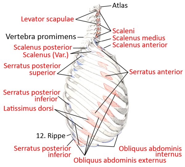
Insertions of the chest and neck muscles from the ventral side (image links to linkmap)
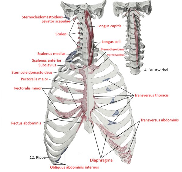
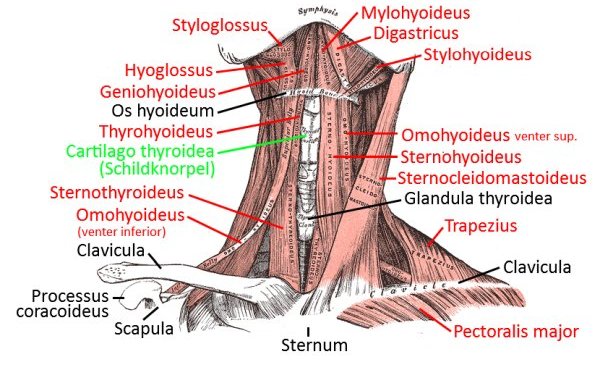
ventral neck muscles (image links to linkmap)
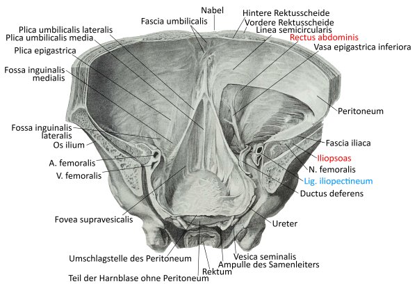
Pelvis from the inside, dorsolateral view
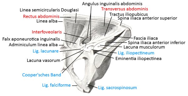
Abdominal wall from diagonally inside
