yogabook / muscles / popliteus

Linkmap
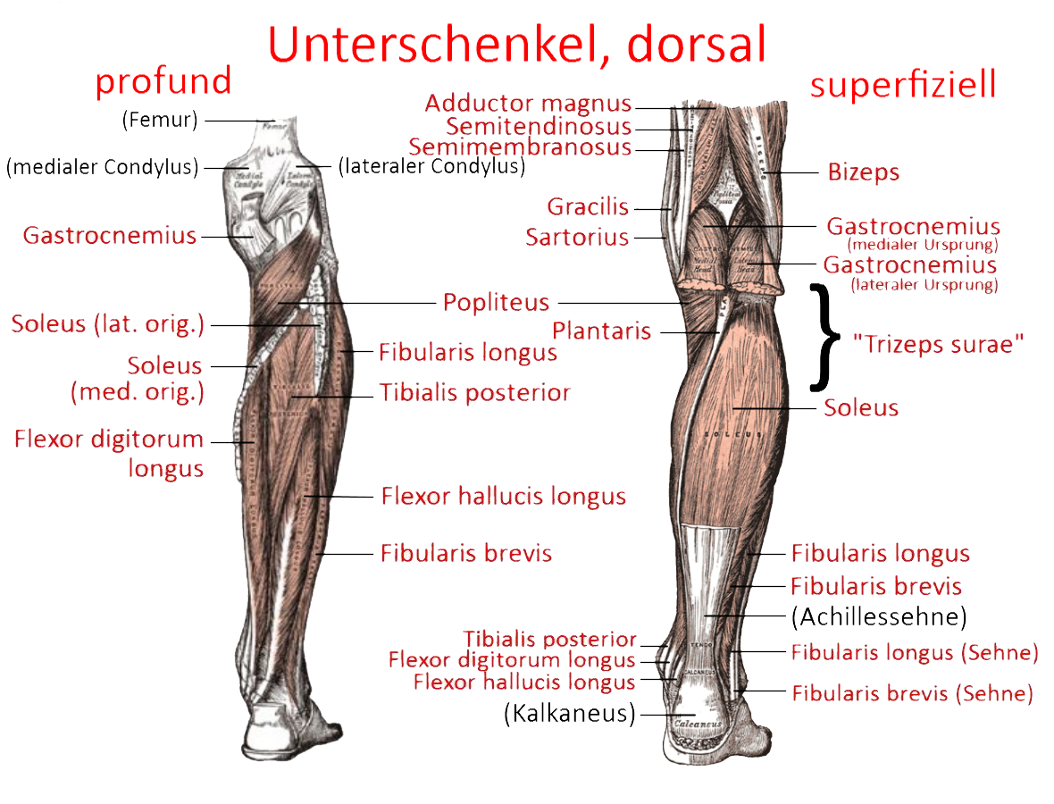
M. popliteus
The elimination of the „physiological dislocation“ called final rotation allows the knee joint to bend and relaxes the collateral ligaments. Its tendon of origin passes through an impression of the lateral femoral condyle. The popliteus is connected to the lateral meniscus and pulls its posterior horn backwards, thus protecting it from entrapment. After the tendon of origin has passed through the approximately 1.3 cm long popliteal hiatus, fibers from the head of the fibula, the lateral meniscus and dorsolateral joint structures are added. The muscle belly and the attachment are then located on the dorsal side of the tibia.
When the knee joint is approximately extended, it has a very weak flexion effect. It pulls on the popliteus arcuate ligament to stabilize the knee joint dorsolaterally. It also stabilizes against varus forces up to 90° of flexion.
Origin: Condylus lateralis femoris of the femur, ventrocaudal to the proximal insertion of the lig. collaterale laterale
Attachment: upper posterior surface of the tibia
Innervation: Nervus tibialis from Plexus sacralis from L4 bis S1
Antagonists:
Movement: with knee joint already strongly flexed: flexion; weak extension with knee joint almost extended. Mainly, however, to stop the final rotation by endorotation of the lower leg in the knee joint.