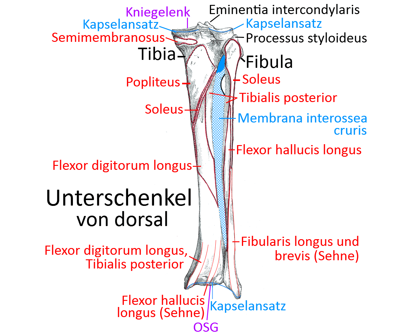
Image: Fibula
Fibula
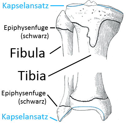
the fibula, one of the two lower leg bones. The fibula lies proximal to the tibia with its „fibular head“, but is not in direct articulation with the femur and is therefore not involved in the knee joint. Distally with the talus, it forms the OSG, in which it surrounds the talus from above and laterally as the lateral part of the malleolar fork. The inner part of the malleolar fork, which is formed by the distal tibia, surrounds the talus from the medial and upper sides.
Fibular head / Caput fibulae
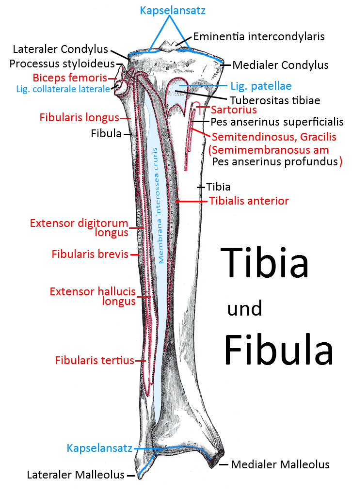
The proximal end of the fibula is known as the fibular head (caput fibulae). This is where the biceps femoris attaches and where the fibularis longus originates. It forms an apex (apex capitis) at the proximal tip. In the medially located facies articularis capitis fibulae, the fibula articulates with the tibia in its facies articularis fibularis at the lateral condyle of the tibia in the proximal tibiofibular joint. The interosseous membrane is extremely important for the transmission of force from the talus via the lateralmalleolus and the rest of the fibula towards the femur. In particular, it prevents lateral dislocation of the caput fibulae onto the medial caput tibiae.
Collum fibulae
The neck of the fibula lies between the caput and the shaft (corpus).
Corpus fibulae
The corpus of the fibula can be divided into three sharp edges: Margo anterior, posterior and interosseus. The interosseous membrane stretches between the interosseous fibular margin and the interosseous tibial margin. On the posterior surface of the fibular shaft, the crista medialis divides the origins of the posterior tibialis (more ventral) from that of the flexor hallucis longus (more dorsal).
Lateral malleolus / Malleolus lateralis
The lateral malleolus is the lateral border of the talus , also known as the „lateral malleolus“. With its medially located facies articularis malleolaris lateralis, it articulates with the tibia before articulating with the talus further inferiorly as the malleolus lateralis.
Joints
Pictures
Tibia and fibula from ventral view (image links to linkmap)
Tibia and fibula from the dorsal side (image links to linkmap)