yogabook / muscles / tibialis posterior
Linkmap
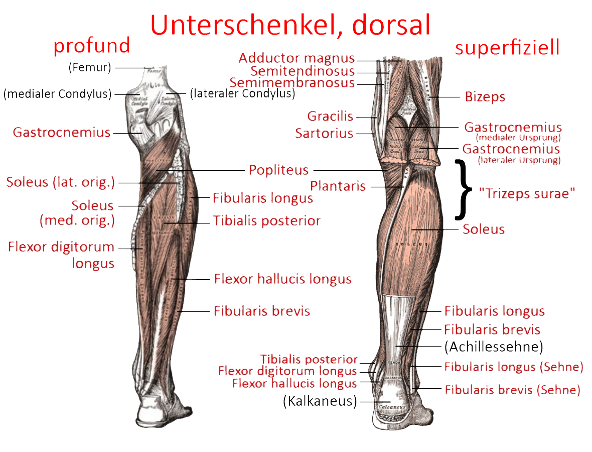
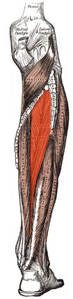
Tibialis posterior
Tibial muscle at the back of the calf, relatively deep, which causes lifting of the foot(dorsal flexion) and supination. The function of the tibialis poterior must be differentiated according to stance and rolling movement.
- Stance: Stabilization of the subtalar joint by inversion and centring of the calcaneus under the talus. Block of the Chopar joint due to the tensile forces coming from the medial side, thus tensioning the transverse arch of the foot and the medial longitudinal arch of the foot.
- Rolling: Elevation of the calcaneus (see Single heel rise test); distal/ventral running tendon parts hold the longitudinal arch of the foot and thus stabilize the forefoot-rearfoot lever arm
The tendon of the posterior tibialis runs through the tarsal tunnel between the calcaneus and the medial malleolus alongside the flexor digitorum longus and flexor hallucis longus muscles. The posterior tibial artery and the tibial nerve also run from there, which can lead to tarsal tunnel syndrome if it becomes constricted. Dysfunction or insufficiency of the posterior tibialis may also be involved in the development of plantar fasciitis. Furthermore, this muscle is probably the one that is responsible for the symptoms in most cases of tibial tunnel syndrome. Insufficiency of the tibialis posterior can also be involved in the development of acquired flat feet and fallen arches as tibialis posterior syndrome: if the tendon, which only has a stroke of approx. 2 cm, is stretched, this leads to correspondingly less tension on the insertion areas, resulting in a hindfoot valgus due to the supinating effect of the tibialis posterior. This shifts the attachment point of the Achilles tendon laterally, which reinforces the hindfoot valgus.
Origin: Membrana interossea, posterior surface of tibiaand fibula as well as with the ramus sustentacularis and ramus plantaris on the calcaneus
Attachment: Tuberositas ossis navicularis (main attachment), Os cuneiforme mediale, base of metatarsals 2-4, occasionally also Os metatarsale 1
As constant attachment are considered:
- Tuberositas ossis navicularis (Hauptansatz, ca ca. 12 × 7 mm)
- Os cuneiforme mediale plantar/medial
Os cuneiforme laterale: plantar (sehr häufig) - Basis der Metatarsalia 2-4, ggf. auch 1 und 5
- Sustentaculum tali: kleiner, rezidivierender Rezessionsast („recurrent slip“)
Inconstant are considered:
- Os cuneiforme intermedium (ca. 20%)
- Os cuboideum (ca. 40–50%)
- Zusätzliche Metatarsalbasen (I und V)
- Zusätzliche Calcaneus-Anteile jenseits des Sustentaculum tali (selten, je nach Studie 10–30%)
Innervation: N. tibialis
Antagonists:
Movement: Plantar flexion, supination, adduction of the foot. Stabilization and maintenance of the longitudinal arch of the foottogether withfibularis longusand caput transversum of the adductor hallucis longus; slight support of the transverse arch of the foot
Strengthening postures ():
The main exercises for direct strengthening are heel raises, tibialis posterior training on slantboard, tibialis posterior training with resistance band.
As a plantar flexor, it also plays a stabilizing role in many postures with a balancing character and in those that have shifted the centre of gravity from the heel to the toes and in which the foot supports a large load: 1st warrior stance, 3rd warrior stance, 3rd warrior stance: backwards against the wall, 3rd warrior stance: backwards against the wall, 3rd warrior stance: backwards against the wall. Warrior pose: backwards against the wall, parsvottanasana, parivrtta trikonasana, parivrtta ardha chandrasana, parivrtta parsvakonasana, ardha chandrasana, uttanasana: eka pada prasarita, vrksasana, hasta padangusthasana, tadasana: eka pada prasarita, uttanasana: eka pada prasarita, John’s sequence, deadlift, exercise supination of the foot
Stretching poses (): malasana, squat 1, squat 2