Contents
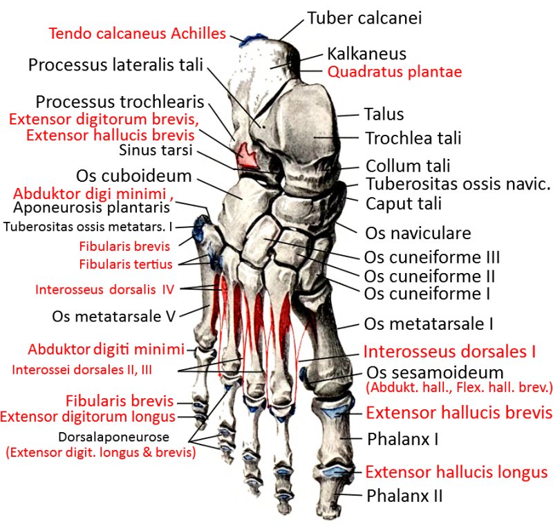
Image: Foot bone from dorsal (image links to linkmap)
The bones of the foot
The bones of the foot consist of the tarsal bones (tarus), the metatarsal bones (metatarsus) and the toes(digiti) with theirphalanges. There is a fundamental similarity to the hand, but there are also some differences, which result, among other things, from the fact that in anatomically zero and many standing everyday postures the body weight rests on the foot, which is at a variable angle of around 90° to the lower leg for stability and rapid locomotion.
Tarsal bone
The calcaneus(heel bone) and talus(ankle bone) form the first row of tarsal bones, while the navicular bone (os naviculare), cuboid bone (os cuboideum) and the three cuneiforms (os cuneiforme mediale, intermedium and laterale) form the distal row. The calcaneus articulates mainly with the cuboid bone and only just touches the navicular bone, which in turn articulates mainly with the talus.
Calcaneus(heel bone)
Tarsal bone of the first row. The calcaneus is the bone on which the talus stands and which, in standard anatomical position, stands on the ground as the dorsal part. The Achilles tendon attaches dorsally to the calcaneus.
Ankle bone (talus)
Tarsal bone of the first row. The talus is located on both sides in the malleolar fork consisting of the lateralmalleolus (part of the fibula) and the medialmalleolus (part of the tibia) and also articulates cranially with the tibia in the ankle joint. Inferiorly, the calcaneus is connected dorsally in the subtalar articulation and the scaphoid and cuboid bone ventrally in the talocalcaneonavicular articulation.
Tarsal bone of the second row. The scaphoid articulates distally with the three cuneiform bones, laterally with the cuboid bone and proximally in the Chopart joint line with the calcaneus and talus.
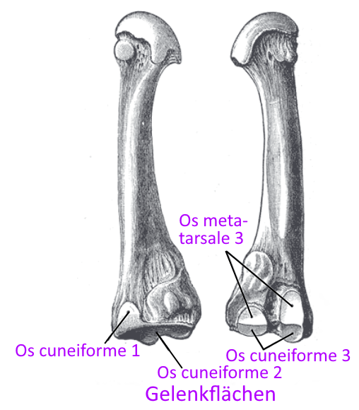
Sphenoid bones (Ossa cuneiformia 1 – 3)
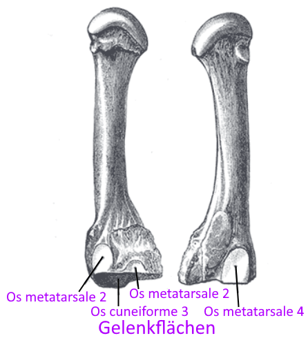
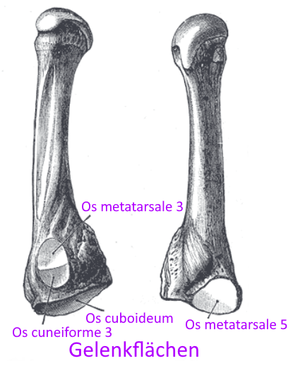
Tarsal bones of the first row, which lie three next to each other and border on the ossa metatarsalia in the Lisfranc joint line.
Cuboid bone (Os cuboideum)
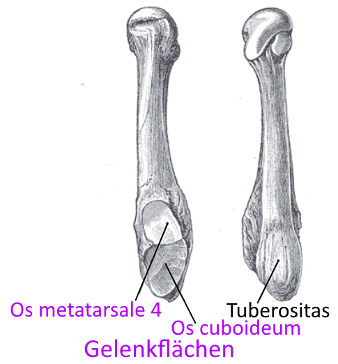
Tarsal bone of the first row, adjacent to the metatarsal bone in the Lisfranc joint.
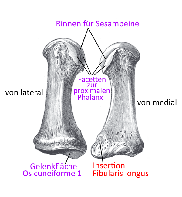
Metatarsals
The metatarsal bones or metatarsals connect the tarsus to the toes. They are aligned longitudinally along the foot like a ray, and each metatarsal bone is connected to a toe via the metatarsophalangeal joint (MTP).
Digiti (toes)
Like the fingers, the toes, apart from the hallux, consist of three limbs(phalanges) that are connected to each other by the interphalangeal joints. The proximal phalange is connected to the metatarsal bones in the MTP (metatarsophalangeal joint).
Phalanges
In the case of toes 2-5, thephalanges are subdivided into
- proximal phalanges, which articulate with the metatarsals in the metatarsophalangeal joint and with the medial phalanges in the proximal interphalangeal joint
- medial phalanges that articulate with the proximal and distal phalanges in the(proximal and distal) interphalangeal joints
- distal phalanges, which connect to the medial phalanges in the distal interphalangeal joint and contain the end of the fingers in the form of the fingertip with nail
Like the pollex, the hallux consists of only two phalanges.
Hallux
Like the pollex (thumb), the hallux (big toe) consists of only two phalanges.
Tapes
For the ligaments, see the ankle.
Anatomical varieties: Accessory foot bones
Os tibiale externum
The external tibial bone is an additional scaphoid bone, located on the medial side of the foot, proximal to the navicular bone with contact to the insertion tendon of the posterior tibialis, which is usually fused to it, predisposing to the development of a flat foot. The connection to the navicular bone is usually fibrous or cartilaginous, but often becomes completely or partially bony by the end of the growth phase and is then called the navicular bone. If the Os tibiale externum is removed, this usually improves the flat foot, which, however, is not an indication for this operation if there are no symptoms.
Supernumerary bone located dorsally on the proximal edge of the navicular bone.
Os supratalar
Supernumerary bone located dorsally on the talus towards the navicular bone, which is usually located somewhat further laterally than the supranavicular bone.
Os trigonum
Supernumerary bone located in the dorsal talocalcaneal angle, which must be differentiated from a torn posterior talar process.
Os vesalianum
Supernumerary bone located at the base of the Os matatarsale 5, usually lateral to the Os cuboideum, which must be differentiated from a torn Os metatarsale 5 and can fuse with it.
Os fibulare
Often several bone fragments in the tendon of the fibularis longus, seen laterally at the level or plantar of the cuboid bone.
Anatomical varieties of the toes (common)
Polydactyly
Presence of a sixth toe. If a corresponding metatarsal bone is also present in excess, the entire ray can be resected.
Syndactyly
Two toes growing together
Pictures
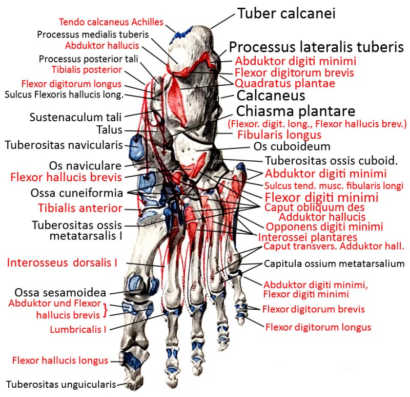
Foot bones from plantar (image links to linkmap)
Bones from dorsal (image links to linkmap)
Bones from plantar (image links to linkmap)
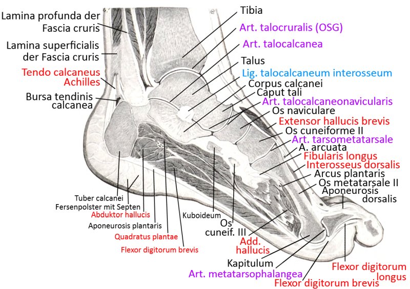
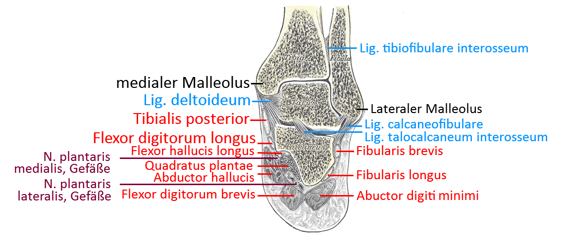
Section in the frontal plane at the level of the talocrural joint
Foot from medial (image links to linkmap)