yogabook / bones / hand bones, finger
Contents
- 1 Images: Joint lines of the hand
- 2 The bones of the hand
- 3 Images
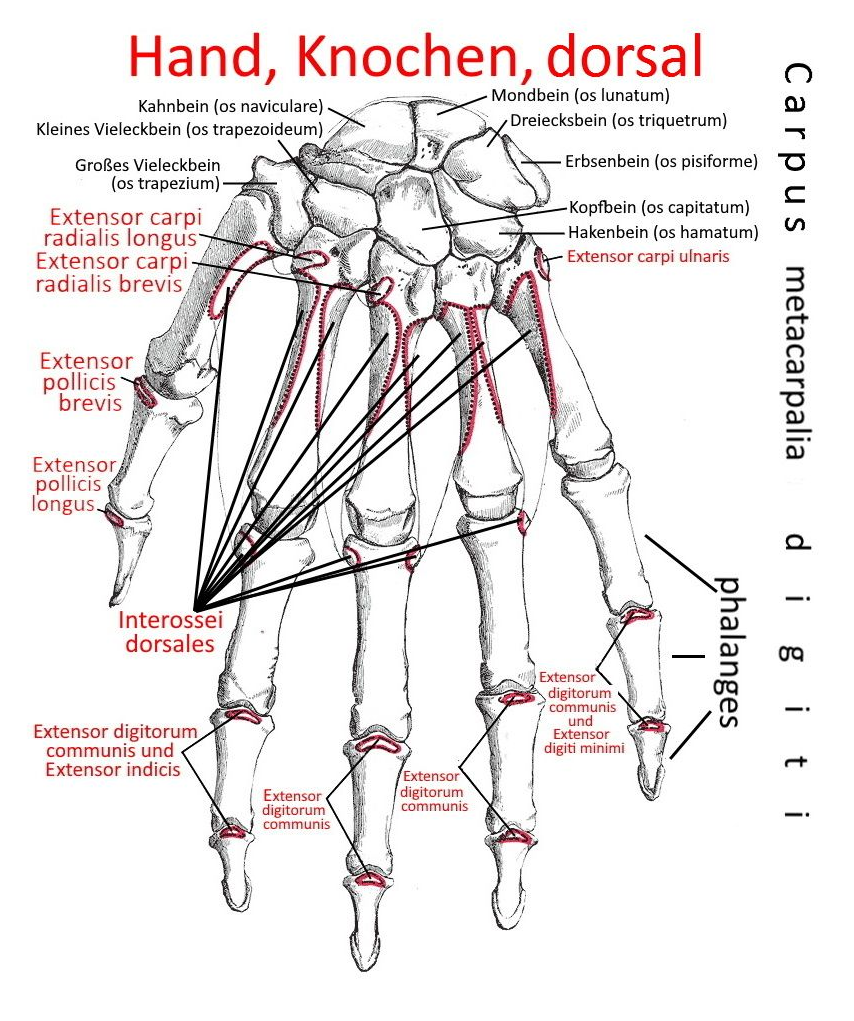
- 3.1 Hand, dorsal (image links to linkmap)
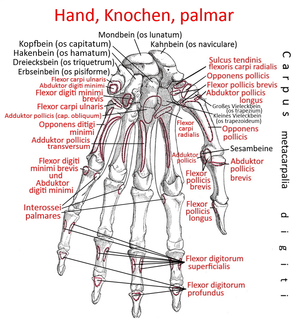
- 3.2 Hand, palmar (image links to linkmap)
- 3.3 Ligaments (image links to linkmap)
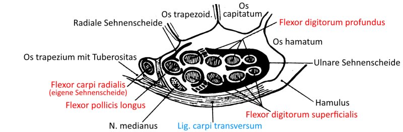
- 3.4 Carpal tunnel (image links to linkmap)
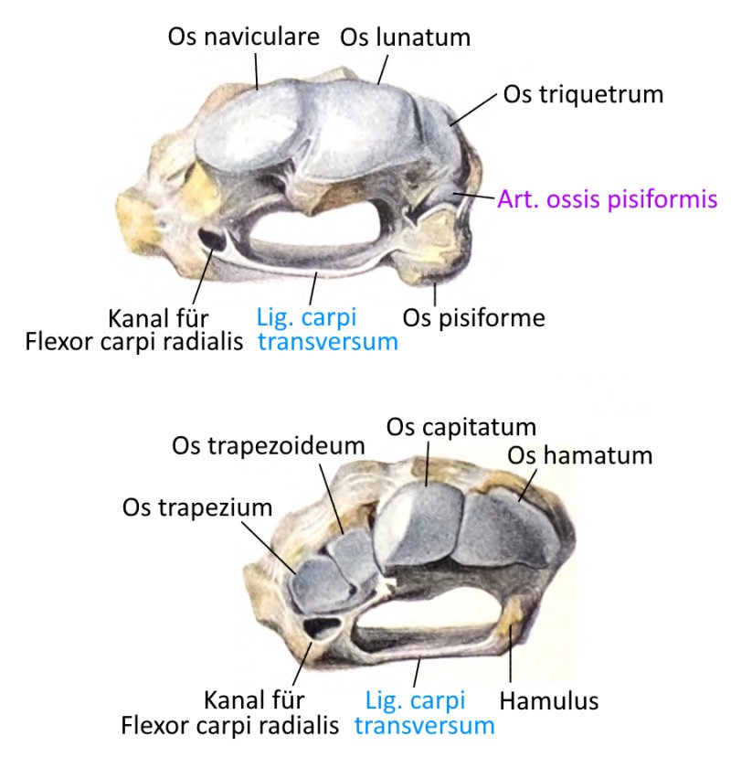
- 3.5 Carpal tunnel (other view)
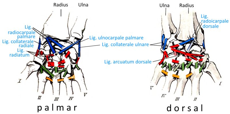
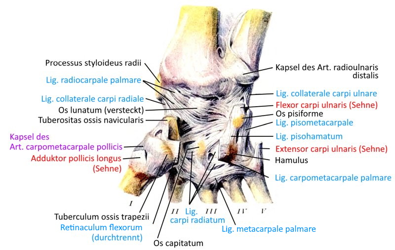
- 3.6 Ligaments, palmar (image links to linkmap)
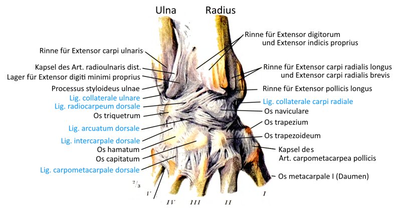
- 3.7 Ligaments, dorsal (image links to linkmap)
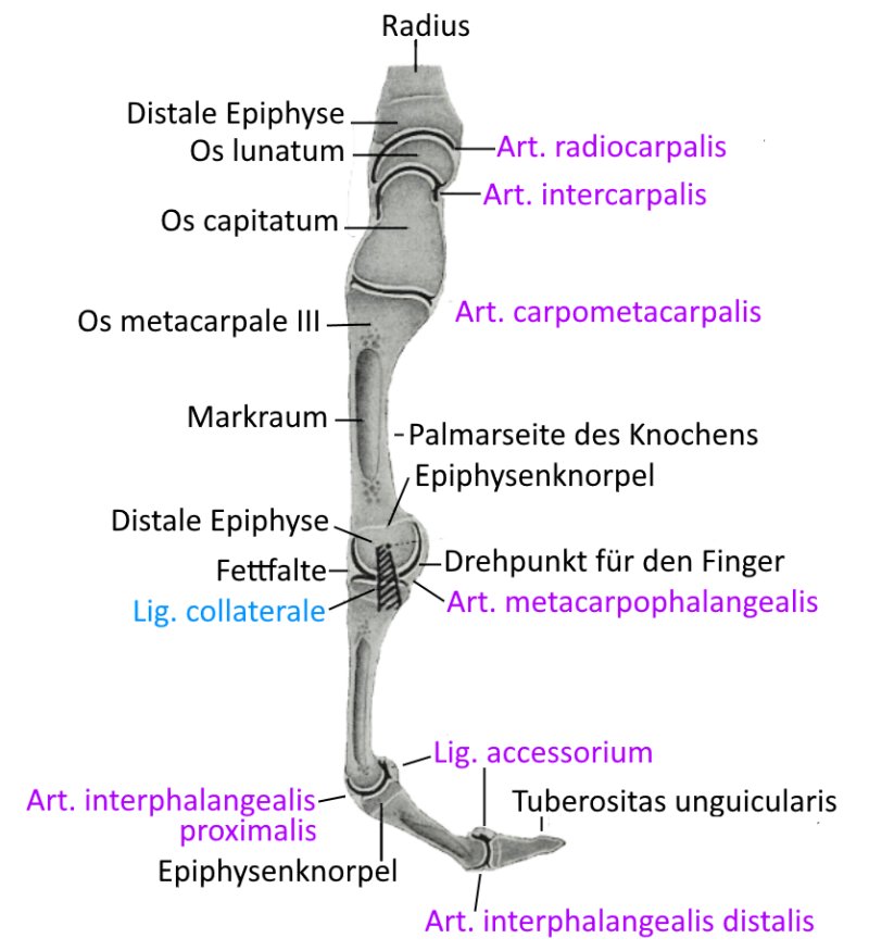
- 3.8 Section through the middle finger
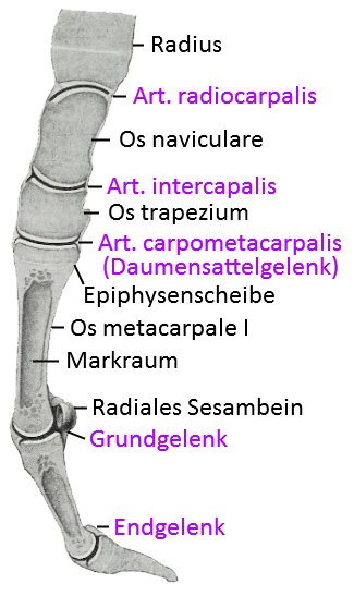
- 3.9 Thumb, sagittal cut
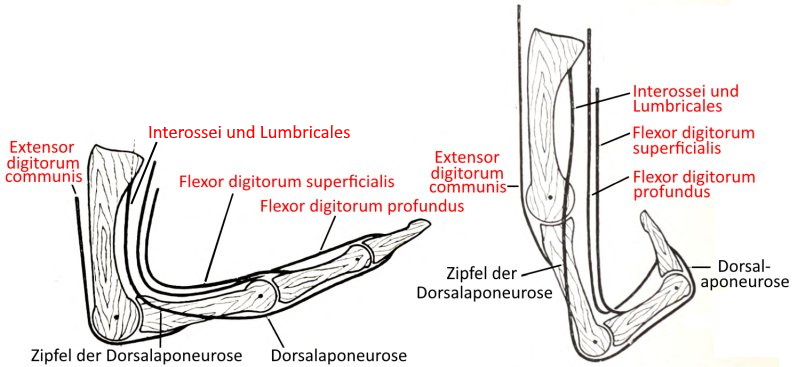
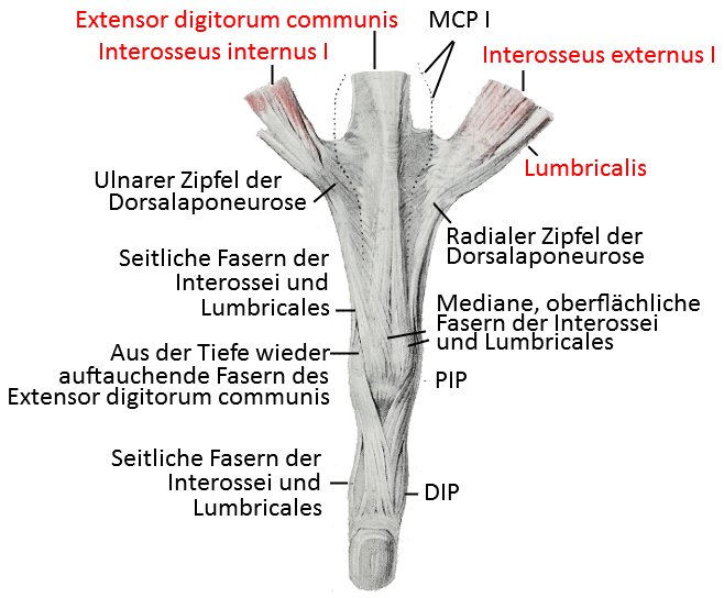
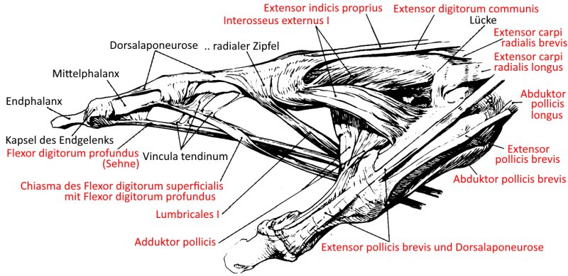
- 3.10 Dorsal aponeurosis of the index finger
- 3.11 Tendons of the hand (image links to linkmap)
- 3.12 Axes of movement of the carpal bones (Henkesche Achsen)
- 3.13 Tendons in the fingers (image links to linkmap)
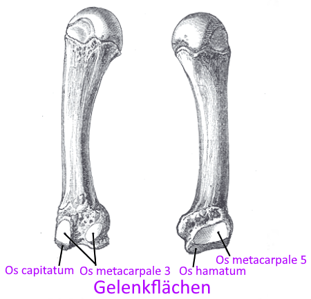
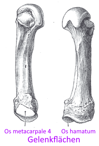
Images: Joint lines of the hand
The bones of the hand
The bones of the hand consist of the carpal bones (carpus), the metacarpal bones (metacarpus) and the bones of the fingers(digiti) with theirphalanges.
Carpal bone
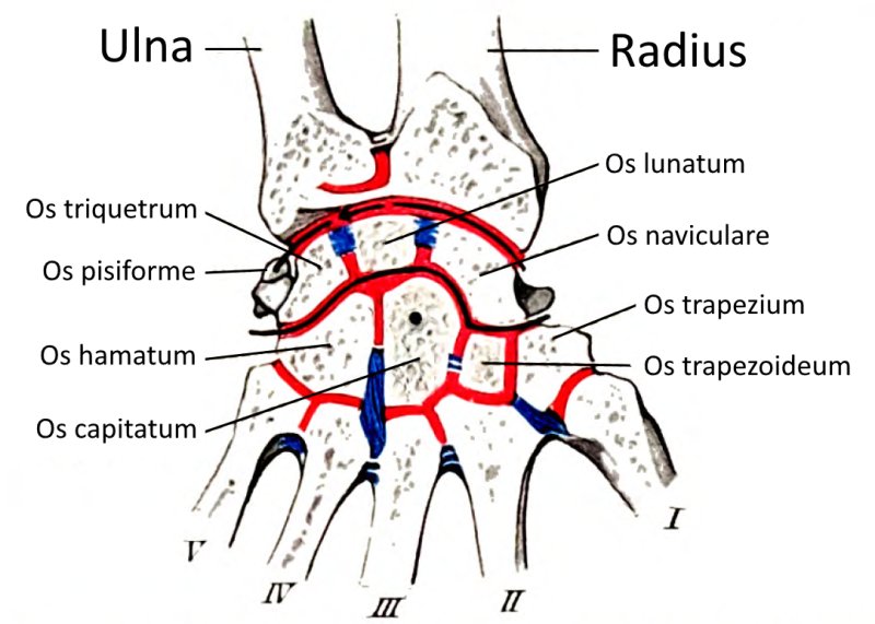
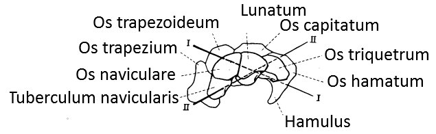
The entirety of the carpal bones, i.e. all bones between the forearm bones ulna and radius on the one hand and the metacarpal bones on the other. The 8 carpal bones are arranged in two rows. In the proximal row, from radial to ulnar, are the scaphoid bone (Os scaphoideum), the lunate bone (Os lunatum) and the triangular bone (Os triquetrum), which articulate as a curved joint line with the radius and form the wrist joint (Art. radiocarpalis) in the true sense. Ulnarly attached in the proximal row is the pea bone (Os pisiforme).
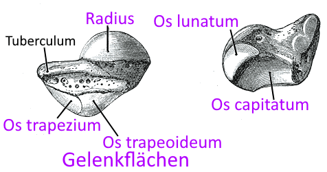
The distal row of carpal bones consisting of the large polygonal bone (Os trapezium), small polygonal bone (Os trapezoideum), capitate bone (Os capitatum) and hook bone (Os hamatum) is connected from radial to ulnar with a curved joint line (Art. mediocarpalis). This joint line corresponds to the Chopart joint line in the foot.
Scaphoid bone (Os scaphoideum)
The first carpal bone of the proximal row when viewed from radial to ulnar. The scaphoid bone is involved in the radiocarpal joint with the lunate bone and the triangular bone by articulating the radius.
Lunate bone (Os lunatum)
The second carpal bone of the proximal row when viewed from radial to ulnar. The lunate bone participates in the radiocarpal joint with the scaphoid and the triangular bone by articulating the radius.
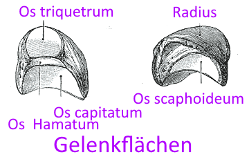
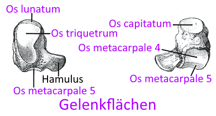
Triangular bone (Os triquetrum)
The third carpal bone of the proximal row, seen from radial to ulnar. The triangular bone is involved in the radiocarpal joint with the scaphoid and the lunate bone by articulating the radius.
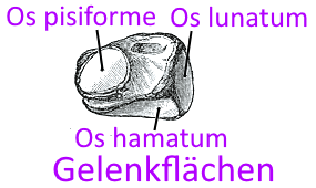
Pea bone (Os pisiforme)
The fourth carpal bone of the proximal row seen from radial to ulnar

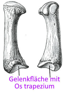
Greater multangular bone (Os trapezium)
The first carpal bone of the distal row seen from radial to ulnar
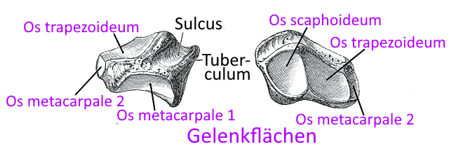
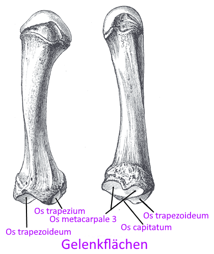
lesser multangular bone (Os trapezoideum)
The second carpal bone of the distal row seen from radial to ulnar
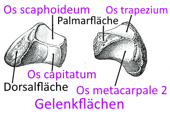
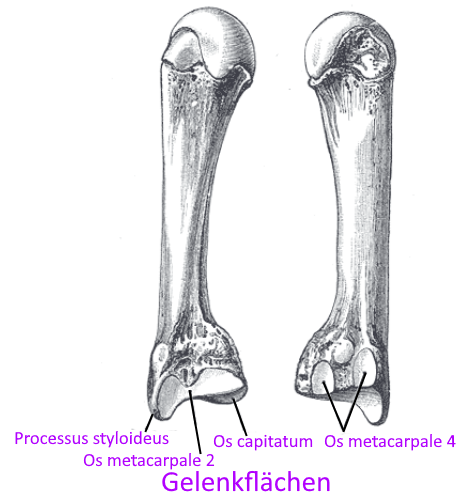
Capitate bone (Os capitatum)
The third carpal bone of the distal row seen from radial to ulnar
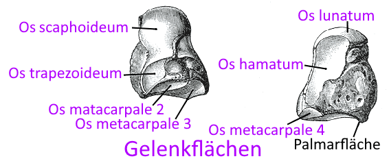
Hamate bone (Os hamatum)
The fourth carpal bone of the distal row seen from radial to ulnar
Carpal tunnel
Palmar depression in the carpus (entirety of the carpal bones) through which the median nerve, the tendons of the profund finger flexors (flexor digitorum profundus muscle), superficial finger flexors (flexor digitorum superficialis muscle) and long thumb flexors (flexor pollicis longus muscle) run. Some authors also include the flexor carpi radialis muscle. The carpal tunnel is covered by a retaining ligament, the flexor retinaculum. Swelling of the tendons can lead to pressure on the nerve and thus to carpal tunnel syndrome, a nerve compression syndrome.
Metacarpal bone
The five metacarpal bones are elongated tubular bones that connect the carpus to the fingers. A multiple curved joint line (artt. carpometacarpales) is present to the carpus (carpal bone), which corresponds to the Lisfranc joint line of the foot; the fingers are connected via the individual metacarpophalangeal joints (artt. metacarpophalangealis).
 |  |  |  |  |
Finger bones
With the exception of the thumb (2 joints or phalanges), all fingers („long fingers“) have three joints(phalanges), one proximal, one medial and one distal. The proximal phalange connects proximally to the carpus (the carpal bones) in the matacarpophalangeal joint (metacarpophalangeal joint) and distally to the medial phalange in the proximal interphalangeal joint. This in turn connects to the distal phalange in the distal interphalangeal joint.
Finger (Digiti)
The fingers consist of two gliders(phalanges) in the case of the thumb and three phalanges in the case of the long fingers, which are connected to interphalangeal joints that allow flexion and extension, so that together with the flexion in the base joint, up to around 270° of flexion relative to the palm is possible. The ability of the fingers to flex thus represents one important part of the fist closure, the other being the opposition and adduction of the thumb.
Phalanges (phalanges)
The two (in the case of the thumb) or three (in the case of the long fingers) phalanges (phalanges) of the fingers(digiti) allow flexion and extension in the interphalangeal joints. Collateral ligaments protect against rotation and ulnar/radial abduction.
Pollex
Like the hallux, the pollex consists of only two phalanges.
Images
Hand, dorsal (image links to linkmap)
Hand, palmar (image links to linkmap)
Ligaments (image links to linkmap)
Carpal tunnel (image links to linkmap)
Carpal tunnel (other view)
Ligaments, palmar (image links to linkmap)
Ligaments, dorsal (image links to linkmap)
Section through the middle finger
Thumb, sagittal cut
Dorsal aponeurosis of the index finger
Tendons of the hand (image links to linkmap)
Axes of movement of the carpal bones (Henkesche Achsen)
Tendons in the fingers (image links to linkmap)