Contents
- 1 Linkmap: Wrist from dorsal
- 2 Articulating bones
- 3 The movements and the muscles involved
- 4 Ligaments, overview
- 5 The ligaments in detail
- 5.1 Lig. capitohamatum
- 5.2 Lig. carpi arcuatum
- 5.3 Lig. carpi palmare
- 5.4 Lig. carpi radiatum (Lig. deltoideum)
- 5.5 Lig. carpi transversum
- 5.6 Ligg. carpometacarpalia palmaria
- 5.7 Dorsal carpometacarpal ligament
- 5.8 Lig. collaterale carpi radiale
- 5.9 Lig. collaterale carpi ulnare
- 5.10 Ligg. intercarpalia dorsalia
- 5.11 Ligg. intercarpalia interossea
- 5.12 Ligg. intercarpalia palmaria
- 5.13 Dorsal metacarpal ligament
- 5.14 Ligg. metacarpalia interossea
- 5.15 Ligg. metacarpalia palmaria
- 5.16 Lig. metacarpale transversum superficiale
- 5.17 Lig. metacarpal transversum profundum
- 5.18 Ligg. palmaria
- 5.19 Lig. pisohamatum
- 5.20 Pisometacarpal ligament
- 5.21 Dorsal radiocarpal ligament
- 5.22 Lig. radiocarpale palmare
- 5.23 Dorsal radiotriquetral ligament
- 5.24 Dorsal radioulnar ligament
- 5.25 Lig. radioulnare palmare
- 5.26 Retinaculum extensorum
- 5.27 Retinaculum flexorum
- 5.28 Lig. trapezoideocapitatum
- 5.29 Lig. ulnocapitatum
- 5.30 Ulnocarpal ligament
- 5.31 Dorsal ulnocarpal ligament
- 5.32 Lig. ulnolunatum
- 5.33 Lig. ulnotriquetrum
- 5.34 Vaginal ligament
- 5.35 Triangular fibrocartilaginous complex (TFCC, ulnocarpal complex)
- 6 Further structures
- 7 Bursa (bursae)
- 8 Pathology
- 9 Tests
- 10 Pictures
- 10.1 Distal radiocarpal joint
- 10.2 Ligaments (image links to linkmap)
- 10.3 Hand, dorsal wrist, profound (image links to linkmap)
- 10.4 Hand, wrist dorsal, superficial (image links to linkmap)
- 10.5 Hand palmar superficial (image links to linkmap)
- 10.6 Hand palmar, semi-superficial (image links to linkmap)
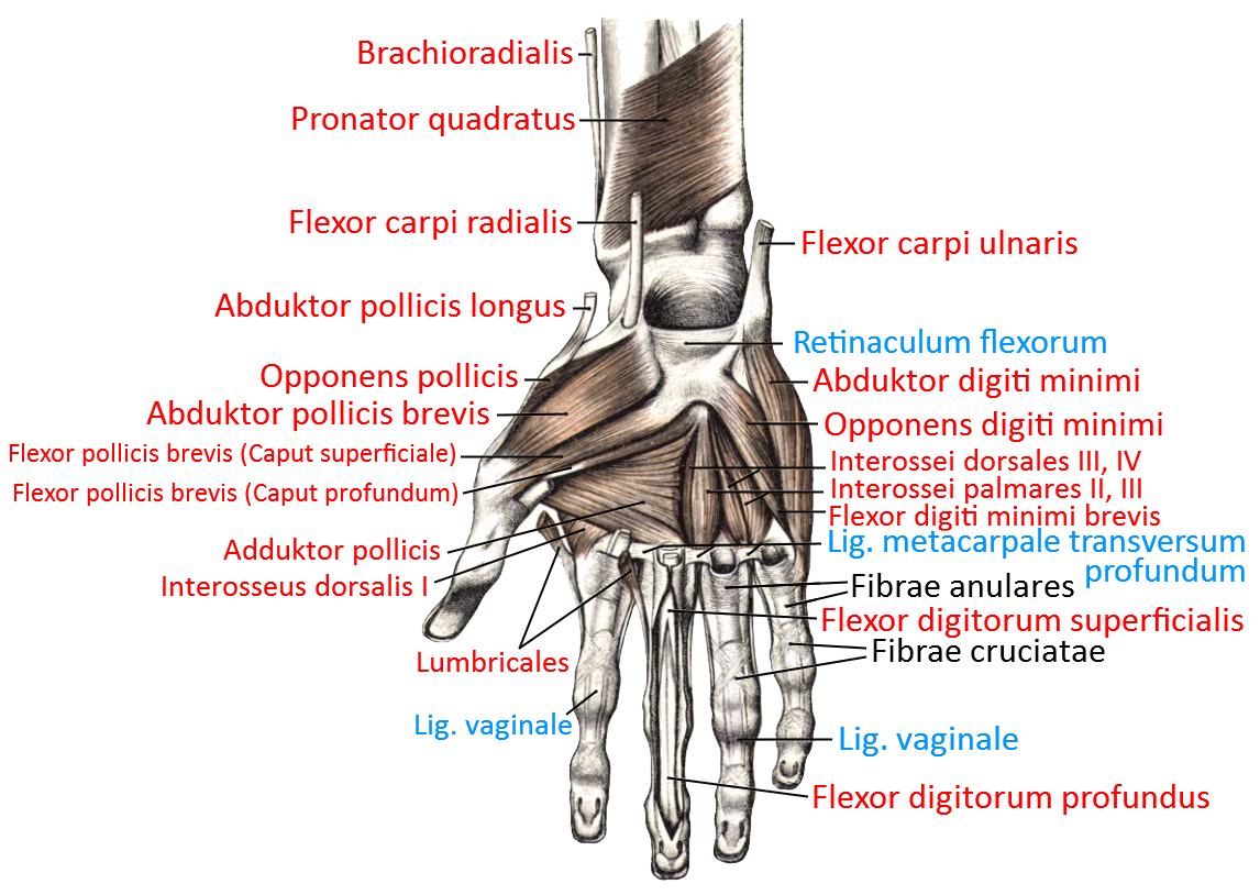
- 10.7 Hand palmar, semi-profound (image links to linkmap)
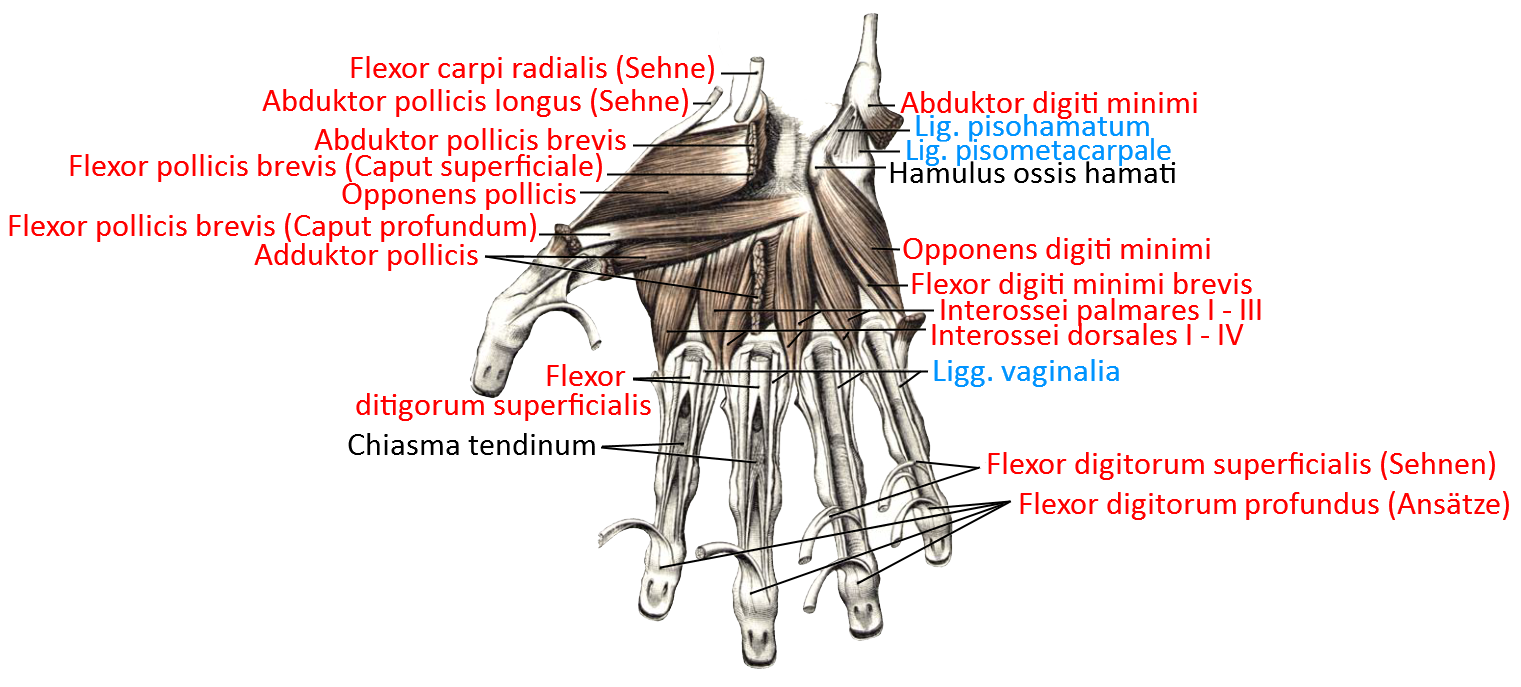
- 10.8 Hand palmar, deep (image links to linkmap)
- 10.9 forearm bones from distal
- 10.10 Bone in radial and ulnar abduction (schematic)
- 10.11 Dorsiflexion and palmar flexion (diagram)
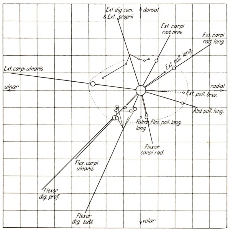
- 10.12 Torques of the muscles
Linkmap: Wrist from dorsal
The wrist is the colloquial term for the radiocarpal joint (art. radiocarpalis), in which the hand can be moved relative to the forearm. However, the first row of carpal bones also forms a joint with the second row, the mediocarpal joint. The radiocarpal articulation is an ovoid joint that enables „flexion“(palmar flexion, i.e. palm to forearm) and „extension“(dorsal flexion, i.e. back of hand to forearm), as well as ulnar abduction (thumb away from forearm) and radial abduction (thumb to forearm), so its range of motion is two-dimensional. If the overturning movement pronation/supination of the forearm is added, the result is a three-dimensional ability to move from the elbow joint with a relatively small moving mass, which has certainly contributed to the success of the evolutionary model „primate“ and humans in particular. It allows fine motor skills as well as a relatively large exertion of force in combination with the muscles of the upper arm and trunk, which move the latter.
See also the entry Carpal bones.
Depending on the position of the arm, over 360° of rotation can be achieved by combining the rotational ability of the upper arm in the shoulder joint with the overturning movement of the forearm.
In particular, the extent of possible dorsiflexion and palmar flexion depends heavily on the position of the finger joints: if the finger joints (all three per ray: MCP, PIP and DIP) are widely flexed, palmar flexion is limited by the mobility limit of the finger extensors. The same applies to dorsiflexion: if the finger joints (all three per ray: MCP, PIP and DIP) are extended, dorsiflexion is limited by the mobility limit of the finger flexors. In the case of ulnar abduction and radial abduction, there is no such dependency with average mobility. The ranges of motion are specified, depending on the position of the finger joints with:
Dorsiflexion: 40-60°, max. 85°
Palmar flexion: 50-60°, max. 80°
Ulnar abduction: 30-40°
Radial abduction: 20-30°
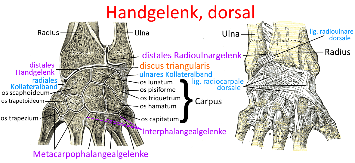
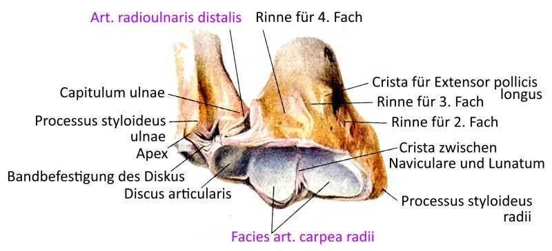
In the radiocarpal joint, the radius and, via an intervertebral disc located between the radius and the ulnar styloid process, the ulna also articulate with three carpal bones:
Scaphoid bone Os scaphoideum, lunate bone Os lunatum and triangular bone Os triquetrum. The joint capsule is lax and contains capsular ligaments.
The mediocarpal articulation is a „toothed hinge joint“ with low mobility
Articulating bones
The movements and the muscles involved
Two movement dimensions are available: dorsiflexion / palmar flexion and radial abduction / ulnar abduction
The movements and their executing muscles:
Dorsiflexion: M. extensor digitorum, M. extensor carpi radialis longus, M. extensor carpi radialis brevis, Extensor carpi ulnaris, M. extensor indicis, M. extensor pollicis longus, M. extensor digiti minimi
Palmar flexion: M. flexor digitorum superficialis, M. flexor digitorum profundus, M. flexor carpi ulnaris, M. flexor carpi radialis, M. flexor pollicis longus, M. palmaris longus,
M. abductor pollicis longus
Radial abduction: M. extensor carpi radialis longus, M. extensor carpi radialis brevis, M. abductor pollicis longus, M. extensor pollicis longus,
M. extensor pollicis brevis, M. flexor carpi radialis, M. flexor pollicis longus
Ulnar abduction: M. extensor carpi ulnaris, M. flexor carpi ulnaris, M. extensor digiti minimi
Ligaments, overview
Alphabetical
- Lig. capitohamatum
- Lig. carpi arcuatum
- Lig. carpi palmare
- Lig. carpi radiatum
- Ligg. carpometacarpalia dorsalia
- Ligg. carpometacarpalia palmaria
- Lig. collaterale carpi radiale
- Lig. collaterale carpi ulnare
- Ligg. intercarpalia dorsalia
- Ligg. intercarpalia interossea
- Ligg. intercarpalia palmaria
- Lig. pisohamatum
- Lig. pisometacarpale
- Lig. radiocarpale palmare
- Lig. radiocarpale dorsale
- Lig. radiotriquetrum dorsale
- Lig. radioulnare dorsale
- Lig. radioulnare palmare
- Lig. trapezoideocapitatum
- Lig. ulnocapitatum
- Lig. ulnocarpale palmare
- Lig. ulnocarpale dorsale
- Lig. ulnolunatum
- Lig. ulnotriquetrum
- Lig. vaginale
- Retinaculum extensorum
- Retinaculum flexorum
between the forearm bone and the carpal bone
- Lig. collaterale carpi ulnare
- Lig. collaterale carpi radiale
- Lig. radiocarpale palmare
- Lig. radiocarpale dorsale
- Lig. ulnocarpale palmare
- Lig. ulnocarpale dorsale
between carpal bones
- Lig. carpi radiatum
- Ligg. intercarpalia palmaria
- Ligg. intercarpalia dorsalia
- Ligg. intercarpalia interossea
- Lig. pisohamatum
between carpal bone and metacarpal bone
more
The ligaments in detail
Lig. capitohamatum
Ligament connecting the head bone to the hook bone.
Pictures: (still without)
Lig. carpi arcuatum
The carpi arcuatum ligament runs with proximal fibers from the scaphoid bone to the triquetrum and with distal fibers from the triquetrum to the trapezium (Os trapezium and Os trapetoideum). It stabilizes the joint row of the mediocarpal joint in the transverse direction.
Images: (still without)
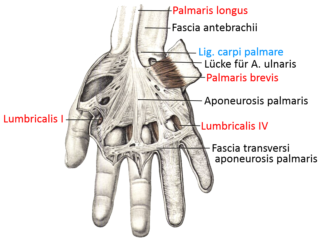
Lig. carpi palmare
Along with the flexorretinaculum, the carpi palmare ligament is part of the superficial palmar layer. It connects the tendons of the flexor carpi ulnaris and the palmaris longus, surrounds the tendon of the flexor carpi ulnaris and is connected to the palmar aponeurosis. It serves to tense the palmar aponeurosis and protect the tendons.
Images:
Linkmap: Hand, palmar, superficial
Linkmap forearm, hand, palmar, superficial
Lig. carpi radiatum (Lig. deltoideum)
The carpi radiatum ligament is a ligament of the middle palmar layer. It is also called the deltoid ligament because it connects the capitate bone to the hamate bone, the triquetrum, the scaphoid bone, the trapezoid bone, the triapezoideum and, rarely, the lunate bone. Fiber tracts of the flexor carpi radialis radiate into the ligament.
Images:
Linkmap: Wrist, ligaments of the carpus
Linkmap: Ligaments, palmar
Lig. carpi transversum
other name for retinaculum flexorum
Ligg. carpometacarpalia palmaria
The carpometacarpal ligaments are short, tight palmar ligaments that connect the bases of the metacarpal bones Ossa metacarpalia II-V with the distal carpal bones.
Images:
Linkmap: Wrist, ligaments of the carpus
Linkmap: Ligaments, palmar
Dorsal carpometacarpal ligament
The carpometacarpal dorsal ligaments are short, taut dorsal ligaments that connect the bases of the metacarpal bones Ossa metacarpalia II-V with the distal carpal bones.
Images: (still without)
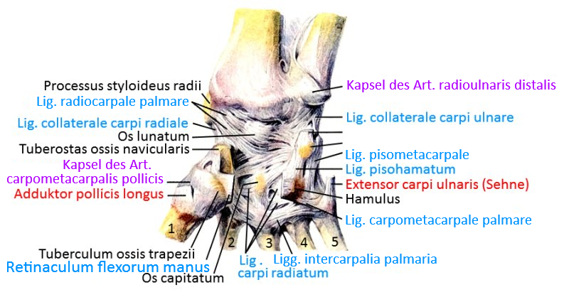
Lig. collaterale carpi radiale
The lig. collaterale carpi radiale runs from the styloid process of the radius to the radial side of the scaphoid bone and its tuberosity. It is connected to the tendon sheath of the flexor carpi radialis and limits the ulnar abduction of the hand.
Images:
Linkmap: Wrist, ligaments of the carpus
Linkmap: Wrist, ligaments, dorsal
Linkmap: Ligaments, palmar
Lig. collaterale carpi ulnare
The carpal collateral ligament runs with dorsal fibers from the styloid process of the ulna and the discus to the triquetrum. A palmar fiber tract runs from the palmar radius to the pisiform pea bone. It is fused with the discus and the meniscus of the TFCC (triangular fibrocartilaginous complex) of the ulnocarpal joint. It limits the radial abduction of the hand.
Images:
Linkmap: Wrist, ligaments of the carpus
Linkmap: Wrist, ligaments, dorsal
Linkmap: Ligaments, palmar
Ligg. intercarpalia dorsalia
The dorsal intercarpal ligaments are profound dorsal ligaments made of strong, firm fibers. They belong to the intrinsic or interosseous ligaments.
Images: (still without)
Ligg. intercarpalia interossea
The intercarpal interosseous ligaments connect the carpal bones profoundly in a transverse direction.
Images: (still without)
Ligg. intercarpalia palmaria
The palmar intercarpal ligaments are deeper interosseous ligaments that connect the carpal bones to each other. They are fused to the joint capsule and ensure good stability against translation of the bones.
Images:
Linkmap: Wrist, ligaments of the carpus
Dorsal metacarpal ligament
Short ligaments that connect the bases of the metacarpals dorsally.
Ligg. metacarpalia interossea
Short ligaments that connect the bases of the metacarpals in depth.
Ligg. metacarpalia palmaria
Short ligaments that connect the palmar bases of the metacarpals.
Lig. metacarpale transversum superficiale
The metacarpal transverse superficial ligament is a fiber tract that runs transversely over the bases of the proximal proximal proximal phalanges and connects to the tendon sheaths of the finger flexors.
Images:
Linkmap: Forearm, hand, palmar, superficial
Lig. metacarpal transversum profundum
The metacarpal transverse ligament is a narrow band that covers the palmar side of metatarsophalangeal joints 2 to 5 (MCP) at the metatarsal bases. The palmar ligaments radiate into the metacarpal transverse ligament.
Images:
Linkmap: forearm, hand, palmar, profundum
Ligg. palmaria
The palmar ligaments are palmar capsular ligaments of the metacarpophalangeal joints, the proximal interphalangeal joints and the distal interphalangeal joints of fingers II to V. They form a sliding support for the tendons of the finger flexors and connect to the transverse metacarpal ligament
Images: (still without)
Lig. pisohamatum
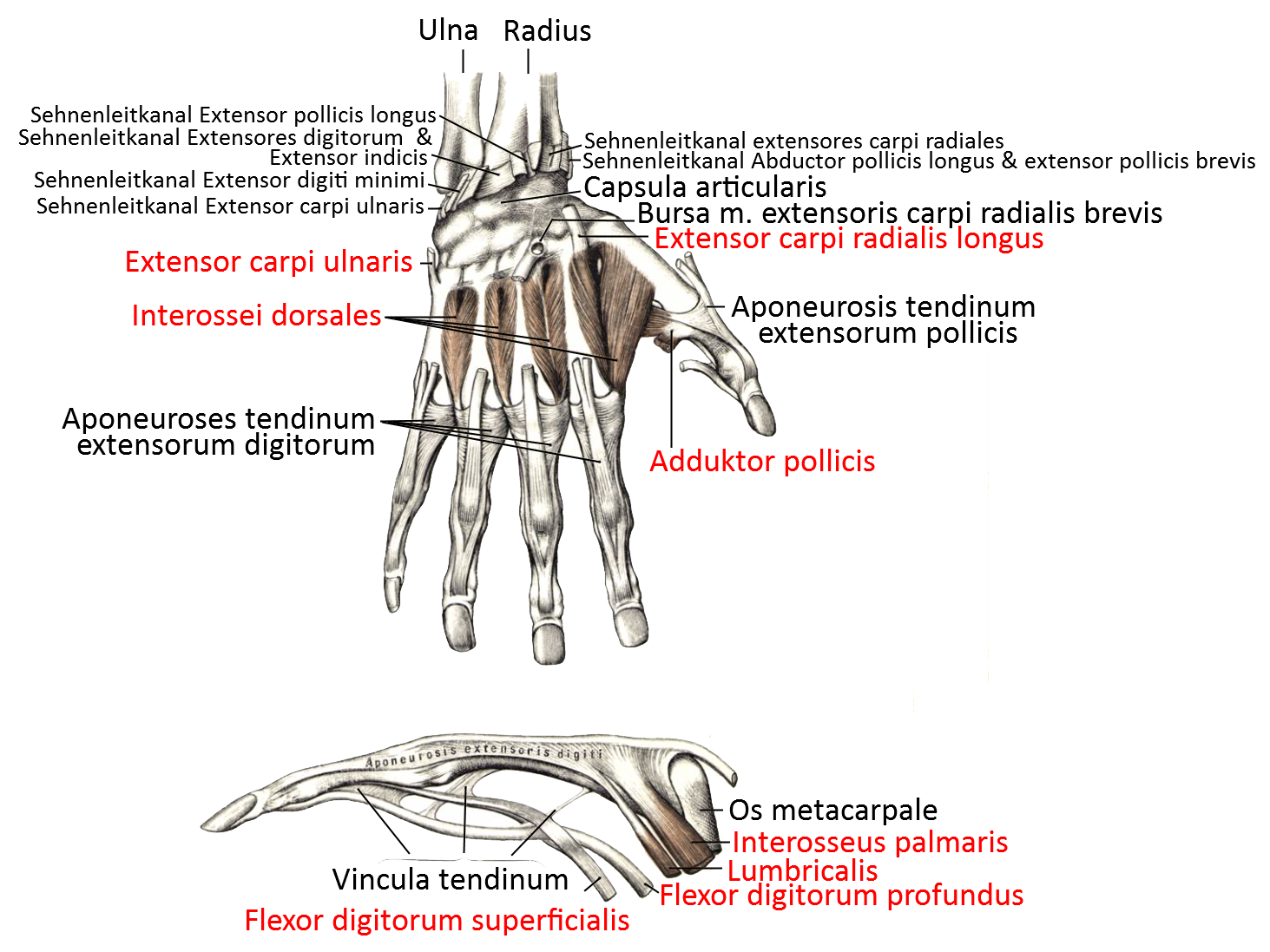
The pisohamate ligament is a short ligament that connects the pisiform bone to the hamulus of the hamate bone. Together with the retinaculum (musculorum) flexorum, it delimits the Guyon’s box dorsally, through which the ulnar artery and ulnar nerve run.
Images:
Linkmap: Forearm, hand, palmar, profound
Linkmap: Hand, palmar, profound
Linkmap: Wrist, ligaments of the carpus
Linkmap: Ligaments, palmar
Pisometacarpal ligament
The pisometacarpal ligament is a palmar ligament between the pisiform bone and the base of the 5th metacarpal bone.
Images:
Linkmap: Forearm, hand, palmar, profound
Linkmap: Wrist, ligaments of the carpus
Linkmap: Ligaments, palmar
Dorsal radiocarpal ligament
The dorsal radiocarpal ligament, along with the arcuate carpal ligament, belongs to the middle dorsal layer. It runs from the radius with three separate fibrous tracts to the respective carpal bones of the distal radiocarpal joint and stabilizes it.
Images:
Linkmap: Wrist, ligaments, dorsal
Lig. radiocarpale palmare
The radoicarpal ligament is a ligament of the middle palmar layer. It consists of a superficial and a deep layer, both of which are connected to the joint capsule. The superficial layer runs from the styloid process of the radius to the capitate bone and the triquetrum, while the deep layer runs from the radius to the scaphoid bone and the lunate bone.
Images:
Linkmap: Wrist, ligaments of the carpus
Linkmap: Ligaments, palmar
Dorsal radiotriquetral ligament
The dorsal radiotriquetral ligament is an approximately 2 cm long ligament that narrows distally between the dorsal tuberosity of the radius and the dorsal side of the triquetrum. It covers the proximal part of the scaphoid bone (Os scaphoideum) and the lunate bone (Os lunatum). It is part of the TFCC.
Images: (still without)
Dorsal radioulnar ligament
The dorsal radioulnar ligament is an extracapsular ligament of the distal radioulnar joint, articulatio radioulnaris distalis, which connects the radius and ulna dorsally. Together with the radioulnar palm ligament, it forms a ring that holds the bones together and is fused with the ulnocarpal articular disc. The ligament stretches during pronation. The dorsal radioulnar ligament is part of the so-called TFCC.
Images:
Linkmap: Wrist, ligaments, dorsal
Lig. radioulnare palmare
The dorsal radioulnar ligament is an extracapsular ligament of the distal radioulnar joint, the articulatio radioulnaris distalis, which connects the palmar radius and ulna. Together with the dorsal radioulnar ligament, it forms a ring that holds the bones together and is fused to the ulnocarpal articular disc. The ligament stretches during supination. The dorsal radioulnar ligament is part of the so-called TFCC,
Images: (still without)
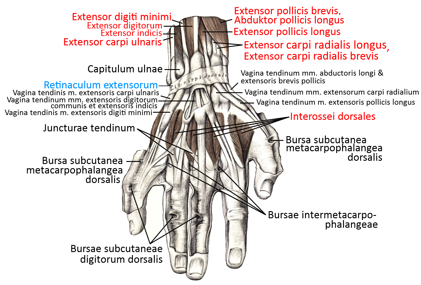
Retinaculum extensorum
The extensor retinaculum is the superficial dorsal layer with a total of 6 compartments for one tendon each. It is connected to the radius and ulna via intermediate septa.
Images:
Linkmap: Wrist, dorsal, superficial
Linkmap: Hand, dorsal, tendon sheaths and muscles
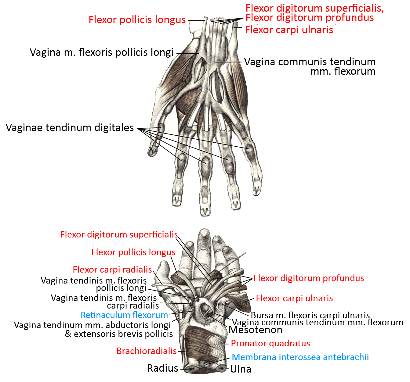
Retinaculum flexorum
The flexor retinaculum is sometimes referred to as the transverse carpal ligament. It limits the carpal tunnel palmarward and is connected to both ulnar and radial carpal bones.
Linkmap: Forearm, hand, palmar, profound
Linkmap: Hand, wrist, palmar, semi-superficial
Linkmap: Hand, palmar, semi-profound
Linkmap: Forearm, palmar, very deep
Linkmap: Wrist, ligaments of the carpus
Linkmap: Hand, section through distal carpus
Linkmap: Hand, dorsal, tendon sheaths and muscles
Lig. trapezoideocapitatum
Ligament connecting the small trapezoid bone to the hook bone.
Pictures: (still without)
Lig. ulnocapitatum
The ulnocapitate ligament is part of the ulnocapitate palmar ligament and the TFCC and runs from the ulna to the palmar side of the capitate bone.
Images: (still without)
Ulnocarpal ligament
The ulnocarpal palmar ligament is a ligament of the middle palmar layer. It runs from the styloid process of the ulna to the discus and the lunate bone (os lunatum), the triangular bone (os triquetrum) and the capitate bone (os capitatum).
Images: (still without)
Dorsal ulnocarpal ligament
The dorsal ulnocarpal ligament runs from the styloid process of the ulna to the dorsal surfaces of the carpal bones.
Images: (still without)
Lig. ulnolunatum
The ulnolunate ligament is part of the ulnocaraple ligament and the TFCC and runs from the ulna and the radioulnar palmar ligament to the anterior horn of the lunate bone.
Images: (still without)
Lig. ulnotriquetrum
The ulnotriquetrum ligament is part of the ulnocaraple ligament and the TFCC and runs from the ulna and the radioulnar palmar ligament to the palmar side of the triquetrum.
Images: (still without)
Vaginal ligament
Ligament that fixes the tendon sheath of the finger flexors.
Some authors differentiate the anular ligaments into
Lig. vaginale accessorium (MCP), Lig. caginale 1 (PIP) and Lig. vaginale 2 (DIP).
Images:
Linkmap: Hand, palmar, semi-profound
Linkmap: Hand, palmar, profound
Linkmap: Forearm, palmar, very profound
Linkmap: Dorsal hand, tendon sheaths and muscles
Triangular fibrocartilaginous complex (TFCC, ulnocarpal complex)
The triangular fibrocartilaginous complex (TFCC) contains the following structures:
- Dorsal radioulnar ligament
- Lig. radioulnare palmare
- Lig. ulnolunatum
- Lig. ulnotriquetrum
- Lig. ulnocapitatum
- Lig. collaterale carpi ulnare
- Lig. radiotriquetrum dorsale
- Meniscus ulnocarpalis
- Discus ulnocarpalis
Further structures
Meniscus ulnocarpalis
The ulnocarpal meniscus is a small disc of connective tissue covered with synovial fluid that lies between the medial edge of the ulnocarpal disc and the triquetrum, the hamate bone, the metacarpal bones 4 and 5 and the styloid process of the ulna. The meniscus is part of the TFCC.
Discus ulnocarpalis (Discus articularis ulnocarpalis, Discus triangularis)
Cartilaginous articular body made of tight connective tissue with parallel fibers between the distal ulna and the concave articular surface of the lunate bone (Os lunatum ) and partly also of the triangular bone (Os triquetrum). The discus is part of the TFCC and lies between the dorsal radioulnar ligament and the palmar radioulnar ligament. The meniscus ulnocarpalis lies between the discus and the triquetrum.
Bursa (bursae)
Bursa extensoris carpi radialis brevis
Bursa between the insertion tendon of the extensor carpi radialis brevis and the base of the metacarpal bone 3.
Images:
Linkmap: Hand, wrist, dorsal, profound
Bursa flexoris carpi radialis
Bursa located on the scaphoid tuberosity, which is useful because of the hypomochlion function of the tuberosity.
Images: (still without)
Bursa flexoris carpi ulnaris (distalis)
Bursa between the insertion tendon of the flexor carpi ulnaris and the base of the pisiform bone.
Images:
Linkmap: Hand, wrist, palmar, semi-superficial
Bursae intermetacarpophalangealae
Bursae between the bases of the ossae metacarpaliae
Images: (still without)
Radial bursa
Bursa of the tendon sheath for flexor pollicis longus and flexor pollicis brevis, which extends into the carpal tunnel. Sometimes it communicates there with the ulnarbursa.
Images: (still without)
Bursae subcutaneae digitorum dorsales
Bursae located between the skin and the dorsal sides of the interphalangeal joints
Images: (still without)
Bursae subcutaneae metacarpophalangealae dorsales
Rare, inconstant bursae between the skin and the dorsal sides of the MCP, usually occurring on the little finger.
Images:
Linkmap: Hand, wrist, dorsal, superficial
Ulnar bursa
Bursa of the tendon sheath for flexor digiti minimi, which continues into the carpal tunnel. Sometimes it communicates there with the radialbursa.
Images: (still without)
Pathology
Less common: impingement, carpal tunnel syndrome
Tests
Tests of the wrist
Carpal tunnel syndrome
Tendosynovitis
Tendosynovitis of the thumb
Paratendinitis
- Linburg-Zeichen (between flexor pollicis longus und flexor indicis)
Luno-triquetary instability
radiocarpal, intercarpale scaphoidal instability
scapholunare Instabilität
Tests of the movement directions
Tests of the spanning muscles
Differentiation of contractures
Strength tests
- definite yoga strength test for palmar flexion of the wrist
- definite yoga strength test for dorsiflexion of the wrist
- definite yoga strength test for radial abduction of the wrist
- definite yoga strength test for ulnar abduction of the wrist
Pictures
Distal radiocarpal joint
Ligaments (image links to linkmap)
Hand, dorsal wrist, profound (image links to linkmap)
Hand, wrist dorsal, superficial (image links to linkmap)
Hand palmar superficial (image links to linkmap)
Hand palmar, semi-superficial (image links to linkmap)
Hand palmar, semi-profound (image links to linkmap)
Hand palmar, deep (image links to linkmap)
forearm bones from distal

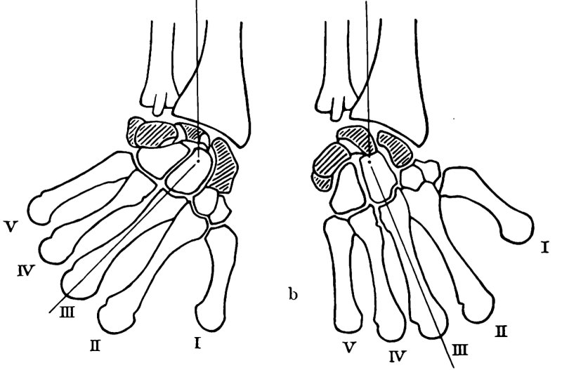
Bone in radial and ulnar abduction (schematic)
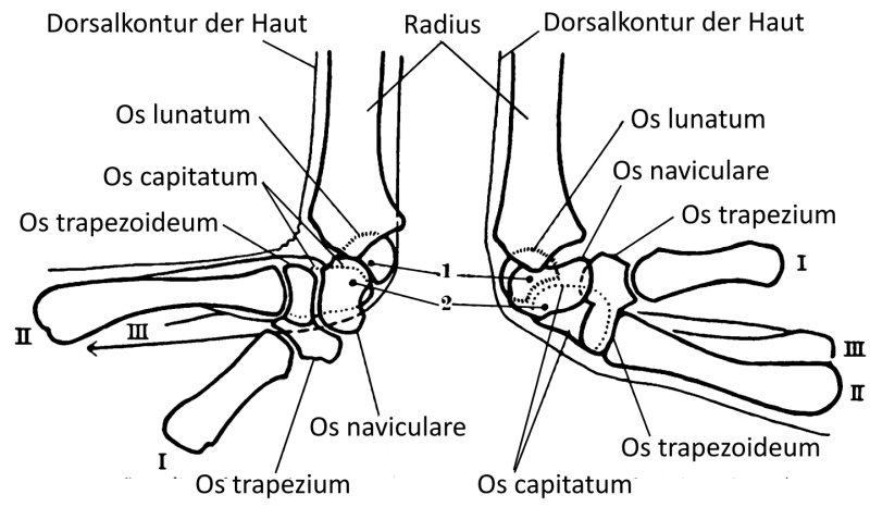
Dorsiflexion and palmar flexion (diagram)
Torques of the muscles