Contents
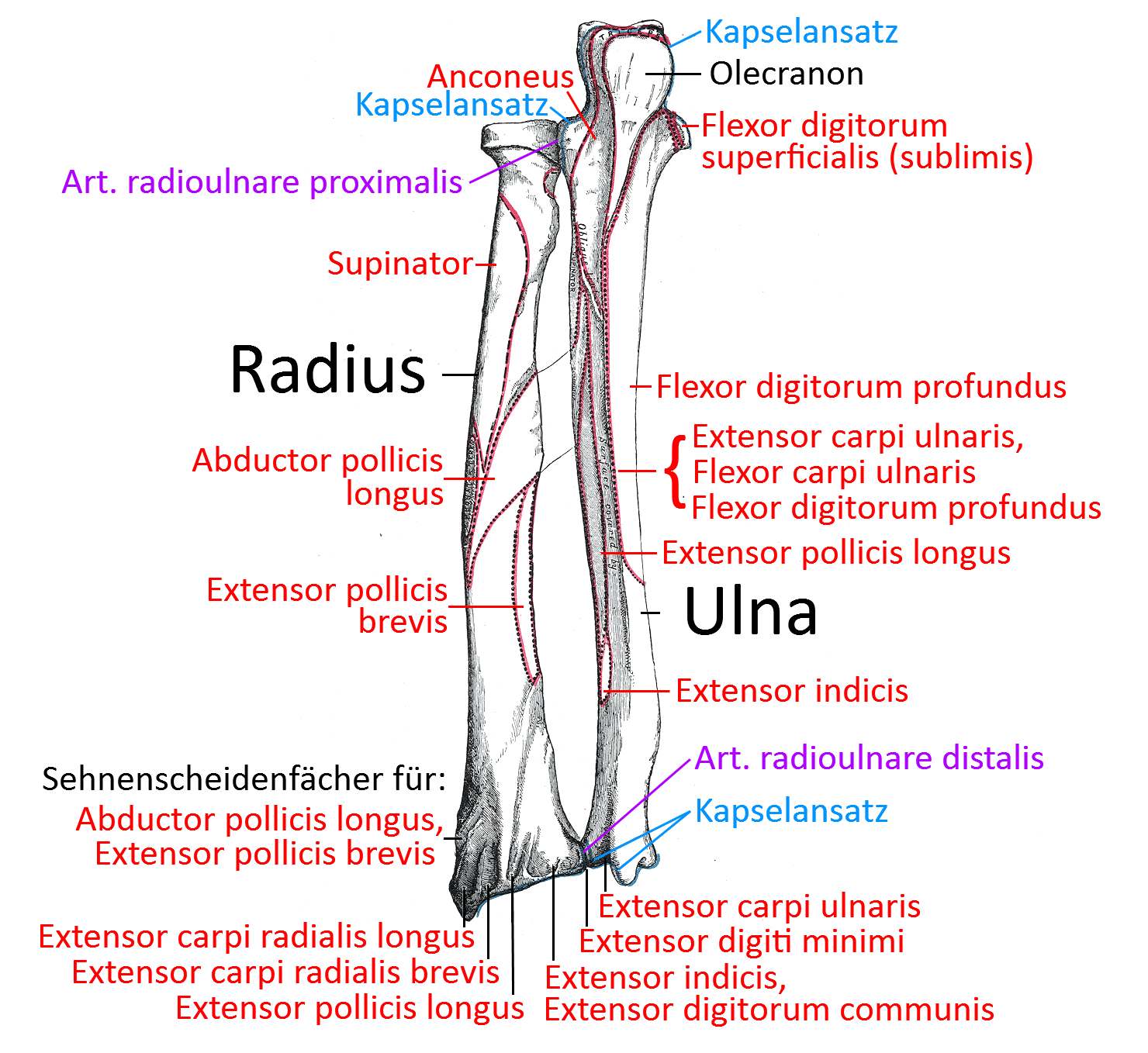
Image: Ulna from dorsal
Ulna
The ulna is the bone of the forearm, which is connected proximally to the humerus, the elbow joint, and distally to the carpal bones of the hand via a discus in the wrist. The ulna forms two joints with the neighboring radius: the proximal radioulnar joint and the distal radioulnar joint.
Extremitas proximalis
Ulnar sulcus (ulnar nerve sulcus)
Bone groove in which the ulnar nerve runs protected. Compression of the nerve can lead to sulcus ulnaris syndrome (cubital tunnel syndrome), which typically causes irritation or numbness in fingers 4 and 5.
Olecranon
Bony process that engages in the olecranon fossa between the condyles of the humerus and limits the movement of the elbow joint in the direction of extension. In addition, the only extensor of the elbow joint with the triceps attaches to the olecranon. Although the olecranon is located in a comparable position to the patella of the leg, the free movement of the bone is lacking here, probably because human movement patterns do not generally include movements and activities that involve the elbows, meaning that a protective ganglion is hardly necessary. In addition, the olecranon limits the extension of the elbow joint. A construction in which the elbow joint was given a ROM of around 270° would be extremely complex and vulnerable and also unnecessary, as objects can be grasped from the same position by endorotation of the arm in the shoulder joint with an overhand grip (instead of an underhand grip).
As the dorsal surface of the olecranon lies directly under the skin, it has abursa olecrani. The triceps attaches proximally to the olecranonbursa. The smooth, concave anterior/palmar opposite side, the incisura trochlearis, has a cartilage cover for articulation with the humerus. Some fibers of the flexor carpi ulnaris attach medially to the olecranon. The anconeus attaches to the lateral edge.
Coronoid process
The coronoid process is a bony projection distal to the incisura trochlearis, to which anterioinferior fibers of the brachialis attach. The lateral surface is covered by cartilage and articulates with the radius. The flexor digitorum superficialis and the pronator teres originate on the medial side. In wide flexion of the elbow joint, the coronoid fossa of the humerus takes up the coronoid process.
Incisura trochlearis (Incisura semilunaris ulnae)
The large, crescent-shaped depression covered with cartilage between the olecranon and the coronoid process articulates with the trochlea of the humerus.
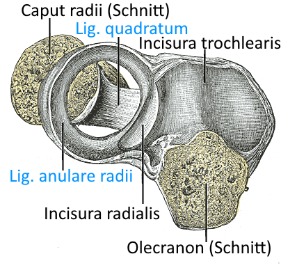
Radial incisura
The incisura radialis is a lateral retraction of the bone covered with cartilage, in which it articulates with the radius in the proximal radioulnar joint. The lig. anulare radii attaches to its slightly protruding edges.
Corpus
As with the humerus, the shaft (corpus) is divided into three surfaces by three edges( anterior, posterior and interosseous): Facies anterior, Facies posterior, Facies medialis.
Margo anterior
The palmar edge separates the anterior facies from the medial fac ies and extends distally from the coronoid process. The flexor digitorum profundus originates proximally and centrally and the pronator quadratus in the distal quarter.
Margo posterior
The posterior margo separates the posterior facies from the medial fac ies and runs from the dorsal side of the olecranon to the ulnar styloid process. An aponeurosis attaches to the proximal three quarters, which is the origin of the flexor carpi ulnaris, extensor carpi ulnaris and flexor digitorum profundus muscles
Margo interosseus (Crista interossea ulnae)
The margo interosseus separates the posterior facies from the anterior facies. From the incisura radialis, two lines converge distally and include an area of origin, the crista musculi supinatoris of the supinator. The Membrana interosseus antebrachii attaches to the Margo interosseus.
Facies anterior
In the anterior facies, the flexor digitorum profundus originates in the proximal three quarters and the pronator quadratus in the distal quarter.
Posterior facies
A bone ridge runs diagonally across the proximal part, at which the anconeus inserts. Further distally, a bony ridge separates the medial origin of the extensor carpi ulnaris from the lateral origins of the muscles
Facies medialis
The flexor digitorum profundus originates in the proximal three quarters.
Extremitas distalis
The extremits distalis, the distal end of the ulna, is considerably narrower than the proximal end, which is also due to the fact that there is no direct articulation with a weight-bearing bone, as the hand is connected to the radius. The two prominent distal structures are the ulnar caput and the ulnar styloid process.
Caput ulnae
The caput ulnae is the distal end, which is connected to the hand via the discus articularis. The lateral side is covered by cartilage and forms the distal radioulnar joint as the ulnar incisura with the distal radius.
Ulnar incisura
The cartilage-covered lateral surface of the ulna, which together with the radius forms the distal radioulnar joint.
Ulnar styloid process
The ulnar styloid process protrudes medially slightly beyond the ulnar caput and is the insertion for the ulnar collateral carpal ligament.
Joints
Radioulnar joints
The ulna articulates both proximally and distally with the radius, i.e. in the proximal proximal radioulnar joint (PRUG), a swivel joint, and in the distal distal radioulnar joint, also a swivel joint. This allows the radius to rotate around the fixed ulna and enables supination and pronation of the forearm. This is one of the ways in which the structure of the forearm differs from that of the lower leg, even though both have two bones each.
Humeroulnar joint
The humeroulnar joint is the hinge joint in which the ulna moves one-dimensionally in terms of flexion and extension of the elbow joint relative to the humerus. The overturning movement of the radius around the ulna in the sense of supination and pronation is completely independent of this.
Pictures
Radius and ulna: anterior view (image linked to linkmap)
Proximal end of ulna
Epiphyseal plates
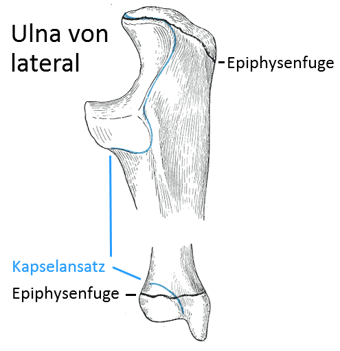
Proximal ulna
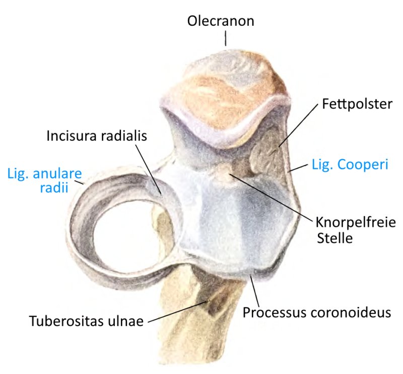
Lig. anulare radii and lig. quadratum