Contents
Image: Humerus from ventral view (Linkmap)
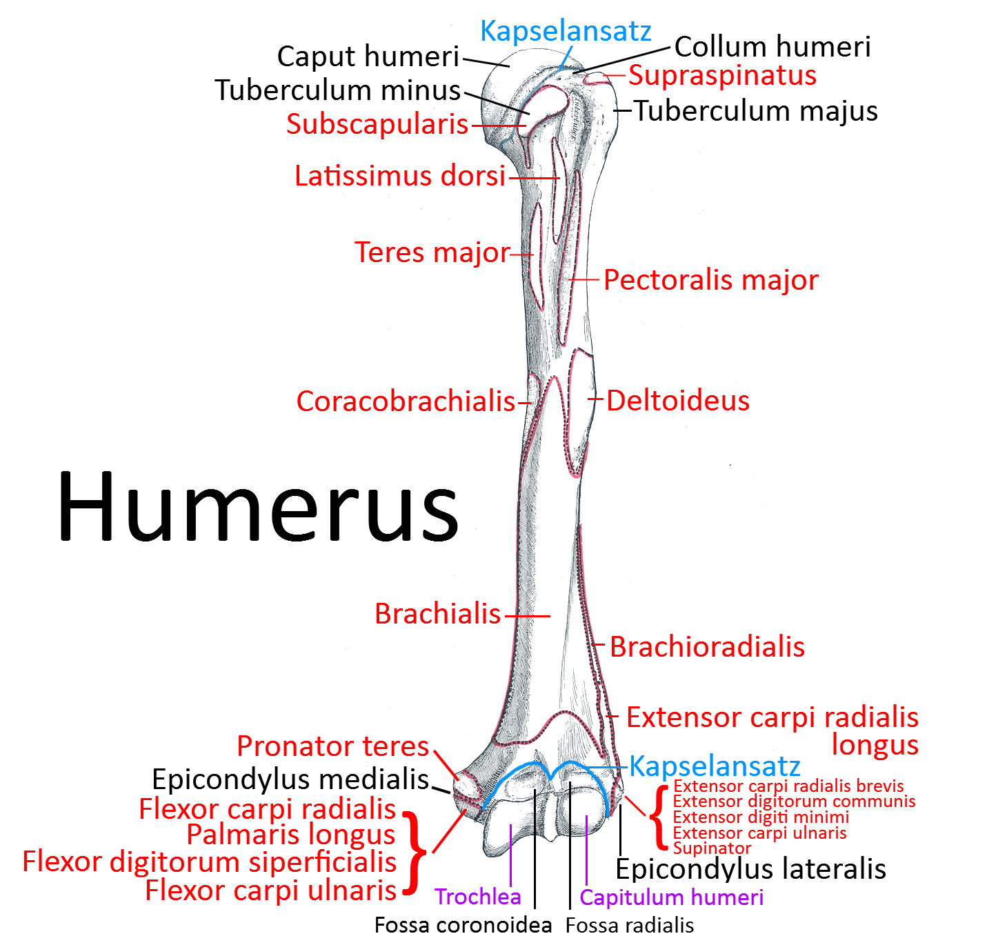
Humerus
Upper arm bone that forms the glenohumeral joint („shoulder joint“) proximally in the glenoid with the shoulder blade and the elbow joint distally with the radius and ulna. The humerus is the longest bone in the upper limb and the only one in the upper arm.
Extremitas proximalis
Caput humeri
The head of the humerus is inclined slightly cranial-medial-dorsal in standard anatomical position. There is an angle of 120 – 140 against its shaft and a retrotorsion angle of 25-40°. It articulates with the glenoid (cavitas glenoidalis) of the scapula in the glenohumeral joint, commonly referred to as the shoulder joint. Ventrally on the shaft lies the lesser tuberosity, which extends clearly ventrally for a good lever arm. The greater tuberosity lies laterally, roughly opposite the head.
Collum humeri
The neck (collum humeri, collum anatomicum) of the humerus is directly adjacent to the head. The collum chirurgicum, on the other hand, is an area below the tuberosity that is predisposed to fractures.
Tuberculum majus
Bone thickening roughly opposite the head of the humerus, on which three muscles of the so-called rotator cuff insert, from cranial to caudal:
Tuberculum minus
Bone thickening clearly extending ventrally, where the subscapularis attaches.
Intertubercular sulcus
The intertubercular sulcus is the bone groove between the bony ridges/lips that extend distally from the greater and lesser tuberosities. The sulcus is lost in the direction of the proximal third point. The tendon of the long head (caput longum) of the biceps and a branch of the anterior circumflex humeral artery run through the sulcus, which is covered by the transverse humeral ligament.
Corpus humeri
The corpus can be subdivided into three surfaces by three margins ( margo anterior, margo lateralis, margo medialis): Facies anterior lateralis, Facies anterior medialis, Facies posterior.
Facies anterior lateralis
The deltoid tuberosity, on which the deltoid muscle inserts, is located approximately in the middle of the anterior lateral facies. The brachialis originates in the distal anterior lateral facies.
Facies anterior medialis
The latissimus dorsi inserts into the upper part of the anterior medial facies. The coracobrachialis inserts halfway in the middle. The brachialis arises in the distal area.
Posterior facies
The medial and lateral heads of the triceps insert over a large area in the anterior posterior facies.
Margo anterior
The margo anterior separates the facies anterior medialis from the facies anterior lateralis and runs from the front of the greater tuberosity to the coronoid fossa. The crista tuberculi majoris, a bony projection located in the cranial region, serves as the base of the pectoralis major. It borders on the ventral border of the deltoid tuberosity for about half of its length. The brachialis originates in the distal area.
Margo lateralis
The margo lateralis, which runs from the back of the greater tuberosity to the epicondylus lateralis humeri, separates the facies anterior lateralis from the facies posterioris. The teres minor attaches in the cranial region, distally from which the lateral head of the triceps arises. The dorsal supracondylar lateral crest serves as the origin of the brachioradialis.
Margo medialis
The margo medialis extending from the lesser tuberosity to the epicondylus medialis humeri has a crista tuberculi minoris in the upper third, to which the teres major inserts. The coracobrachialis attaches sinewy in a slight depression approximately in the middle. As it moves closer to the epicondyle, the medial supracondylar crest becomes increasingly prominent.
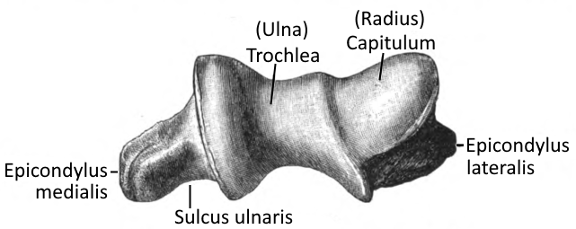
Extremitas distalis
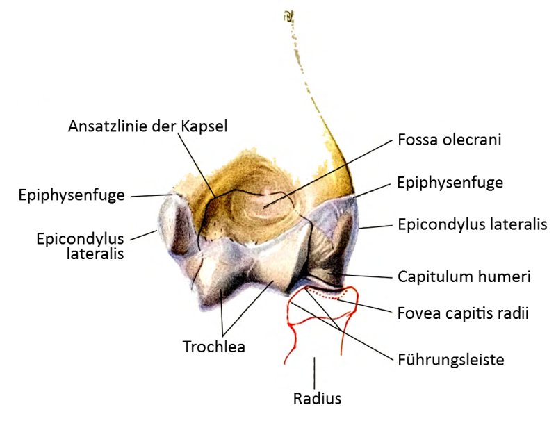
The distal end of the humerus has two articular surfaces: laterally the capitulum humeri(radius) and medially the trochlea humeri(ulna).
Capitulum humeri
The capitulum is the humeral geniculate surface of the concave articular surface of the radius. It is hemispherical with a clearly bulging cartilage surface.
Trochlea
The trochlea resembles a transverse cylinder between the front and back of the humerus.
Radial fossa
Proximal to the capitulum is the radial fossa, which receives the anterior edge of the radial head when the elbow joint is strongly flexed.
Coronoid fossa
Ventral proximal to the trochlea is the coronoid fossa, which receives the coronoid process when the elbow joint is flexed.
Fossa olecrani
The olecranon fossa is a triangular depression proximal to the trochlea that receives the olecranon when the elbow joint is extended.
Epicondylus lateralis humeri
Muscle origin at the lateral condyle of the humerus, to which the muscles of the dorsiflexors (extensors) of the wrist and the finger extensors primarily attach:
- Extensor carpi radialis brevis
- Extensor carpi ulnaris
- Extensor digitorum (communis)
- Extensor digiti minimi
- Supinator.
Epicondylus medialis humeri
Muscle origin at the medial condyle of the humerus, to which the muscles of the palmar flexors(flexors) of the wrist and the finger flexors located in the forearm attach:
- M. flexor carpi radialis
- M. flexor carpi ulnaris
- M. palmaris longus
- M. flexor digitorum superficialis
- M. pronator teres
Ulnar sulcus
The ulnar nerve runs in the ulnar sulcus on the dorsal side of the medial epicondyle.
Joints
- Shoulder joint (glenohumeral joint, humeroscapular articulation, glenohumeral articulation)
- Elbow joint (art. cubiti, with partial joints art. humeroulnaris, art. humeroradialis)
Pathology
The most common disorders of the humerus are of a traumatic nature: fractures of the shaft, proximal and distal fractures.
Images
Humerus from dorsal view (image links to linkmap)
distal humerus
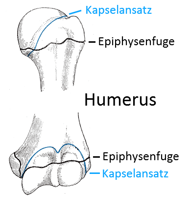
Epiphyseal joints
distal humerus from inferior