yogabook / joints / elbow joint
Contents
- 1 image: Elbow joint (Linkmap)
- 2 Elbow joint (also: elbow joint)
- 3 Hyperextension
- 4 Partial joints
- 5 The movements and the muscles involved
- 6 Ligaments
- 6.1 Lig. anulare radii
- 6.2 Outer ligament complex (LCLC)
- 6.3 Chorda obliqua
- 6.4 Lateral collateral accessorius ligament
- 6.5 Lig. collaterale radiale (RCL: radial collateral ligament, LCL: lateral collateral ligament)
- 6.6 Lig. collaterale ulnare (medial collateral ligament, MCL: medial collateral ligament, UCL: ulnar collateral ligament)
- 6.7 Lateral ulnar collateral ligament (LUCL)
- 6.8 Lig. collaterale ulnare mediale (MUCL: medial ulnar collateral ligament)
- 6.9 Lig. quadratum
- 7 Bursa (bursae)
- 7.1 Bursa anconei
- 7.2 Bicipitoradial bursa
- 7.3 Bursa brachialis
- 7.4 Bursa cubitalis interossea
- 7.5 Bursa extensoris carpi radialis brevis proximalis
- 7.6 Bursa extensoris carpi ulnaris
- 7.7 Bursa extensoris carpi radialis longus
- 7.8 Bursa intertendinea olecrani
- 7.9 Bursa subcutanea epicondyli medialis
- 7.10 Bursa subcutanea epicondyli lateralis
- 7.11 Bursa subcutanea olecrani
- 7.12 Bursa subtendinea tricipitis brachii
- 8 Pathology
- 9 Tests
- 10 Images
- 10.1 Bones (image links to linkmap)
- 10.2 Ligaments from the inside/flexion side (image links to linkmap)
- 10.3 Ligaments from ulnar (image links to linkmap)
- 10.4 Ligaments from radial (image links to linkmap)
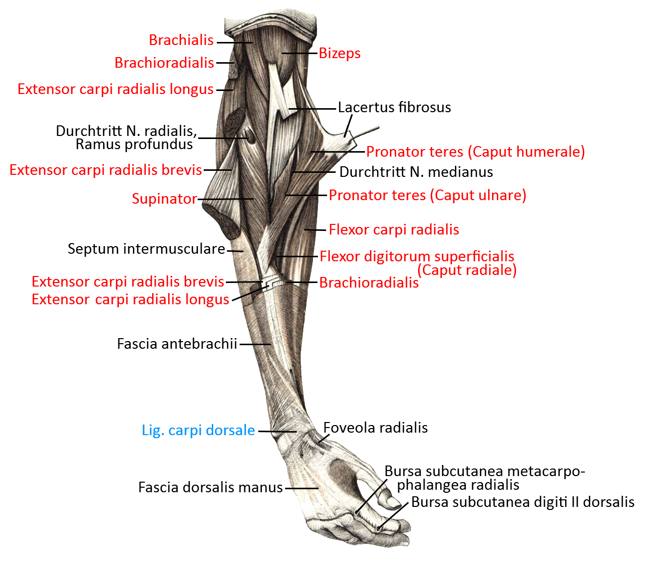
- 10.5 Muscles: from medial (image links to linkmap)
- 10.6 Muscles: Inner elbow, profound, exploded(image links to linkmap)
- 10.7 Muscles: Inner elbow, superficial, exploded(image links to linkmap)
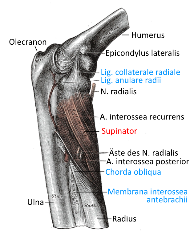
- 10.8 Elbow joint from lateral with supinator muscle (image links to linkmap)
image: Elbow joint (Linkmap)
Elbow joint (also: elbow joint)
The elbow joint (art. cubiti) is the joint in the upper limb in which the forearm moves relative to the upper arm. The main movement is flexion/extension; in addition, the forearm can apparently rotate on its axis, but this is a crossing of the ulna and radius, which retain their position at the elbow and can rotate the wrist by approx. 180°(pronation/supination) by crossing over, or from neutral zero by 80-90° each in the direction of pronation or supination. In standard anatomical position, the two bones ulna and radius run parallel, so that just under 180° of pronation is still available from this widely supinated position. Important movements such as screwing and unscrewing(pronation) and closing(supination) are performed in this way. The two movements – performed here with both arms in opposite directions – also play a central role when wringing out fabrics.
Hyperextension
Quite a few people can overstretch (hyperextend) the elbow joint to a greater or lesser extent. In the case of the elbow joint, end-degree hyperextension leads to compression of the articular cartilage of ulna and humerus and must therefore be avoided. In addition, in some cases this causes discomfort in the mediodorsal elbow joint in the transition to the ulcus ulnaris.
Cubitus valgus
In the elbow joint, a slight valgus position (deviation of the forearm towards the radial side relative to the upper arm) of at least 5° is physiological; in men, up to 10° is considered physiological, and in women, up to 15°. Angles exceeding this are considered cubitus valgus. Cubitus valgus and its varus counterpart, cubitus varus, usually have a traumatic background or result from chronic dislocation tendency. The non-acquired form occurs significantly more frequently and is more pronounced in females than in males and is also often associated with more or less pronounced hyperextensibility.
Partial joints
The elbow joint consists of three partial joints:
1. humeroulnaris joint (humeroulnar joint, humeral-ellar joint)
The humeroulnar joint is the one-dimensionally movable hinge joint between the humerus and ulna, in which flexion and extension take place. Supination/pronation of the forearm is not affected by this movement.
2nd humeroradial articulation (humeroradial joint, humeral-salivary joint)
The humeroradial joint is the two-dimensional mobile ball-and-socket joint in which the radius moves relative to the humerus. As the radius rotates during the overturning movement of the forearm(supination / pronation), a second dimension of movement is required here in addition to flexion and extension.
Both the interosseous membrane and the chorda obliqua (for supination) limit the overturning movement. Together with the ability of the humerus to rotate in the glenohumeral joint(shoulder joint), the overturning movement of the forearm provides an angular range of up to approximately 360° when the arm is in the lateral position, whereby the overturning movement of the forearm is used far more frequently for a specific purpose in everyday activities than the rotation of the upper arm, which is certainly also due to the fact that the overturning movement requires much less effort and energy input and is also more finely rotated. It also takes up far less space in confined spaces.
3. proximal radioulnar joint (proximal radioulnar joint/salivary ulnar joint)
The radioulnar joint is the one-dimensionally movable wheel joint in which the proximal radius rotates relative to the ulna during pronation and supination. The distal radioulnar joint is located at the distal end of the ulna and radius, in which a corresponding movement must also take place in the proximal joint during movement.
The movements and the muscles involved
Flexion: biceps, M. brachialis, M. brachioradialis, M. extensor carpi radialis longus, M. extensor carpi radialis brevis, M. pronator teres, M. flexor digitorum superficialis, M. palmaris longus, other less important
Extension: triceps, M. anconeus (hardly, stated differently in the literature)
Pronation: M. pronator quadratus, M. pronator teres, M. flexor carpi radialis, M. brachioradialis from a supinated position, M. extensor carpi radialis longus (weak when the arm is bent)
Supination: M. supinator, biceps, M. extensor pollicis longus, M. extensor pollicis brevis, M. brachioradialis, M. extensor indicis, M. extensor carpi radialis longus (weak with outstretched arm)
Ligaments
The elbow joint is stabilized by many intracapsular ligaments:
Lig. collaterale radiale (RCL), Lig. collaterale ulnare laterale (LUCL), Lig. collaterale ulnare mediale (MUCL), Lig. anulare radii. The outer ligament complex consisting of the radial collateral ligament (RCL) and the lateral collateral ligament (LUCL) stabilizes against varus movement and the inner ligament complex with the medial collateral ligament (MUCL) stabilizes against valgus movement. The medial ulnar collateral ligament (MUCL) consists of an anterior and a posterior area, the former of which stabilizes more strongly against valgus stress. The RCL primarily stabilizes the proximal radioulnar joint and radiates into the anular ligament. All collateral ligaments originate from the epicondyles: RCL and LUCL from the lateral epicondyle of the humerus and MUCL from the medial epicondyle of the humerus. As they are located intracapsularly, they also lie deeper than the extensor and flexor muscles of the forearm, which originate from the condyles.
- Lig. anulare radii
- Outer ligament complex
- Chorda obliqua
- Lateral collateral accessorius ligament
- Lig. collaterale radiale (RCL, LCL)
- Ulnar collateral ligament (MCL, UCL)
- Lateral ulnar collateral ligament (LUCL )
- Lig. collaterale ulnare mediale
- Lig. quadratum
The bands in detail, see also the diagrams below:
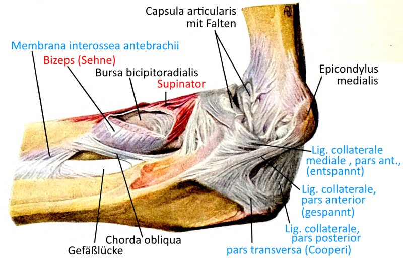
Lig. anulare radii
A capsular ligament extending from both ends of the radial incisura ulnae, which holds the proximal end of the radius (radial head) and is also cartilaginous on the inner side. The two legs of the radial collateral ligament radiate into the radial anular ligament. Limited tensile strength of the elbow joint in children sometimes leads to entrapment of the radial head in the pronated position.
Images:
Linkmap: Lateral view of theelbow joint with ligaments and supinator
Linkmap: Muscles, supinators and pronators of the forearm
Linkmap: Muscles, forearm and hand, palmar, profound
Linkmap: Muscles, inner elbow, profound
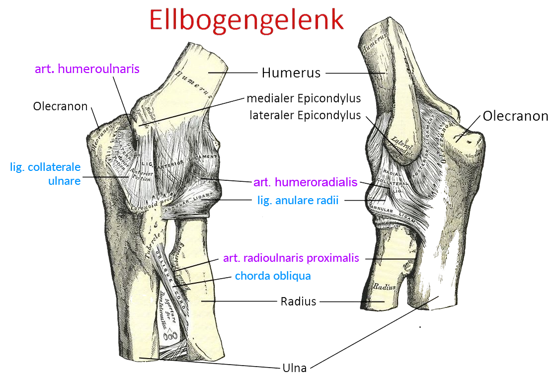
Linkmap: Elbow joint, medial and lateral
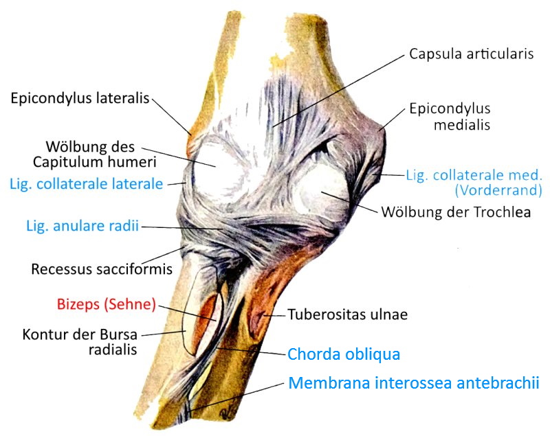
Linkmap: Elbow joint, ligaments from flexor side
Linkmap: Elbow joint, ligaments from radial side
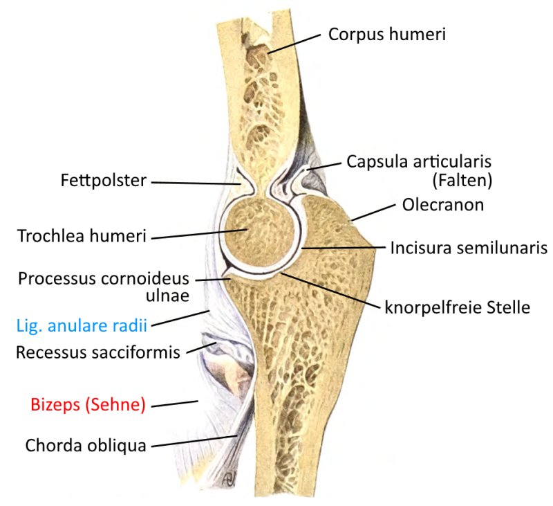
Linkmap: Elbow joint with lig. anulare radii and lig. quadratum
Linkmap: Supinator muscle
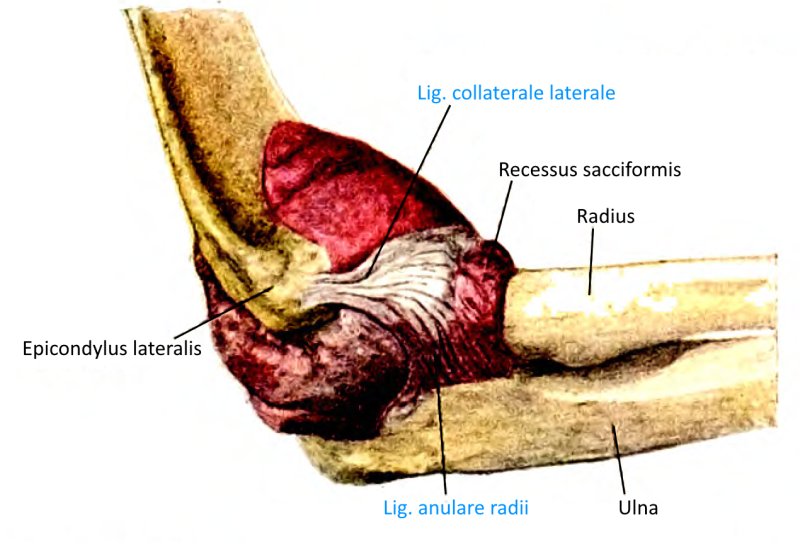
Outer ligament complex (LCLC)
The following four ligaments are sometimes grouped together under the term lateral collateral ligament complex:
- Lig. anulare radii
- Lig. collaterale radiale
- Lig. collaterale ulnare laterale
- Lateral collateral accessorius ligament
Chorda obliqua
The chorda obliqua is a ligament that extends from the ulnar tuberosity distal to the radial tuberosity to limit supination of the forearm. The fibers of the chorda obliqua run in the opposite direction to those of the interosseous membrane of the antebrachial ligament.
Images:
Linkmap: Elbow joint lateral with ligaments and supinator
Linkmap: Elbow joint, ligaments from the flexor side
Linkmap: Elbow joint from medial and lateral side
Lateral collateral accessorius ligament
The lateral collateral accessorius ligament runs from the anular radial ligament via the lateral radial head to its insertion immediately distal to the posterior edge of the radial incisura of the ulna. It stabilizes the anulare radii ligament .
Images: (still without images)
Lig. collaterale radiale (RCL: radial collateral ligament, LCL: lateral collateral ligament)
This ligament radiates from the lateral humeral epicondyle into the radial anular ligament in an approximately delta-shaped manner via two fiber tracts, once medially and once laterally. Further fibers run to the radial incisura ulnae. It is weaker than its ulnar counterpart and contributes to varus stability.
Images:
Linkmap: Elbow joint lateral with supinator
Linkmap: Muscles, forearm, pronators and supinators
Linkmap: Elbow joint, ligaments from radial
Linkmap: Elbow joint, ligaments from flexor side
Linkmap: Muscles, forearm and hand, palmar profund
Lig. collaterale ulnare (medial collateral ligament, MCL: medial collateral ligament, UCL: ulnar collateral ligament)
The ulnar collateral ligament is a simplified term for the medial ulnar collateral ligament, see there. In addition to the medial ulnar collateral ligament, there is another ulnar ligament, the lateral ulnar collateral ligament.
Images:
Linkmap: Elbow joint, ligaments, flexor side
Linkmap: Medial and lateral view of theelbow joint
Linkmap: Elbow joint, ligaments from ulnar side
Lateral ulnar collateral ligament (LUCL)
The ligament located on the lateral elbow joint, it runs as a loop from the epicondylus humeri radialis dorsally around the posterolateral head of the radius to the tuberosity of the crista supinatoria ulna. It is more frequently involved in radial head fractures, and its damage then causes varus instabilityof the elbow joint. It is a very important stabilizer against varus movement.
Pictures: (still without pictures)
Lig. collaterale ulnare mediale (MUCL: medial ulnar collateral ligament)
The MUCL is often referred to simply as the ulnar collateral ligament (UCL or MCL). It runs from the epicondylus medialis humeri to the medial side of the incisura trochlearis of the ulna. Some fibers of the anconeus attach to this ligament. Around 55% of valgus stress is absorbed by this ligament. Especially throwing sports such as javelin throwing, in which significant valgus stress occurs, strain this ligament. It consists of three parts:
- Pars anterior (anteromedial collateral ligament, AMCL), insertion is the ulnar surface of the coronoid process of the ulna, about 18 mm dorsal to the tip of the coronoid process. It is the most important stabilizing structure against valgus movement.
- Pars posterior (posteromedial collateral ligament, PMCL), insertion is the ulnar surface of the olecranon. It has a less stabilizing function than the anterior pars.
- Pars transversa (transverse bundle, Cooper ligament), the weakest of the three parts, it connects the ulnar parts of the other two ligaments. No significant contribution against valgus movement is attributed to this ligament.
Lig. quadratum
The quadratum ligament is a thin capsular ligament that runs from the lower edge of the radial incisura of the ulna to the collum of the radius and limits supination and, to a lesser extent, pronation.
Images:
Linkmap: Elbow joint with lig. anulare radii and lig. quadratum
Bursa (bursae)
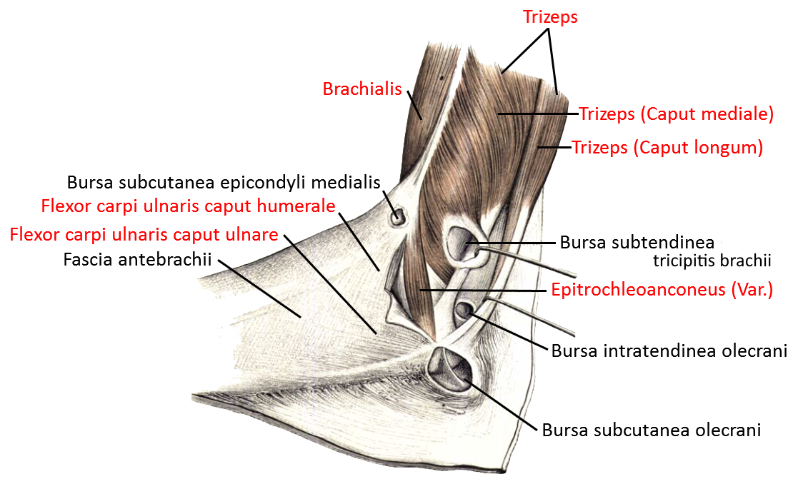
Bursa anconei
Bursa located between the lateral epicondyle and the origin of the anconeus. This bursa can communicate with the joint space.
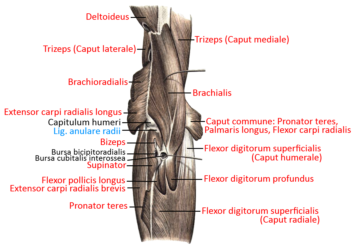
Bicipitoradial bursa
between the insertion tendon of the biceps brachii and the insertion of the biceps on the anterior part of the radial tuberosity. The bursa is primarily important as a buffer during pronation and supination of the arm, whereby it is compressed during pronation. An inconstant bursa cubitalis interosseus is sometimes located in its vicinity between the attachment tendon of the biceps and the ulna or the chorda obliqua.
Images:
Linkmap: Inner elbow, profound
Bursa brachialis
Inconstant, rarely occurring submuscular bursa between the lateral edge of the brachialis and the joint capsule.
Bursa cubitalis interossea
less common (20%) inconstant bursa between the insertion tendon of the biceps and the proximal ulna. It is one of two bursae in the cubital fossa and is particularly important for low-friction gliding during pronation and supination.
Images:
Linkmap: Inner elbow, profound
Bursa extensoris carpi radialis brevis proximalis
Subtendinöse Bursa profund der Ursprungssehne des Extensor carpi radialis brevis.
Bursa extensoris carpi ulnaris
Subtendinous bursa deep to the tendon of origin of the extensor carpi ulnaris
Bursa extensoris carpi radialis longus
Submuscular bursa of the extensor carpi radialis longus to the deeper lying lig. anuare radii.
Bursa intertendinea olecrani
in the attachment tendon of the triceps at the olecranon or between this and the proximal ulna .
Images:
Linkmap: Medial view of the elbow
Bursa subcutanea epicondyli medialis
Also known as bursa subcutanea epicondyli ulnaris, an inconstant bursa that occurs rarely and usually only in the dominant arm.
Images:
Linkmap: Medial view of the elbow
Bursa subcutanea epicondyli lateralis
Inconstant subcutaneous bursa over the lateral epicondyle of the humerus, occurs rather rarely and usually only in adults.
Bursa subcutanea olecrani
a non-communicating bursa between the tuber olecrani at the upper end of the ulna and the skin. It extends to the base of the triceps. Bursitis develops here primarily as a result of mechanical stress such as prolonged, frequent leaning on the olecranon, which is why it is also known as Student’s elbow.
Images:
Linkmap:. Elbow from the medial side
Bursa subtendinea tricipitis brachii
eine auch als Bursa subtendinea olecrani bezeichnete profunde Bursa zwischen der Ansatzsehne des Trizeps und dem Olecranon am proximalen Ende der Ulna
Bilder:
Linkmap: Ellbogen von medial
Pathology
Some diseases of the joint:
- Epicondylitis humeri radialis (tennis elbow)
- Epicondylitis humeri ulnaris (golfer’s elbow)
- Cubital tunnel syndrome (sulcus ulnaris syndrome, ulnar groove syndrome)
- Ligament stretching
- Impingement syndrome
- Olecranon bursitis
- Radialhead fracture
Tests
Tests of the elbow joint
Tennis elbow (medial epicondylitis)
Golfers elbow (lateral epicondylitis)
Cubital tunnel syndrome
Medial collateral ligament
Instability
Extension deficit
Tests of the movement directions
Supination and pronation
Tests of the spanning muscles
Biceps
- Popeye-sign (rupture of origin tendon)
- Reverse Popeye-Sign (rupture of insertion tendon)
- Ludington-Test (rupture of origin tendon)
Triceps
Images
Bones (image links to linkmap)
Ligaments from the inside/flexion side (image links to linkmap)
Ligaments from ulnar (image links to linkmap)
Ligaments from radial (image links to linkmap)
Muscles: from medial (image links to linkmap)
Muscles: Inner elbow, profound, exploded(image links to linkmap)
Muscles: Inner elbow, superficial, exploded(image links to linkmap)
Elbow joint from lateral with supinator muscle (image links to linkmap)