Contents
Image: Knee joint with patella, sagittal section
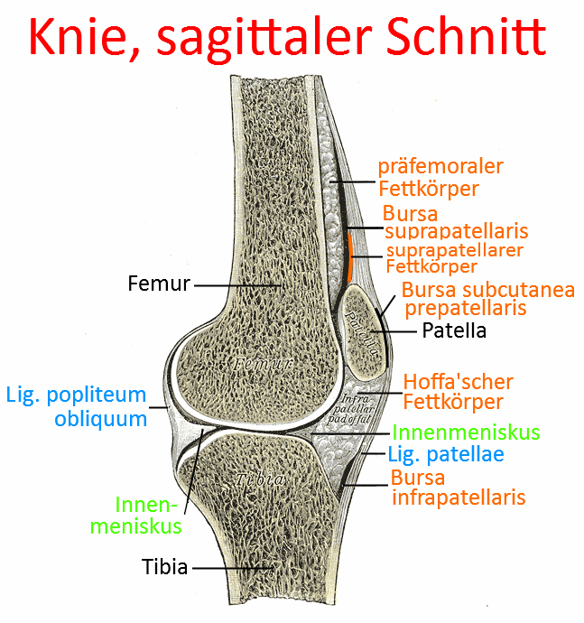
Patella
Patella, the largest ganglion and sesamoid bone or hypomochlion of the human body. The patella is a bony disk located in front of the femur; it slides over the femoropatellar sliding bearing (articulatio femoropatellaris) of the distal femur during extension and flexion of the knee joint. The patellar ligament attaches to the caudal end of the patella(caudal patellar pole) and around 50% of the tendon fibers of the quadriceps attach to the cranial end(cranial patellar pole), so that its tendon force can be transmitted to the tibial tuberosity, the bone roughening of the proximal ventral tibia, and extension in the knee joint can be achieved. As a hypomochlion, the patella causes an increased torque.
The quadriceps attach to the caudal pole of the patella according to their position: Vastus medialis more medial, Vastus intermedius more medial, and Rectus femoris and Vastus lateralis more medial to lateral, Vastus medialis and Vastus lateralis thus form a kind of patella restraint system. This can result in a displacement of the patella (typically the pull of the vastus lateralis is too high in relation to the vastus medialis rather than the other way around, i.e. laterally) in the case of markedly unequal traction, as well as a rotational moment of the patella(caudal to lateral) and a tilt(lateral to dorsal), which poses a particular risk to the lateral part of the retropatellar cartilage. Chondropathia patellae (PFPS) can later lead to retropatellar arthrosis. As the retropatellar cartilage is relatively cell-poor and consists largely of connective tissue, self-healing is only possible to a very limited extent. In addition, painful patellar dislocations occur more easily under these conditions, which usually tear the medial retinaculum, leading to renewed patellar dislocations all the more quickly.
The patella has an average minimum lever arm of 36 mm; without the patella, it would be around 25 mm. This alone is a serious factor against patellectomies, unless they are absolutely necessary, especially since the tendon of the quadriceps appears too long afterwards, which means an unfavourable shift in terms of the force-length function. This effect is multiplied by the deterioration of the lever arm, which alone represents a factor of 1.44. The most common disruptive factor in the patellofemoral joint is an inappropriate geometry of the trochlea femoris. The sulcus angle of the trochlea is the single factor that correlates best with the symptoms of patellofemoral complaints. Physiologically, the medial half of the patella is less pronounced than the lateral half, which accounts for the biomechanically determined force distribution of 40 to 60. Near the extension of the knee joint, the retropatellar contact pressure decreases; when the knee joint is almost extended, it can be said that no articulation is taking place. The patella is then very mobile and its stability depends greatly on the soft tissues connected to it. Even at 20 degrees flexion, normal articulation and stability can be assumed. Due to the variable curvature of the femoral condyles, the patella does not always form a congruent joint with the femur at every angle of flexion of the knee joint at every angle of flexion of the knee joint. While at 20° flexion it is mainly the distal part of the patella that is in contact with the femur; with increasing flexion, the contact surface shifts in the cranial direction. Up to 90° flexion, every part of the retropatellar cartilage, with the exception of the odd facet, was in contact with the trochlea femoris. From 90° flexion, the quadriceps tendon also comes into direct contact with the trochlea femoris. The odd facet comes into contact with the medial femoral condyle at approximately 130 degrees flexion. The contact area of the patella with the femur increases from 20 to 90°, while the retropatellar contact pressure also increases.
Patellar poles
the two „poles“ (viewed vertically) of the patella (kneecap), the caudal patellar pole and the cranial patellar pole. The quadriceps attaches tendinously to the cranial patellar pole, and the patellar ligament, which is sometimes incorrectly referred to as the „patellar tendon“, attaches to the lower pole. As the patellar ligament is taut and inflexible in a taut state, i.e. whenever there is a minimum amount of tension on the quadriceps that exceeds the resting tension, the caudal patellar pole is at a constant distance from the tibial tuberosity, the area where the patellar ligament attaches to the tibia.
Cranial patellar pole
Around 50% of the fibers of the quadriceps insertion tendon attach to the cranial patellar pole in order to transmit its contraction force to the tibial tuberosity via this hypomochlion to increase torque. The remaining fibers of the insertion tendon cover the patella in the direction of the tibial tuberosity or run medial or lateral to the patella as the medial patellarretinaculum or lateral patellarretinaculum.
Caudal patellar pole
The patellar ligament attaches to the caudal patellar pole to transfer the movement of the patella to the tibial tuberosity. The patellar ligament has no significant elasticity and therefore guarantees a fixed distance between the caudal patellar pole and the tibial tuberosity when it is tensed, which means that when the quadriceps contract concentrically, its contraction force causes the patella and consequently the tibial tuberosity to move.
Retropatellar cartilage
The retropatellar cartilage, i.e. the cartilage layer that slides over its joint partner, the femur or its cartilage, in the femoropatellar sliding bearing, is the thickest in the human body for good reason. Depending on the angle of the knee joint and the contraction force of the quadriceps, high pressures are quickly applied here. Damage to the cartilage is known as chondropathia patellae (PFPS) or, more advanced, as retropatellar arthrosis.