yogabook / muscles / quadrizeps
Contents
Linkmap
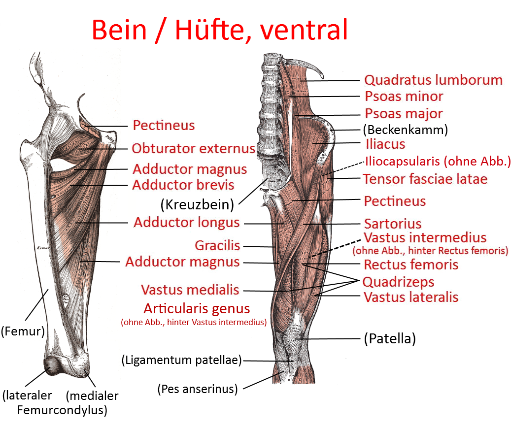
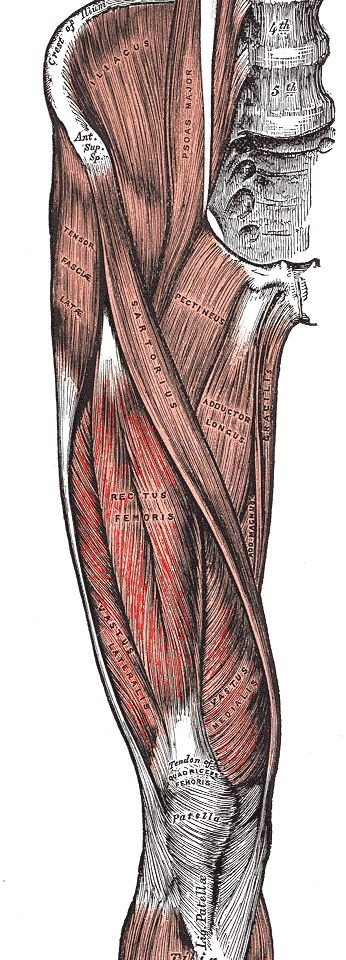
M. quadriceps
large knee extensor consisting of four parts, including
- three monoarticular heads: vastus lateralis, vastus medialis, vastus intermedius and
- a biarticular head: the rectus femoris muscle
The biarticular rectus femoris muscle is involved in flexion in the hip joint and tends to spasm in postures with an extended knee joint and wide flexion in the hip joint. The vastus intermedius has a separation, also known as the articularis genus muscle, which attaches to the suprapatellar bursa and tightens it so that it cannot be squeezed. If the quadriceps are too weak, the femur will slide ventrally in relation to the tibia, especially when walking downhill and descending stairs after the foot has touched down, which can lead to the menisci becoming locked and later to osteoarthritis. If its tone is too high, the retropatellar cartilage will be damaged, resulting in chondropathia patellae and possibly later retropatellar arthrosis. Excessive tension also predisposes to damage to the patellar ligament(patellar tip syndrome/jumper’s knee, which usually occurs at the caudal patellar pole, but more rarely also affects the attachment of the patellar ligament to the tibial tuberosity, or damage to the tibial tuberosity in the form of Osgood-Schlatter’s disease in athletic adolescents.
The quadriceps are connected to local structures via regional proprioceptors and reflexes. For example, externally induced varus stress to test for lateral gapping triggers increased traction of the vastus lateralis on the patella and, conversely, valgus stress increases traction of the vastus medialis. This mechanism contributes to centralization of the patella and reduces the tendency to dislocation. The frequently changing tensile components of the two quadriceps components during more complex movements therefore simultaneously change the pressure conditions on the retopatellar cartilage and thus serve to nourish it. However, this structure can be permanently disrupted by disorders such as axial misalignment, which is one of the reasons for the resulting tendency to disrupt the patellar cartilage.
Origin: Origin of the rectus femoris: Spina iliaca anterior inferior (SIAI), upper edge of the acetabulum (acetabulum). Origin of the Vastii: anterior and lateral proximal femoral surface, linea aspera, lateral surface of the greater trochanter, linea intertrochanterica, gluteal tuberosity, linea aspera
Attachment: outer surface of the patella, tibial tuberosity of the tibia
Innervation: Nervus femoralis (L2-4)
Antagonists:
Movement: Extension, exorotation in the hip joint, the biarticular, rectus femoris also: Flexion in the hip joint
Strengthening postures (812): utkatasana, 1st warrior, 2nd warrior, caturkonasana, dvi pada viparita dandasana, eka pada viparita dandasana, hip opener 1, hip opener 2, navasana, parsvakonasana, theke, ustrasana
Stretching poses (811): virasana, supta virasana, quadriceps stretch 1, quadriceps stretch 2, ardha supta krouncasana
Rectus femoris
The biarticular head of the quadriceps described in more detail here, which not only stretches in the knee joint but also flexes in the hip joint.
Vastus medialis
The medial monoarticular head of the quadriceps, which attaches between the cranial patellar pole and the medial-caudal edge.
Many authors identify a caudal portion within the vastus medialis with significantly more fibers running in a transverse direction (50° instead of 17° in the cranial area) as the vastus medialis obliquus, especially since tendon fibers of the adductor magnus radiate to the patella together with this portion. The vastus medialis appears to be of greater importance for knee health, as its atrophy (possibly one of the causes) is found in older meniscus lesions, insufficiency of the anterior cruciate ligament and a number of other disorders of the knee joint.
Vastus medialis obliquus
The distal part of the vastus medialis, which pulls towards the medial cranial patellar pole at an angle of approx. 50° instead of 17° in the cranial part and joins with tendon fibers of the adductor magnus.
Vastus intermedius
The head of the quadriceps lying more profoundly behind the rectus femoris.
Vastus lateralis
The lateral monoarticular head of the quadriceps, which attaches between the cranial patellar pole and the lateral caudal edge. The caudal parts are quite variable and arise partly from the back of the iliotibial tract. Because of their more transverse orientation, they are referred to as the lateral obliquebranch. In females they insert at the lateral cranial patellar pole, in males this insertion is somewhat wider, more towards the medial cranial patellar pole, and on the lateral side of the patella more caudally. If the medial parts of the quadriceps(vastus medialis, in particular the vastus medialis obliquus ) are not in sufficient proportion to the lateral parts, this favors a tendency towards lateral patellar dislocation.
Quadriceps tendon
The quadriceps tendon is the common attachment tendon of the quadriceps to the cranial patellar pole. Some fibers of the tendon run medially and laterally past the patella and form the medial retinaculum patellae (fibers of the vastus medialis) or lateral retinaculum patellae(vastus lateralis and rectus femoris), which protect the patella from dislocation alongside the transverse lateralretinaculum patellae and, if present, the medial transverseretinaculum patellae. Insertional tendinopathies of the quadriceps tendon are rather rare, but possible in principle.
On closer inspection, the quadriceps tendon has three layers or strata (laminae) separated by very thin fatty tissue: the uppermost layer originates from the tendon of the rectus femoris, the middle layer from the vastus medialis and vastus lateralis, and the deepest layer from the vastus intermedius. The number of layers can vary between 2 and 4. The vastus lateralis radiates into the tendon at an angle of approximately 21°, the vastus medialis at approximately 41°. Several of the fibers of the rectus femoris do not attach to the cranial patellar pole, but rather cover the patella in order to attach to the tuberositas tibiae with the fibers of the ligamentum patellae originating at the caudal patellar pole. The width of the quadriceps tendon varies between approximately 27 and 33 mm, and its length between 38 and 49 mm.
Tests
Quadrizeps-Dehnungstest, Dreyer-Test (quadriceps tendon)