Contents
- 1 Image: Hip bone with its parts pubic bone, ischium, ilium (link to linkmap)
- 2 Hip bone from lateral with insertions (linkmap)
- 3 Hip bone
- 3.1 Acetabulum
- 3.2 Crista iliaca
- 3.3 Linea glutea
- 3.4 Linea arcuata
- 3.5 SIAS (Spina iliaca anterior superior, ASIS: anterior superior iliac spine)
- 3.6 SIAI (Spina iliaca anterior inferior, AIIS: anterior inferior iliac spine)
- 3.7 SIPS (Spina iliaca posterior superior, PSIS: posterior superior iliac spine)
- 3.8 SIPI (Spina iliaca posterior inferior, PIIS: posterior inferior iliac spine)
- 3.9 Incisura ischiadica major
- 3.10 Incisura ischiadica minor
- 3.11 Acetabular labrum
- 3.12 Foramen obturatum
- 3.13 Facies glutea / Facies glutealis
- 3.14 Iliac tuberosity
- 3.15 Eminentia iliopubica
- 3.16 Facies auricularis
- 4 Joints
- 5 Images
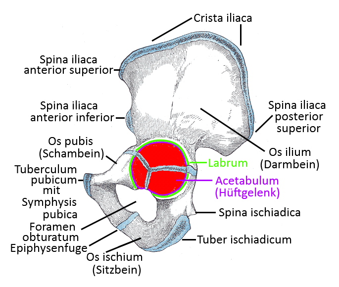
Image: Hip bone with its parts pubic bone, ischium, ilium (link to linkmap)
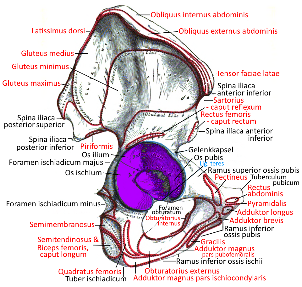
Hip bone from lateral with insertions (linkmap)
Hip bone
The hip bone is the junction of the three bones ischium, pubis and ilium after the epiphyseal joints have closed at the end of the growth phase. These three bones converge in a Y-shape in the acetabulum .
Acetabulum
the joint socket in which the head of the femur articulates with the hip bone. The term literally means „little pot of vinegar“ because its shape is reminiscent of this. The acetabulum is where the ilium, ischium and pubic bone meet. Around the 15th year of life, the bones have grown together to such an extent that one can speak of a single bone, the hip bone.
Crista iliaca
the bony upper edge of the hip bone, which extends forward from the sacrum on both sides. The crista iliaca extends from the SIAS to the PSIS and is the insertion for the quadratus lumborum and some abdominal muscles:
Linea glutea
There are three different lines on the dorsal surface of the ilium, between which glutei are attached:
- Linea glutea anterior
- Posterior gluteal line
- Linea glutea inferior
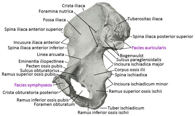
Linea arcuata
The arcuate line is a laterally located rounded bony edge on the ventral side of the ilium. It lies directly caudal to the iliac fossa, where the iliac muscle originates. It is the lateral part of the linea terminalis, which separates the large pelvis from the small pelvis. It is followed caudally-medially by the pecten ossis pubis and cranial-medially by the sacrum.
SIAS (Spina iliaca anterior superior, ASIS: anterior superior iliac spine)
the anterior superior iliac spine, commonly known as the „hip bone“, a bony protrusion on the hip bone that sticks out forwards, one on the left and one on the right
SIAI (Spina iliaca anterior inferior, AIIS: anterior inferior iliac spine)
the anterior inferior iliac spine, origin of the rectus femoris and part of the iliacus, one on the left and one on the right.
SIPS (Spina iliaca posterior superior, PSIS: posterior superior iliac spine)
the posterior superior iliac spine, another important landmark on the pelvis, located sagittally approximately at the upper end of the sacrum and transversely approximately at the level of the SI joint.
SIPI (Spina iliaca posterior inferior, PIIS: posterior inferior iliac spine)
Bony protrusion below the SIPS. Both protrusions are separated by a bony hollow, the posterior inferior iliac spine borders the greater sciatic notch cranially.
Incisura ischiadica major
Bone indentation in the posteromedial edge of the hip bone, directly above the ischial spine.
Incisura ischiadica minor
Crescent-shaped bony depression on the posteromedial edge of the ischium, caudal of the ischial spine, cranial of the ischial tuberosity of the ischial bone. The surface is smooth and covered with cartilage over which the tendon of the internal obturator muscle can glide with little friction, buffered by the internal obturator musclebursa ischiadica. The incisura has a hypomochlion function.
Ligamentum sacrospinale and ligamentum sacrotuberale close the incisura ischiadica minor, which appears as foramen ischiadicum minus.
Acetabular labrum
The cartilage lip that provides the hip joint with additional support: due to the wide opening (small circular section) required for the comprehensive mobility of the hip joint and the additional small incongruence present, the hip joint requires additional stabilizing factors for sufficient stability. These are, on the one hand, good ligament guidance and, on the other, the cartilage lip labrum acetabuli.
Foramen obturatum
The obturator foramen is a largely circular recess in the lower hip bone, which is largely covered by the obturator internus and obturator externus muscles in addition to a coarse connective tissue membrane, the obturator membrane. The obturator nerve and the obturator vein and artery run through the remaining obturator canal. Multiple births in particular can lead to a hernia here at an advanced age.
Facies glutea / Facies glutealis
The facies glutea is the dorsal outer surface of the ilium, where the gluteal ducts originate.
Iliac tuberosity
The iliac tuberosity is a roughening of the bone bordered by the facies auricularis ossis ilii, the posterior superior iliac spine and the iliac crest, to which the sacroiliac ligament inserts.
Eminentia iliopubica
The iliopubic eminence is a bony prominence in the hip bone, marking the junction between the ilium and the pubis. It is the dorsal attachment of the arcus iliopectineus (fascia iliopectinea), which runs to the inguinal ligament.
Facies auricularis
The facies auricularis is the articular surface of the ilium with the sacrum in the SI joint.
Joints
- Hip joint (art. coxae)
- Sacroiliac joint(SIJ, sacroiliac articulation)
Images
Hip bone from medial
Ossification of the hip bone (newborn, 7th and 14th year)
