Calcaneus with insertions
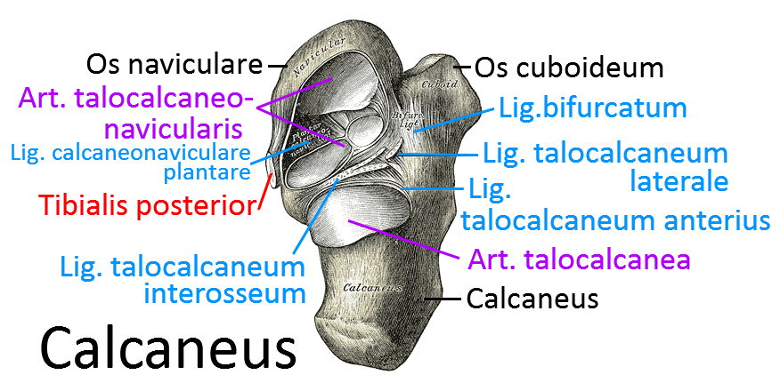
The calcaneus, located below the talus and, like the talus, part of the tarsal bones. In the joint with the proximally adjacent talus (talocalcanea), inversion and eversion mainly take place.
In the joint to the cranially adjacent talus, the talocalcaneal joint (art. talocalcanea), there is hardly any plantar flexion and dorsiflexion, but also almost exclusively pronation and supination. The calcaneus is the attachment point for the triceps surae, which causes the plantar flexion required for walking.
Sustentaculum tali
The calcaneus has a distal-medial bony prominence, the sustenaculum tali, which supports the talus and limits its movement. The sustenaculum serves as a hypomochlion for the tendon of the flexor hallucis longus. The tendon of the tibialis posterior, the flexor digitorum longus and the tibial nerve run through its medial groove(tarsal tunnel), which runs in the longitudinal direction of the foot.
The interosseous talocalcaneal ligament, the most important ligament between the talus and calcaneus, lies in the canalis tarsi, which ends at the suspensory sac and separates the two chambers of the lower ankle joint. The calcaneonavicular plantar ligament and the deltoid ligament insert into the suspensory sac. The suspensory sac is palpable about 1.5 cm inferior to the medial malleolus.
Trochlea fibularis / peroneal tubercle
Bony projection located on the lateral calcaneus, below which the tendon of the fibularis longus runs in the sulcus tendinis musculi fibularis longi. Above it runs the tendon of the fibularis brevis.
Tuber calcanei
pronounced bony protrusion at the dorsal end of the calcaneus, where the Achilles tendon attaches.
Bursa tendinis calcanei
Bursa between calcaneal tuberosity and Achilles tendon
Processi of the Tuber calcanei
The two projections Processus medialis tuberis calcanei and Processus lateralis tuberis calcanei are located on the underside of the tuber calcanei.
Processus medialis tuberis calcanei
This protrusion is the origin of the abductor hallucis and flexor digitorum brevis muscles.
Processus lateralis tuberis calcanei
The abductor digiti muscle attaches to and between the two processes. The aponeurosis plantaris also originates here.
Articulated surfaces
Facies articularis talaris anterior
Forms the vUSG with the talus.
Facies articularis talaris media
Forms the vUSG with the talus.
Facies articularis talaris posterior
Forms the subtalar joint with the talus Art. subtalaris