yogabuch / joints / acromioclavicular joint
Contents
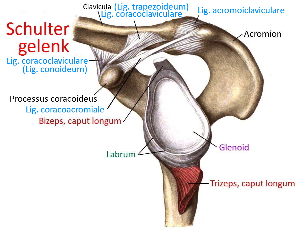
Image: Acromioclavicular joint in the context of the shoulder

Acromioclavicular joint (acromioclavicular joint, AC joint)
Joint between the clavicle and the shoulder blade. This joint contains the most wear-prone intervertebral disc in the human body, which is often no longer detectable after the age of 45, although this usually remains asymptomatic.
In terms of the joint surfaces, the acromioclavicular joint is a planar joint, but functionally a ball joint due to the disc.
Articulating bones
Movements
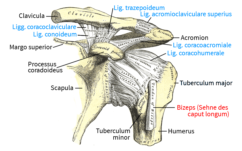
Movements in the acromioclavicular joint are always accompanied by movements in the sternoclavicular and scapulothoracic joints. Stable ligaments secure the joint horizontally, primarily through the acromioclavicular ligament, and vertically through coracoclavicular ligaments(coracoclavicular ligament: Lig. trapezoideum ventral lig. Lig. conoideum dorsal).
The clavicle moves passively with the scapula in three degrees of freedom, so the movement of the scapula is mainly caused by trunkoscapular muscles.
Pathology
The pathology of the joint mainly involves the (often traumatic) weakening of the ligament structure with dislocation, which is known as acromioclavicular joint dislocation.
If the ligamentous structure is sufficiently damaged, the scapula descends caudally, which acts like a cranially dislocated clavicle. It can then be manually repositioned caudally (or the scapula can be repositioned cranially), but this is not permanent, rather the clavicle acts more like a piano key that is pressed down, whereupon it rises again of its own accord, which has led to the term piano key phenomenon.
Ligaments
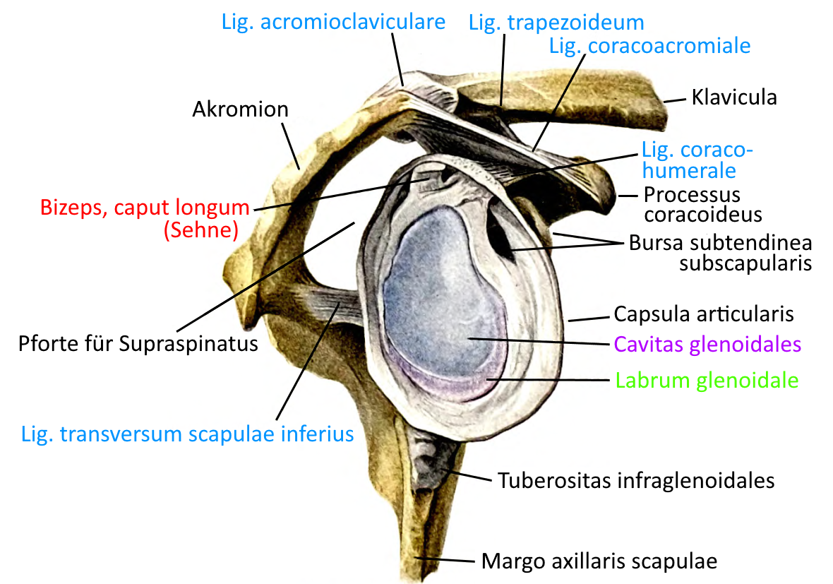
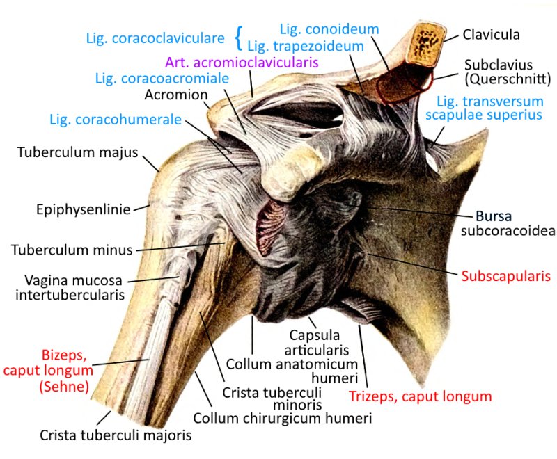
The acromioclavicular joint is secured by three ligaments, for more details see the following links on the page about the shoulder joint:
Bursae
The bursae of the acromioclavicular joint are described in the shoulder joint, see there.
Tests
Tests of the acromioclavicular joint
Instability of the joint
Tests of the movement directions
Tests of the spanning muscles
Charts
Glenoid (image links to linkmap)

Ligaments (image links to linkmap)

Ligaments (image links to linkmap)

