yogabuch / joints / finger joint
Contents
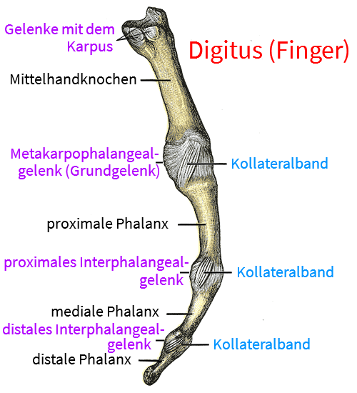
Image: Finger with joints from the side
Finger joints in general (metacarpophalangeal joint, proximal, distal interphalangeal joint)
The fingers consist of three phalanges in the case of the long fingers and only two in the case of the thumb. The proximal phalanx connects to the metacarpal bones, so the finger joints include the metacarpophalangeal joints (MCP) and the interphalangeal joints (interphalangeal joints), of which there is one PIP(proximal interphalangeal joint) and one DIP(distal interphalangeal joint), with the exception of the thumb. In the interphalangeal joints there is only the flexion/extension dimension of movement, in the metacarpophalangeal joints there is also radial and ulnar abduction/adduction, which together allow the fingers to spread and spread apart by means of the palmar and dorsal interossei.
Articulating bones
- Metacarpalia
- proximal phalanx
- medial phalanx
- distal phalanx
Joints
Metacarpophalangeal joint (MCP)
(See also the separate page on the metacarpophalangeal joints)
The metacarpophalangeal joint (MCP) is the joint at the transition from the palm of the hand (palmar) to the fingers. It allows two-dimensional mobility: flexion up to approx. 90° and extension, possibly up to slight hyperextension. Overextension in the order of 10° is still considered physiological, but from 90° (in relation to the little finger) at the latest, a point is awarded in the Bighton score for hypermobility.
CCM countries
and in the case of the fingers 2-5 as well:
Interphalangeal joints
Proximal interphalangeal joint (PIP)
The proximal interphalangeal joint lies between the proximal and medial phalanges and, like the distal interphalangeal joint (DIP), only allows one-dimensional movement: Flexion and extension (possibly into slight hyperextension). The PIP has collateral ligaments that absorb movement in the varus or valgus direction.
Distal interphalangeal joint (DIP)
The distal interphalangeal joint is the last on the respective ray and moves the last phalanx relative to the middle one. Like the proximal interphalangeal joint, it only allows one-dimensional movement: Flexion and extension (possibly into slight hyperextension). The DIP also has collateral ligaments that absorb movement in the varus or valgus direction.
Additional ligaments of the interphalangeal joints
- Ligg. anularia
- Ligg. obliqua
- Ligg. collateralia
- Ligg. collateralia accessoria
Tapes
Ligg. anularia
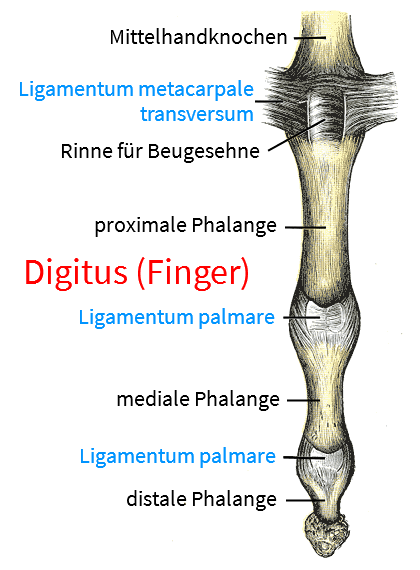
The anular ligaments are attached palmarly at the level of the respective interphalangeal joints with fiber tracts to the shafts of the proximal and medial phalanges. Their main purpose is to attach the tendon sheaths so that they do not become detached.
Images: (still without)
Ligg. collateralia
As the collateral ligaments of fingers 2-5 run from proximal/dorsal to distal/palmar, they are tense when the metacarpophalangeal joint is flexed and prevent adduction and abduction. With increasing extension of the metacarpophalangeal joints, the range of motion in this dimension increases.
In the case of the MCP (metacarpophalangeal joint), the collateral l igaments extend obliquely from the dorsolateral side of the metacarpals to the bases of the proximal phalanges . The collateral ligaments of the interphalangeal joints run on the medial and lateral sides of the joints from the head of one phalanx to the base of the next.
Ligg. collateralia accessoria
The collateral ligaments are additional collateral ligaments that are also attached to the fibrocartilage plates on the palmar side.
Images: (still without)
Ligg. metacarpalia transvera profunda
The deep transverse ligaments connect the metacarpal heads and extend into the joint capsule and the palmar aponeurosis.
Images: (still without)
Ligg. metacarpalia transvera superficialia
The superficial transverse ligaments connect the metacarpal heads and extend into the joint capsule and the palmar aponeurosis.
Images: (still without)
Ligg. obliqua
The oblique ligaments are „cruciate ligaments“ that run between the anular ligaments and reinforce the tendon sheath.
Pictures: (still without)
Ligg. palmaria (palmar plate)
The palmar ligaments extend from the root of the collateral ligaments to the fibrocartilage plates at the bases of the phalanges.
Images: (still without)
Vaginal ligament
Ligament that fixes the tendon sheath of the finger flexors. Some authors differentiate the anular ligaments into the vaginal accessory ligament (MCP), the caginal 1 ligament (PIP) and the vaginal 2 ligament (DIP).
A certain degree of hyperextension (beyond the geometric 180°) is considered physiological and is not uncommon, especially in females. Hyperextensions of 90° at the latest are pathological and (in relation to the little finger) a point in the Beighton Score for the diagnosis of hypermobility syndrome. Noticeable varus mobility or valgus mobility in the interphalangeal joints is pathological and a sign of insufficiency of the collateral ligaments(joint instability).
Images:
Linkmap: Hand, wrist, semi-profound
Linkmap: Hand, palmar, profound
Linkmap: Forearm, palmar, very profound
Pathology
- Dupuytren’s contracture
- Rheumatoid arthritis (middle and finger joints)
- Reiter’s disease
- Psoriatic arthritis (thrush)
- Ligament stretching
- Trauma with ligament or capsule injury (especially dorsal hyperextension, lateral deviation, hyperflexion, but also axial impact trauma.
- Osteoarthritis
In rheumatoid arthritis („true rheumatism“), the metacarpophalangeal joints, PIP and DIP are mainly affected, in gout all finger joints can be affected, and in IBDs(ulcerative colitis, Crohn’s disease) the finger joints can also be affected less frequently. Psoriatic arthritis usually shows the classic beam affection, i.e. affecting all finger joints, including the metacarpophalangealand distal joints, as well as a soft tissue distension called sausage finger. Activated osteoarthritis does not usually show any increased inflammatory parameters, except in the high-resolution CRP.
Tests
Tests of the finger joints
Distinction between flexibility restrictions
Tests of the movement directions
Tests of the spanning muscles
- definite yoga strength test for finger flexors
- definite yoga strength test for finger flexors manually
- definite yoga Krafttest für Fingerextensoren
Images
View from volar (palmar)