Contents
Talus from lateral (slightly ventral-cranial)
Talus
The talus is located above the calcaneus and, like the calcaneus, is part of the tarsal bones. In the OSG (talocrural articulation ), the talus provides significant support for the load resting on the tibia; the corresponding joint surface is the largest in the OSG. In addition, the talus is surrounded medially by the medial malleolus, also part of the tibia, and laterally by the lateral malleolus, part of the fibula. Caudally, the talus has articular facets with the calcaneus (art. talocalcanea) and ventrally with the navicular bone (art. talocalcaneonavicularis). The OSG only allows dorsiflexion and plantar flexion of the foot, while the lower joints allow inversion and eversion, but not dorsiflexion and plantar flexion.
The talus does not have a single insertion for a muscle, neither origin nor insertion, but instead has extensive areas of cartilage. This is very atypical for a bone of the locomotor system and makes it appear as a kind of „bony meniscus“ between the tibia and the malleoli and, on the other hand, the calcaneus. As it bears the body weight – or dynamically a multiple of it – when standing and in all forms of locomotion on the feet, it is a particularly important bone. Fractures of the talus account for only 4% of all foot fractures and usually result from falls from a great height; however, a rapid trauma in the direction of plantar flexion with high kinetic energy can lead to a dislocation with accompanying ligament ruptures.
External rotation of the talus in the ankle joint (during dorsiflexion)
The articular surface of the talus facing the tibia is not symmetrical in shape but, in the anteroposterior direction, is laterally longer than medially and ventrally wider than dorsally. This means, on the one hand, that during dorsiflexion, the talus performs an exorotation (ventral) in the longitudinal axis of the tibia, meaning that the foot rotates outwards at the front, which must not be confused with the abduction of the foot, which the foot performs in conjunction with pronation. This is sometimes also described as internal rotation of the tibia relative to the talus during dorsiflexion. Depending on the literature, the extent ranges from 5–6 to 8–9° between the neutral position and 30°
dorsiflexion. On the other hand, the wider anterior surface leads to a slight spreading of the syndesmosis during dorsiflexion of 1–2 mm, which explains its purpose in this context. The movement of the tibia is therefore not purely a rolling movement on the tibia, but rather a sliding component coupled with a slight rotation. Starting from the neutral position and moving towards plantar flexion, the rotational component of the talus-tibia movement decreases significantly and is biomechanically barely detectable.
Thus, the external rotation of the talus during dorsiflexion, which is determined by the shape and alignment of the talocrural joint, helps to functionally complement the movement and stabilisation functions of the knee joint in the context of final rotation. Both mechanisms can be understood as evolutionary optimisations in bipedal gait and stance.
Naturally, this works best when walking on flat ground. As soon as the slope becomes steeper, the foot remains in a state of greater dorsiflexion throughout, meaning that this mechanism no longer functions optimally. Even when walking downhill, the external rotation of the talus compensates for the varus rotation of the knee joint to a lesser extent; however, this is of little consequence because people tend to walk downhill more steeply with greater average knee flexion, i.e. with a knee joint in which the tibia is less externally rotated.
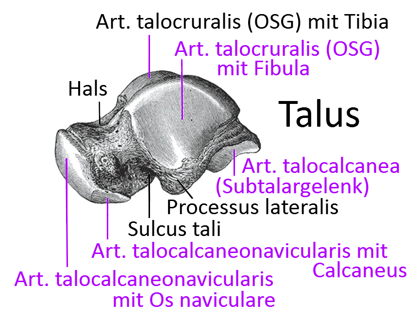
Corpus tali
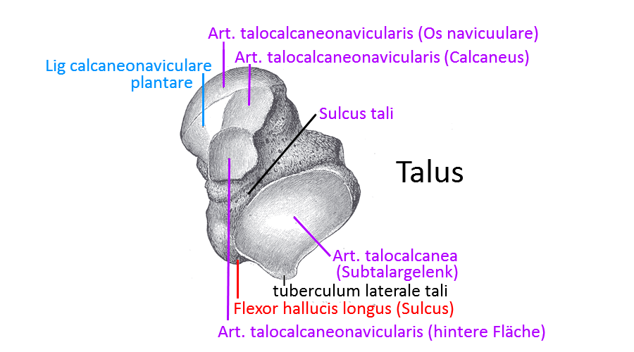
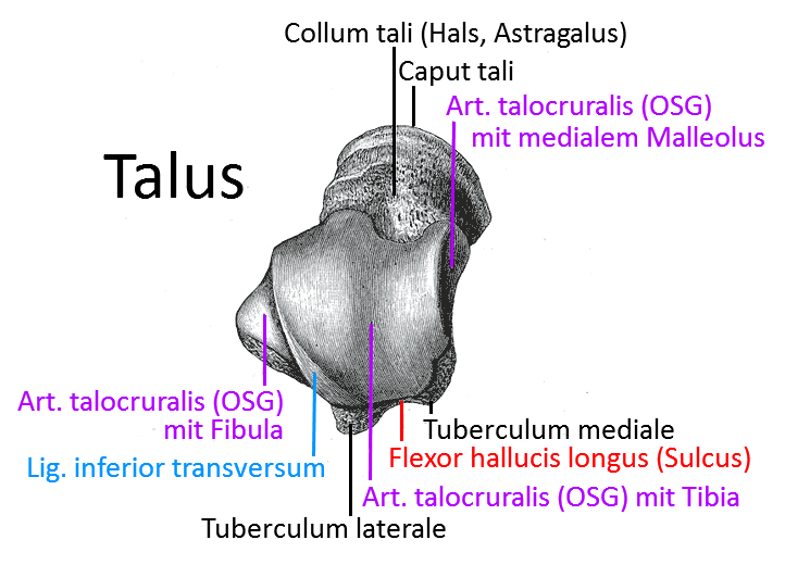
The ventral part of the talus with the trochlea tali, which articulates with the facies articularis inferior of the tibia. The lateral malleolar facet of the talus articulates with the lateralmalleolus of the fibula. The medial malleolar facet articulates with the medialmalleolus of the tibia. These three articular surfaces are the distal part of the talocrural articulation. With the inferior facies, the talus articulates with the posterior calcaneal articularis of the calcaneus. The posterior facies has a groove for the tendon of the flexor hallucis longus.
Lateral tubercle / posterior process
Lateral to this groove is the posterior talar process (also known as the lateral tubercle). The lateral tuberosity, which is much more prominent than the medial tuberosity, is present in 13% of cases as an independent accessory bone of the trigonum.
Medial tuberosity
Medial to the groove in the corpus tali is a smaller medial tubercle.
Collum tali
The collum tali (neck) is the retracted middle part of the talus. The articular surface of the calcanea media lies on the underside of the collum tali.
Sulcus tali
The collum tali has an inferior talar sulcus that separates the anterior articular surfaces from the posterior articular calcaneal facies. In the calcaneus there is a corresponding groove (sulcus calcanei), through which a channel is created in which the talocalcaneal interosseous ligament lies . Ligaments are attached to the roughened medial side of the collum tali.
Caput tali
At the head of the talus lies the articular surface facies articularis calcanea anterior to the calcaneus and the surface facies articularis navicularis aligned with the cannon bone. The calcaneonavicular plantar ligament lies in the cartilaginous surface in between.
Joints
- Art. subtalaris / Art. talocalcanearis / subtalar joint / talocalcaneares joint
- Art. talocruralis(upper ankle joint)
- Art. talonavicularis (part of the anterior lower ankle joint / Chopart joint)
Articulated surfaces
In summary, the talus has articular surfaces with
- of the malleolar fork: trochlea tali, facies malleolaris lateralis, facies malleolaris medialis
- the calcaneus: facies articularis calcanea anterior, facies articularis calcanea media, facies articularis calcanea posterior
- the navicular bone Os naviculare: Facies articularis navicularis
Trochlea tali
The „talus roll“, the cranial articular surface of the talus, in which plantar flexion and dorsiflexion of the foot in the ankle joint occur, surrounded by the malleolar fork.
Pictures
Talus from cranial-ventral
Talus from caudal