Contents
- 1 Vertebral joint
- 2 Ligaments
- 2.1 Fasciculi longitudinales
- 2.2 Lig. alare
- 2.3 Lig. apicis dentis
- 2.4 Atlantoaxial ligament
- 2.5 Anterior atlantoaxial ligament
- 2.6 Posterior atlantoaxial ligament
- 2.7 Atlantooccipital ligament
- 2.8 Lig. atlantooccipital anterius
- 2.9 Lateral atlantooccipital ligament
- 2.10 Lig. atlantooccipitale posterius
- 2.11 Lig. capitis costae intraarticulare
- 2.12 Lig. capitis costae radiatum
- 2.13 Lig. costotransversarium
- 2.14 Lig. cruciatum atlantis
- 2.15 Lig. flavum (interosseous ligament)
- 2.16 Interspinous ligament (interspinosum)
- 2.17 Intertransverse ligament
- 2.18 Lig. longitudinale anterius (anterior longitudinal ligament)
- 2.19 Lig. longitudinale posterius (posterior longitudinal ligament)
- 2.20 Lumbocostal ligament
- 2.21 Lig. nuchae (nuchal ligament)
- 2.22 Supraspinous ligament
- 2.23 Lig. transversum atlantis (transverse ligament of the first cervical vertebra)
- 2.24 Membrana atlantoaxialis anterior (Lig. atlantoaxiale)
- 2.25 Membrana atlantoaxialis posterior (Lig. atlantoaxiale)
- 2.26 Membrana atlantooccipitalis anterior (Lig. atlantooccipitale)
- 2.27 Membrana atlantooccipitalis posterior (Lig. atlantooccipitale posterior)
- 2.28 Membrana tectoria
- 3 Bursa (bursae)
- 4 Further structures
- 5 Movements
- 6 Pathology
- 7 Tests
- 8 Images
- 9 Thoracic vertebrae
- 10 Lumbar vertebrae
- 11 Lumbar vertebrae: median section
- 12 Lumbar vertebrae: ligaments (image links to linkmap)
- 13 Costovertebral joint in sagittal section (image links to linkmap)
- 14 Costovertebral joint with spinal canal and ligaments (image links to linkmap)
- 15 Costovertebral joint schematic (image links to linkmap)
- 16 Costovertebral joint of ventrolateral (image links to linkmap)
- 17 Atlantooccipital joint (image links to linkmap)
- 18 Atlas
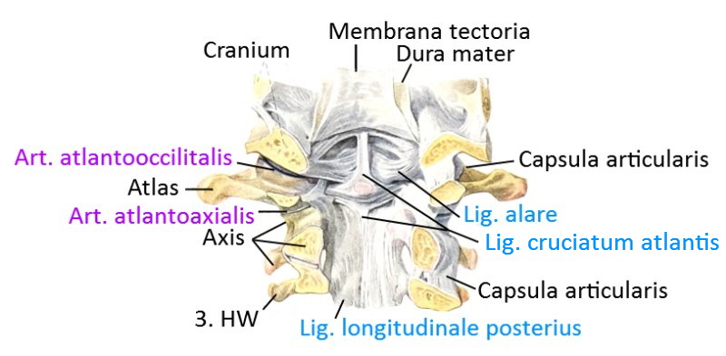
- 19 Atlas and Axis: Ligaments
- 20 Atlas and axis: ligaments, diagram (image links to linkmap)
(see below for further graphics)
Vertebral joint
The autochthonous back musculature is what is commonly referred to as the erector spinae (back extensor), even though its function goes beyond mere extension and includes both lateral flexion and rotation of the spine. The description of the relevant asanas can be found under the entry on the erector spinae. Some muscles perform an isolated movement, but most perform a combined movement. The movements take place simultaneously in many joints of the spine and cannot be differentiated segment by segment. Roughly speaking, the lumbar spine, thoracic spine and cervical spine can be controlled separately. The segments consist of two adjacent vertebral bodies, the intervertebral discs between them and the dorsal facet joints. Basically, all vertebrae are similar, but we differentiate between the cervical spine, thoracic spine and lumbar spine, and the vertebrae differ in some details. The atlas (1st cervical spine) and axis (2nd cervical spine) play a special role: the atlas does not have a vertebral body but a hole into which the ventrally pointing „dens“ of the axis protrudes. The cervical spine is the most mobile, but also the most susceptible to interference, followed by the lumbar spine, and the thoracic spine is the least susceptible to interference. About 50% of the flexion and extension of the cervical spine occurs between the occiput and C1! The remaining 50% is distributed among the other vertebral joints with a focus on C5-C6. Approximately 50% of the rotation also occurs in C1-C2, the rest is evenly distributed. The upper thoracic spine shows little mobility, the lumbar spine shows great flexion/extension capability and lateral tilt capability, but less rotational capability, the cervical spine, like the lumbar spine, tends to degenerative changes in the intervertebral discs, which in the case of rotation of the cervical spine leads to compression of the neighboring nerves and arteries, resulting in cervicocephalalgia, cervical syndrome and, in the case of compression of the vertebral artery, dizziness. verbebralis can also lead to dizziness, nausea, visual disturbances, syncope, nystagmus (Barre-Lieou sign, AI to manual manipulation). Flexion is better in men, other mobility in women. With age, mobility decreases except for rotation in C1-C2. Passive mobility is better when lying down than upright due to relaxed muscles. The „facet joints“ (artt. zygapophysiales) are usually referred to as vertebral joints, which are located as artt. superiores and artt. inferiores on almost all vertebrae in pairs dorsally (left/right). They allow a certain, limited degree of rotation, lateral flexion and extension. The joint formed by two vertebral bodies and the intervertebral discs between them is a symphysis (symphysis intervertebralis, „false joint“). The intervertebral disc (discus intervertebralis), formed from a highly water-binding nucleus (nucleus pulposus) and a fibrous ring (annulus fibrosus) surrounding it, distributes the pressure between the base and top surface largely evenly according to Pascal’s principle thanks to its high water content. As water is pressed out during the day under the load of the partial body weight, the lost water must be absorbed again during the night. Strictly speaking, this is not water but a nutrient solution to supply the metabolism of the intervertebral discs, as the intervertebral discs no longer have any blood vessels after the end of growth, from around the age of 20. The loss of water over the day is approx. 1-2 cm and thus exceeds the loss in size caused by the arch of the foot sinking in (approx. 1 cm). The spinal column is held together by many ligaments. See the following section.
Ligaments
Lig. alare
Lig. apicis dentis
Lig. atlantoaxiale
Lig. atlantoaxiale anterius (to: Membrana atlantooccipitalis)
Lig. atlantoaxiale posterius (to: Membrana atlantooccipitalis)
Lig. atlantooccipitale
Lig. atlantooccipitale anterius
Lig. atlantooccipitale laterale
Lig. atlantooccipitale posterius
Lig. capitis costae intraarticulare
Lig. capitis costae radiatum
Lig. costotransversarium
Lig. costotransversarium laterale
Lig. costotransversarium superius
Lig. flavum (Zwischenbogenband)
Lig. interspinale/interspinosum
Lig. intertransversarium
Lig. longitudinale anterius (anterior longitudinal ligament)
Lig. longitudinale posterius (posterior longitudinal ligament)
Lig. lumbocostale
Lig. nuchae (nuchal ligament)
Lig. supraspinale
Lig. transversum atlantis (transverse ligament of C1)
Membrana atlantoaxialis anterior et posterior (= Lig. atlantoaxiale)
Membrana atlantooccipitalis anterior und posterior (Lig. atlantooccipitale)
Membrana tectoria
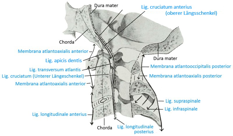
Fasciculi longitudinales
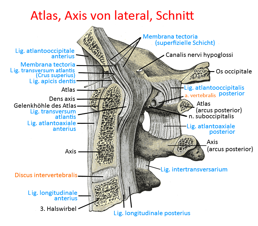
the vertical part of the atlantoaxial cruciate ligament, also known as the longitudinal bundle. The upper part, fasciculus longitudinalis superior, runs to the ventral side of the foramen magnum. The lower part, fasciculus longitudinalis inferior, runs to the dorsal side of the second vertebral body.
Lig. alare
Ligaments from the dorsolateral surface of the apix dens to the ventromedial edge of the condyles of the occipital bone at the edge of the foramen magnum. They hold the skull and restrain its movement; in neutral zero some of these ligaments are taut, others are relaxed. They limit rotation of the head and its flexion in relation to the cervical spine.
Images:
Linkmap: Ligaments of the atlas and axis
Linkmap: Cervical spine from cranial, profound
Linkmap: Axis from dorsocranial
Linkmap: Axis lateral
Lig. apicis dentis
A short ligament from the tip of the dens axis to the margo anterior of the foramen magnum to stabilize the two head joints
Images: linkmap/cranial_spine_from_dorsal_profund.gif
Linkmap: Atlas, axis, ligaments, schematic
Linkmap: Cervical spine from cranial, profound
Linkmap: Atlas, axis from lateral
Atlantoaxial ligament
The atlantooccipital ligament consists of the anterior atlantoaxial ligament and the posterior atlantoaxial ligament. These are often also referred to as the anterior atlantoaxial membrane and the posterior atlantoaxial membrane, see there.
Images:
Linkmap: Atlas, Axis from lateral
Linkmap: Cervical spine from ventral
Linkmap: cranial cervical spine from dorsal, superficial
Anterior atlantoaxial ligament
Synonym of Membrana atlantoaxialis anterior, see there.
Posterior atlantoaxial ligament
Synonym of Membrana atlantoaxialis posterior, see there.
Atlantooccipital ligament
The atlantooccipital ligament consists of the anterior atlantooccipital ligament and the posterior atlantooccipital ligament. These are often also referred to as the anterior atlantooccipital membrane and the posterior atlantooccipital membrane, see there.
Images:
Linkmap: Atlas, Axis from lateral
Linkmap: Cervical spine from ventral
Linkmap: cranial cervical spine from dorsal, superficial
Lig. atlantooccipital anterius
Synonymous with Membrana atlantooccipitalis anterior, see there.
Lateral atlantooccipital ligament
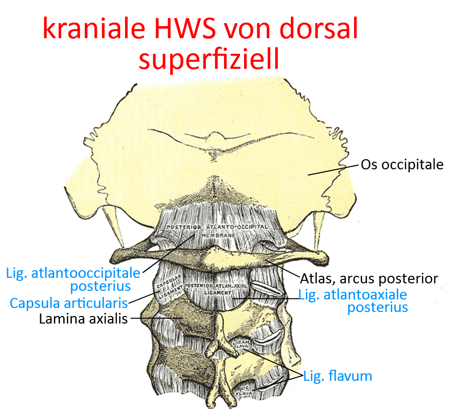
The lateral atlantooccipital ligament runs from the lateral edge of the foramen magnum to the posterior arch of the atlas.
Images:
Linkmap: cranial cervical spine from dorsal, superficial
Lig. atlantooccipitale posterius
Synonym of Membrana atlantooccipitalis posterior, see there.
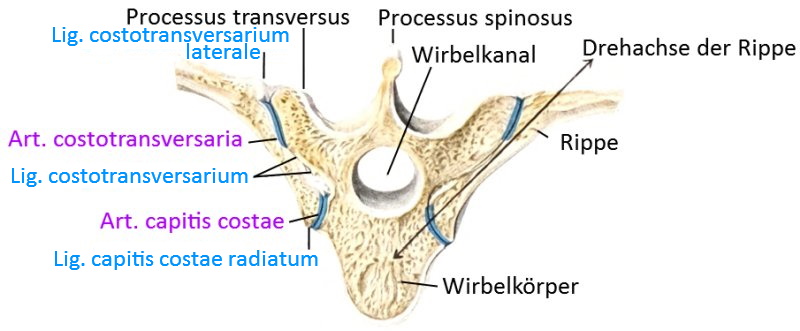
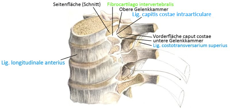
Lig. capitis costae intraarticulare
The capitis costae intraarticular ligament runs from the capitis costae crest through the capsule of the capitis costae to the neighboring intervertebral disc, thus dividing the costavertebral articulation into two chambers. This ligament is missing in the 1st, 11th and 12th ribs.
Images: (still without)
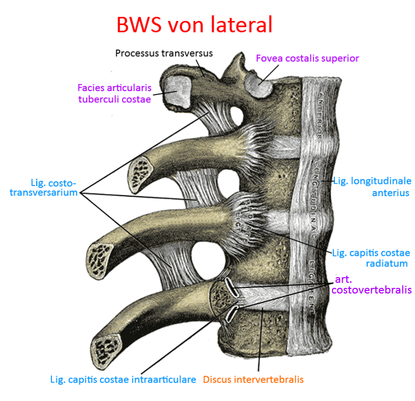
Lig. capitis costae radiatum
Ligament extending radially from the ventral bone surface of the rib head(caput costae) to the two thoracic vertebrae involved in the costoververtebral joint and the intervertebral disc (discus intervertebralis) between them to secure the costovertebral joint.
Image: linkmap/BWS_von_lateral.png
Linkmap: Costovertebral joint, transverse section
Linkmap: BWS, lateral
Lig. costotransversarium
Ligament from the rib to the transverse process. There are two parts:
- lateral: from the transverse process to the outside of the rib of the same segment.
- superius: from the processus transversus to the edge of the bone („crista colli costae“) at the upper edge of the rib below.
Pictures: linkmap/BWS_von_lateral.png
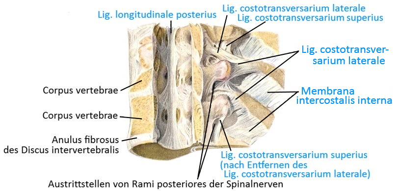
Lateral costotransversal ligament
The lig. costotransversarium laterale is a short ligament that runs from the transverse process to the lateral side of the rib of the same segment.
Images:
Linkmap: Costovertebral joint
Linkmap: Costovertebral joint, schematic
Linkmap: Costovertebral joint with spinal canal and ligaments
Linkmap: Costovertebral joint, transverse section
Linkmap: Torso, back, partial view, profound
Linkmap: Trunk, thoracic spine, cervical spine, dorsolateral view
Lig. costotransversarium superius (tuberculum costale)
The costal tuberosity ligament runs from the tip of the transverse process of a vertebra to the corresponding rib tubercle.
Images:
Linkmap: Costovertebral joint
Linkmap: Costovertebral joint from the ventrolateral side
Linkmap: Costovertebral joint, schematic
Linkmap: Costovertebral joint with spinal canal and ligaments
Linkmap: Dorsal view of the thoracic spine and cervical spine
Lig. cruciatum atlantis
The atlantoaxial cruciate ligament consists of a horizontal part, the transverse atlantoaxial ligament, and the longitudinal fasciculi longitudinales.
Lig. flavum (interosseous ligament)
Ligament formerly known as the interarcual ligament, which runs through the ventral side of the vertebral arches from the axis to the sacrum. The thickness of these ligaments increases caudally. They have a supporting effect when the spine is lifted into flexion and limit flexion; they lie between the laminae arcuum vertebrarum of adjacent vertebral arches.
Images: linkmap/LWS_from_lateral_section.gif
Linkmap: Lumbar spine, ligaments
Linkmap: Lumbar spine from lateral
Linkmap: thoracic spine, ventral, profound
Linkmap: cranial cervical spine from dorsal, superficial
Interspinous ligament (interspinosum)
between the spinous processes of adjacent vertebrae over the entire distance between their base and tip; limit flexion; the strength of the ligaments increases from cervical to caudal. At the tip of the spinous processes they lie against the supraspinous ligament, at the base against the flava ligament.
Images: linkmap/LWS_from_lateral_section.gif
Linkmap: Lumbar spine, ligaments
Linkmap: Trunk, lateral, very deep
Linkmap: Lumbar spine from lateral
Intertransverse ligament
Ligaments between the transverse processes; limit rotation and lateral flexion; they may be absent in the cervical spine; in some cases they are replaced by the intertransverse muscles.
Images: linkmap/Atlas_Axis_from_lateral.png
Linkmap: Lumbar spine, ligaments
Linkmap: Atlas, Axis from lateral
Lig. longitudinale anterius (anterior longitudinal ligament)
anterior longitudinal ligament over the vertebral bodies and intervertebral discs ventrally from the occipital bone (base) to caudal to S1; limited extension; the fiber tracts are made of firm collagenous connective tissue and radiate into the annuli fibrosi of the intervertebral discs. The anterior longitudinal ligament is wider than the dorsal posterior longitudinal ligament. Together with the posterior longitudinal ligament, supraspinal ligament and nuchal ligament, it forms the long spinal ligaments. Hemorrhage in this ligament (Simon’s hemorrhage, triggered by tensile stress) is considered a typical sign of death by hanging.
Images: linkmap/BWS_von_lateral.png
Linkmap: Atlas, Axis, Ligaments, schematic
Linkmap: Costovertebral joint from ventrolateral view
Linkmap: Lumbar spine, ligaments
Linkmap: Trunk, posterior abdominal wall from ventral view
Linkmap: Pelvis, ligaments, ventral
Linkmap: Lumbar spine from lateral
Linkmap: Atlas, Axis from lateral
Linkmap: Cervical spine from ventral
Linkmap: BWS from lateral
Lig. longitudinale posterius (posterior longitudinal ligament)
posterior longitudinal ligament, lies in the spinal canal on the dorsal side of the vertebral bodies. It lies against these and the intervertebral discs and forms the ventral lining of the spinal canal, while the lig. flavum lies ventrally against the vertebral arches and thus forms the dorsal lining of the spinal canal. It is only loosely connected to the vertebral bodies, but firmly connected to the intervertebral discs. It extends from the axis to the sacrum. The posterior longitudinal ligament limits flexion; together with the anterior longitudinal ligament, supraspinous ligament and nuchal ligament, it forms the long spinal ligaments.
Images: linkmap/ligamentum_longitudinale_posterius.gif
Linkmap: Costovertebral joint
Linkmap: Atlas, axis, ligaments, schematic
Linkmap: Ligaments, atlas and axis
Linkmap: Costovertebral joint with spinal canal and ligaments
Linkmap: Lumbar spine, ligaments
Linkmap: Cervical spine from cranial, profound
Linkmap: Lumbar spine from lateral
Linkmap: Atlax, Axis from lateral
Linkmap: BWS, Lig. longitudinale posterius
Lumbocostal ligament
The lumbocostal ligament reinforces the thoracolumbar fascia and runs with strong fibers from the 12th rib to the costal processes of L1 and L2.
Images:
Linkmap: Trunk, dorsal, profound
Linkmap: Trunk, lateral, very deep
Lig. nuchae (nuchal ligament)
Cervical ligament, limited flexion; between the external occipital protuberance over the spinous processes of the cervical spine. The nuchae ligament is the cervical continuation of the supraspinous ligament. Together with lig. longitudinale anterius, lig. supraspinale and lig. nuchae, it forms the long spinal ligaments.
Images: linkmap/LWS_from_lateral_section.gif
Linkmap: Trunk, back, partial view, profound
Linkmap: Torso, thoracic spine, cervical spine from dorsolateral view
Linkmap: Torso, lateral, very profound
Linkmap: BWS and cervical spine from dorsal
Supraspinous ligament
limited flexion; over the tips of the spinous processes from C7 to the sacrum. Fibrocartilage is formed where it touches the spinous processes. It increases in thickness caudally and is fused with neighboring fasciae. Together with the anterior longitudinal ligament, posterior longitudinal ligament and nuchal ligament, it forms the long spinal ligaments. This ligament is the origin of the greater rhomboid. In the area of the cervical spine it merges into the nuchae ligament.
Images: linkmap/LWS_from_lateral_section.gif
Linkmap: Atlas, axis, ligaments, diagram
Linkmap: Lumbar spine, ligaments
Linkmap: Trunk, lateral, very deep
Linkmap: Pelvis, ligaments, dorsal
Linkmap: Lumbar spine from lateral
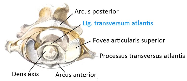
Lig. transversum atlantis (transverse ligament of the first cervical vertebra)
In addition to the longitudinal fasciculi, the transverse atlantis ligament is part of the cruciform atlantis ligament. It connects the two lateral parts of the atlas and can withstand 350 N and can be overstretched by up to 8 mm. In the middle, it connects to the longitudinal fasciculi, which extend from the ventral edge of the foramen magnum to the dorsal edge of the vertebral body of the axis.
Images: linkmap/cranial_spine_from_dorsal_profund.gif
Linkmap: Atlas, Axis, Ligaments, Schema
Linkmap: Atlantooccipital joint
Linkmap: Cervical spine from cranial, profound
Linkmap: Axis, dorsocranial
Linkmap: Atlas, Axis, lateral
Linkmap: Atlas, cranial
Membrana atlantoaxialis anterior (Lig. atlantoaxiale)
the atlantoaxial membrane is the equivalent of the atlantooccipital membrane in the first cervical segment and runs from the anterior arch (arcus anterior) of the atlas to the anterior edge of the body of the axis.
Images: linkmap/cranial_spine_from_dorsal_superficial.png
Linkmap: Atlas, Axis from lateral
Linkmap: Cervical spine from ventral
Linkmap: Atlas, axis, ligaments, diagram
Membrana atlantoaxialis posterior (Lig. atlantoaxiale)
the atlantoaxial membrane is the equivalent of the atlantooccipital membrane in the first cervical segment and runs from the posterior arch (arcus posterior) of the atlas to the upper edge of the laminae of the axis.
Images: linkmap/cranial_spine_from_dorsal_superficial.png
Linkmap: Atlas, Axis, Ligaments, Schema
Membrana atlantooccipitalis anterior (Lig. atlantooccipitale)
The anterior part of the atlantooccipital membrane, which originates ventrally in front of the foramen magnum and extends to the anterior vertebral arch (where the vertebral body is in other vertebrae ).
Image: linkmap/cranial_spine_from_dorsal_superficial.png
Linkmap: Atlas, axis from lateral
Linkmap: Cervical spine from ventral
Membrana atlantooccipitalis posterior (Lig. atlantooccipitale posterior)
The posterior part of the atlantooccipital membrane, which originates behind the foramen magnum and extends to the posterior arch of the atlas.
Image: linkmap/cranial_spine_from_dorsal_superficial.png
Linkmap: Atlas, axis, ligaments, schematic
Membrana tectoria
Ligament in the spinal canal in the region of the upper cervicals and continuation of the posterior longitudinal ligament. It originates on the dorsal side of the body of the second axis and runs to the basal part of the os occipitale. Some fibers connect with the joint capsule of the first upper cervical joint, others on the dorsal side merge with the hard meninges (dura mater) of the brain, and still others connect with the transversum atlantis ligament. The membrana tectoria brakes rotational and flexion movements in the area of the upper cervical joints and has a holding function.
Images: linkmap/cranial_spine_from_dorsal_profund.gif
Linkmap: Atlas, axis, ligaments, diagram
Linkmap: Cervical spine from cranial, profund
Linkmap: Atlas, Axis, lateral
Bursa (bursae)
Bursa apicis dentis
So-called „tip bursa“ dorsal to the dens axis and the longitudinal fasciculus of the cruciform ligament (also: cruciatum) atlantis. This bursa frequently communicates with the joint space of the posterior dentate articulation.
Bursa cruciatotectoria
Bursa between the fasciculus transversalis atlantis of the lig. cruciforme (also: cruciatum) atlantis and the covering membrane.
Bursae interspinaliae lumbales
Bursae between the processus spinosi(spinous processes) of neighboring vertebrae, which usually do not develop until around the 10th year of life. Their frequency increases with age and increases caudally.
Bursa subcutanea coccygea
Subcutaneous bursa over the tip of the coccyx.
Sacral subcutaneous bursa
Inconstant subcutaneous bursa between the crista sacralis mediana of the sacrum and the coccyx.
Further structures
Intervertebral disc
The intervertebral discs(discus intervertebralis) are elastic cartilaginous buffers between bones that optimally distribute the pressure between the vertebral bodies, i.e. their upper and lower plates, evenly according to Pascal’s principle. The nuclei (nucleus pulposus) of the intervertebral discs are highly water-binding. While the intervertebral discs lose fluid during the day due to the pressure of the partial body weight on them, which is reflected in a measurable decrease in body length, they fill up again during the night. With age, the ability to bind water decreases, which reduces the distances between the intervertebral discs and increases disc herniations (protrusion, prolapse).
Nucleus pulposus
The nucleus pulposus is the gel-like core of the intervertebral discs. It is designed in such a way that it can bind a great deal of water, which is partially pressed out during the day under the load of the partial body weight and absorbed again at night. The turnover of the nucleus pulposus is still quite short at two to three weeks, unlike that of the annulus fibrosus (1 – 1.5 years). With age, the water-binding capacity of the nucleus pulposus decreases, which favors the degeneration of the intervertebral discs.
Anulus fibrosus
The fibrous ring around the annulus fibrosus, which surrounds the nucleus pulposus. At 1 to 1.5 years, the turnover is rather long and therefore also the healing time in the event of a herniated disc.
Movements
Rotation in the transverse plane For all areas of the spine: Rotatores. Depending on the section of the spine, acting directly on the vertebral bodies or indirectly via the rotation of the upper body
- Cervical spine: ipsilateral: Splenius; kontralateral: Semispinalis
- Thoracic spine: ipsilateral: Obliquus internus abdominis; kontralateral: Obliquus externus abdominis, Multifidi, Semispinalis
- Lumbar spine: ipsilateral: Obliquus internus abdominis; contralateral: Obliquus externus abdominis
contralateral rotation same muscles of the contralateral side
Lateral flexion Depending on the section of the spine:
- Cervical spine: ispilateral: Semispinalis, Splenius, Iliocostales, Intertransversarii, Longissimus; kontralateral: Multifidi
- Thoracic spine: ipsilateral: Semispinalis, Longissimus, Intertransversarii, Iliocostales, Obliquus internus abdominis (in Verbindung mit dem ispilateralen Obliquus externus abdominis); kontralateral: Multifidi, Levatores costarum
- Lumbar spine: ipsilateral: Psoas major, Psoas minor, Quadratus Lumborum, Teile der Iliocostales, Intertransversarii, Longissimus, Obliquus internus abdominis (in Verbindung mit dem ispilateralen Obliquus externus abdominis); kontralateral: Multifidi
contralateral lateral flexion same muscles of the contralateral side
Extension Some of these muscles only extend when contracted on both sides. Distinguish section of the spine:
- Cervical spine: Intertransversarii, Semispinalis, Iliocostales, Longissimus, Splenius, Rotatores
- Thoracic spine: Intertransversarii, Semispinalis, Iliocostales, Longissimus, Levatores costarum, Rotatores
- Lumbar spine: Intertransversarii, Iliocostales, Longissimus, Rotatores, Quadratus Lumborum, Psoas major
Flexion Depending on the section of the spine:
- Cervical spine:
- Thoracic spine: rectus abdominis, obliquus internus abdominis together with obliquus externus abdominis
- Lumbar spine: rectus abdominis, obliquus internus abdominis together with obliquus externus abdominis
Pathology
Some diseases of the joint: Scheuermann’s disease, intervertebral disc disorders/nerve root compression syndrome(protrusio, prolapse, sequestrum), osteoporosis, ankylosing spondylitis, Reiter’s disease, psoriatic arthritis, spondylolisthesis, vertebral bone metastases, Paget’s disease
Tests
Tests of the joint
Cervical radikulopathie:
- Cluster of Wainner (Cervical Radiculopathy Provocation Tests)
- Perkussionstest HWS
- Spurling-Test
- Cervical spine distraction test
- Schulterkaudalisierungstest
- Elvey-Test (Plexus brachialis Spannungs-Test, Upper Limb Tension Test)
- Bakody Schulterabduktionstest
- Jackson-Kompressionstest
- Foramina-intervertebralia-Kompressionstest
- Flexionskompressionstest
- Extensionskompressionstest
- Lhermite-sign
Space occupying processes:
Compression / lesion of the brachial plexus:
Lumbar spine instability:
Lumbar spine radikulopathy:
Lumbar spinal canal stenosis:
Mixed tests:
- Schepelmann-Test (rib fracture, intercostal neuralgia, pleurisy)
- Stork test (One-legged stand test) (stress fracture, facett jint arthrosis)
Tests of the movement directions
Mobility in general:
Cervical mobility:
- HWS-Rotations-Screening
- Kopfrotationstest bei maximaler Extension
- Kopfrotationstest bei maximaler Flexion
- Segmentaler Funktionstest der HWS
- Soto-Hall-Test
- Perkussionstest HWS
- O’Donoghues Test
Thoracical spine:
LWS-Beweglichkeit:
Tests of the spanning muscles
Cervical spine: autochthonous muscles
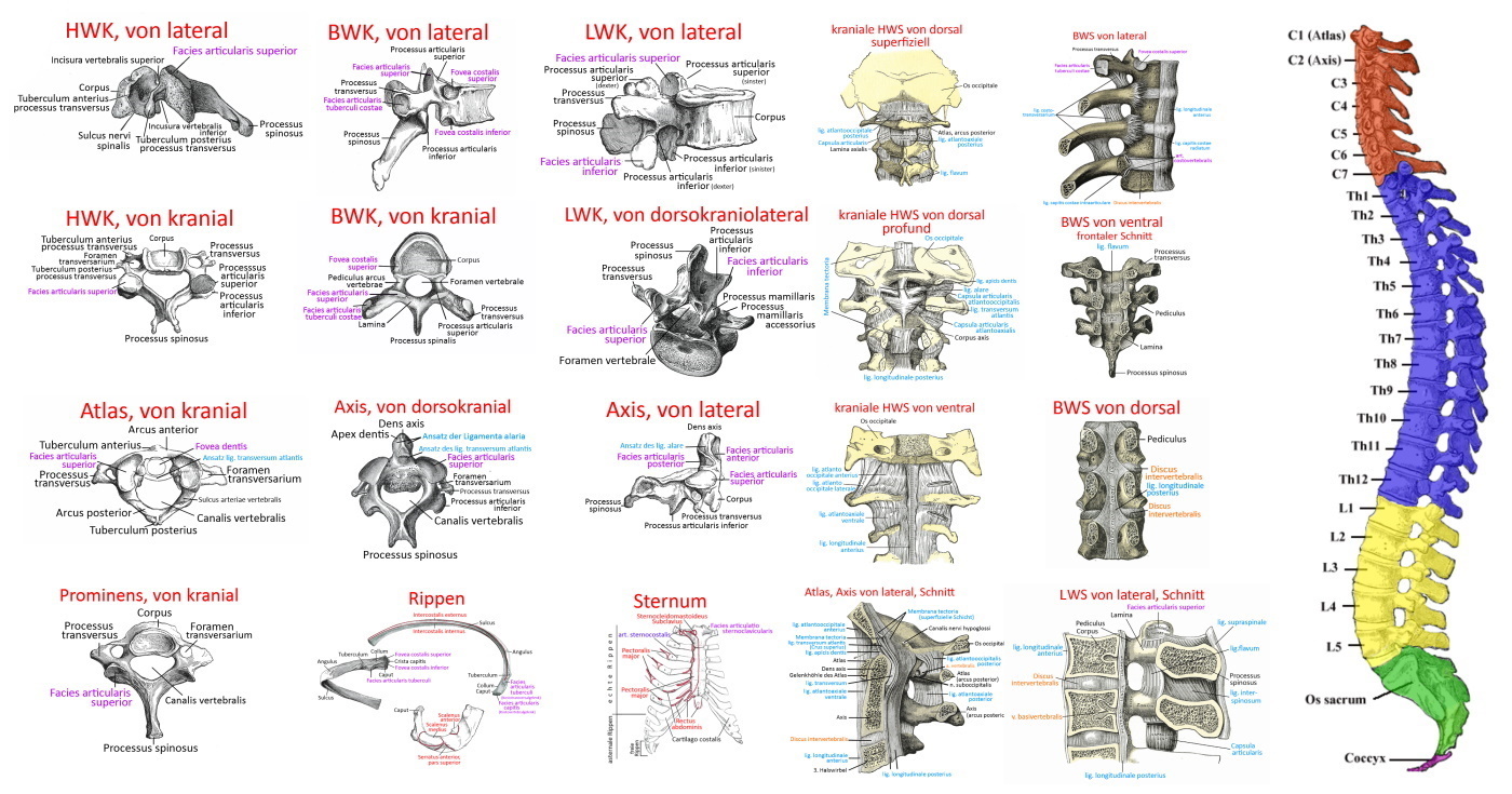
Images
Cervical vertebrae
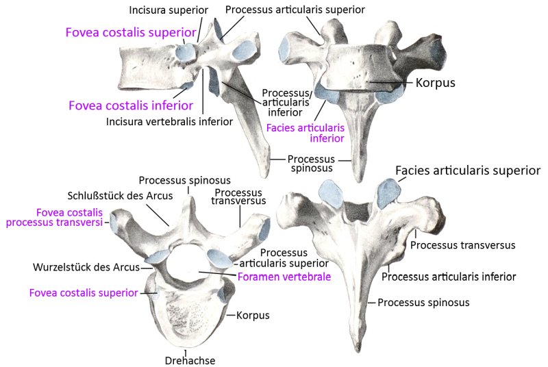
Thoracic vertebrae
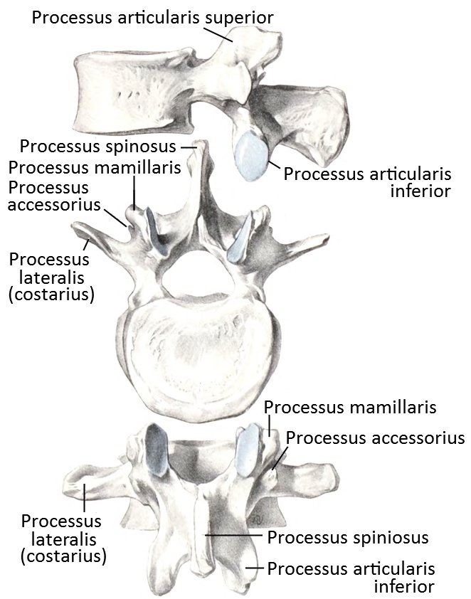
Lumbar vertebrae
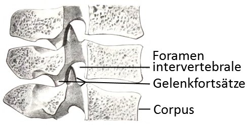
Lumbar vertebrae: median section
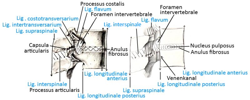
Lumbar vertebrae: ligaments (image links to linkmap)
Costovertebral joint in sagittal section (image links to linkmap)
Costovertebral joint with spinal canal and ligaments (image links to linkmap)
Costovertebral joint schematic (image links to linkmap)

Costovertebral joint of ventrolateral (image links to linkmap)
Atlantooccipital joint (image links to linkmap)
Atlas

Atlas and Axis: Ligaments
Atlas and axis: ligaments, diagram (image links to linkmap)