Contents
- 1 Image: Views of the foot (4 Linkmaps)
- 2 The ankle
- 2.1 Movements of the foot in itself
- 2.2 Bones of the foot
- 2.3 The different joints
- 2.4 Achilles tendon
- 3 The movements and the muscles involved
- 4 Bursae
- 4.1 Burses of the OSG region
- 4.2 Bursing the foot
- 4.2.1 Bursae intermetatarsophalangeae
- 4.2.2 Bursa interossei
- 4.2.3 Bursa lumbricalis
- 4.2.4 Bursa sinus tarsi
- 4.2.5 Bursa subcalcanea
- 4.2.6 Bursa subcutanea basis ossis metatarsalis 5 dorsalis
- 4.2.7 Bursa subcutanea basis ossis metatarsalis 5 plantaris
- 4.2.8 Bursa subcutanea capitis ossis metatarsalis 1 dorsalis
- 4.2.9 Bursa subcutanea capitis ossis metatarsalis 5 dorsalis
- 4.2.10 Bursa subcutanea capitis phalangis
- 4.2.11 Bursa subcutanea capitis tali medialis
- 4.2.12 Bursa subcutanea capitis tali lateralis
- 4.2.13 Bursa subcutanea ossis cuneiformis medialis
- 4.2.14 Bursa subcutanea plantaris capitis ossis metatarsalis 5
- 4.2.15 Bursa subcutanea tuberis ossis navicularis
- 4.2.16 Bursa subcutanea tuberositas ossis metatarsalis 5
- 4.2.17 Bursa subtendinea extensoris hallucis longus
- 4.2.18 Bursa subtendinea fibularis brevis
- 4.2.19 Bursa subtendinea tibialis anterioris
- 4.2.20 Bursa subtendinea tibialis posterioris
- 4.2.21 Bursa synovialis subretinacularis
- 4.2.22 Bursa tendinea extensoris hallucis longus
- 4.2.23 Bursa tendinis fibularis tertius
- 5 Ligaments, overview
- 5.1 alphabetisch sortiert
- 5.2 sorted by location
- 5.3 distal tibiofibular syndesmosis
- 5.4 dorsal ligaments that connect the tibia and fibula to the tarsus
- 5.5 dorsal ligaments that connect the talus to the rest of the tarsus
- 5.6 Other tarsal ligaments
- 5.7 Other dorsal ligaments
- 5.8 plantar ligaments, general
- 5.9 plantar ligaments of the tarsus
- 5.10 Ligaments between the metacarpals
- 5.11 Lig. bifurcatum (Chopart ligament, bifurcate ligament)
- 5.12 Lig. calcaneocuboideum plantare (Ligamentum plantare breve)
- 5.13 Ligg. calcaneocuboidea
- 5.14 Lig. calcaneocuboideum
- 5.15 Dorsal calcaneocuboid ligament
- 5.16 Medial calcaneocuboid ligament
- 5.17 Lig. calcaneocuboideum plantare
- 5.18 Calcaneofibular ligament (CFL)
- 5.19 Calcaneonavicular ligament
- 5.20 Dorsal calcaneonavicular ligament
- 5.21 Plantar calcaneonavicular ligament (SL, spring ligament, glenoid ligament)
- 5.22 Inferior calcaneonavicular ligament (ICNL)
- 5.23 Lig. calcaneonavicular superius (SMCNL, superiomedial calcaneo-navicular ligament)
- 5.24 Lateral collateral ligament („outer ligament“ or „outer ligament complex“)
- 5.25 Medial collateral lig.
- 5.26 Dorsal cuboidonavicular lig.
- 5.27 Lig. cuboideonavicular plantar
- 5.28 Ligg. cuneocuboideae
- 5.29 Ligg. cuneometatarsalia interossea
- 5.30 Ligg. cuneonavicularia dorsalia
- 5.31 Ligg. cuneonavicularia plantaria
- 5.32 Lig. deltoideum (Lig. collaterale mediale, medial collateral ligament)
- 5.33 Anterior fibulotalar ligament (ATFL)
- 5.34 Posterior fibulotalar ligament
- 5.35 Ligg. intercuneiformia dorsalia
- 5.36 Ligg. intercuneiformia interosseosae
- 5.37 Ligg. intercuneiformia plantaria
- 5.38 Laciniatum ligament
- 5.39 Lig. lisfranc
- 5.40 Lig. malleoli lateralis anterius
- 5.41 Lig. malleoli lateralis posterius
- 5.42 Ligg. metatarsalia dorsalia
- 5.43 Ligg. metatarsalia interossea dorsalia
- 5.44 Ligg. metatarsalia interossea plantaria
- 5.45 Ligg. metatarsalia plantaria
- 5.46 Lig. metatarsale transversum profundum
- 5.47 Lig. metatarsale transversum superficiale
- 5.48 Ligg. naviculocuneiforme dorsalia / plantaria
- 5.49 Lig. plantare longum
- 5.50 Lig. talocalcaneum anterius
- 5.51 Lig. talocalcaneum interosseum
- 5.52 Lateral talocalcaneal ligament
- 5.53 Medial talocalcaneal ligament
- 5.54 Lig. talocalcaneum posterius
- 5.55 Lig. talofibulare anterius
- 5.56 Posterior talofibular ligament
- 5.57 Plantar talometatarsal ligament
- 5.58 Lig. talonaviculare (also: talonaviculare dorsale)
- 5.59 Dorsal talonavicular lig.
- 5.60 Anterior talotibial ligament
- 5.61 Posterior talotibial ligament
- 5.62 Dorsal tarsal ligament
- 5.63 Ligg. tarsi interossea
- 5.64 Ligg. tarsi plantaria
- 5.65 Ligg. tarsometatarsalia dorsalia
- 5.66 Ligg. tarsometatarsalia plantaria
- 5.67 Tibiocalcaneal ligament (TCL)
- 5.68 Tibiocalcaneonavicular ligament (TCNL)
- 5.69 Ligg. tibiofibularia (tibiofibular syndsmosis)
- 5.70 Lig. tibiofibulare anterius (inferius) (anterior syndesmosis ligament, AITFL)
- 5.71 Lig. tibiofibulare interosseum
- 5.72 Posterior tibiofibular ligament (posterior syndesmosis ligament)
- 5.73 Lig. tibiofibulare transversum (part of the syndesmosis ligament)
- 5.74 Tibionavicular ligament (TNL)
- 5.75 Tibiospring (TSL)
- 5.76 Anterior tibiotalar ligament (ATTL)
- 5.77 Posterior tibiotalar ligament (PTTL)
- 5.78 Posterior superficial tibiotalar ligament (STTL)
- 5.79 Retinaculi
- 6 Retinaculum flexorum
- 7 Spring Ligament (SL)
- 8 Pathology
- 9 Tests
- 9.1 Tests of the ankle joint
- 9.1.1 Hindfoot valgus
- 9.1.2 Splayfoot
- 9.1.3 Morton-neuralgia
- 9.1.4 Hallux rigidus
- 9.1.5 Metatarsopahlangeal joints (damage)
- 9.1.6 Talipes valgus and posterior tibial muscle/tendon insifficiency (PTTD):
- 9.1.7 baby’s talipes varus (pigeon toe)
- 9.1.8 Lateral ligaments
- 9.1.9 Achilles tendon
- 9.1.10 Tibiofibular syndesmosis
- 9.1.11 Impingement
- 9.1.12 Tarsal tunnel syndrome
- 9.1.13 Plantarfasciitis
- 9.1.14 Hyperpronation
- 9.1.15 Foot torsion
- 9.1.16 Calcaneus Stress fracture
- 9.1.17 Shin splint
- 9.2 Tests of the movement directions
- 9.3 Tests of the spanning muscles
- 9.1 Tests of the ankle joint
- 10 Images
- 10.1 Increase in mobility restrictions with age
- 10.2 Lateral ligaments (image links to linkmap)
- 10.3 Plantar ligaments (image links to linkmap)
- 10.4 Bones from plantar (image links to linkmap)
- 10.5 Bones from dorsal (image links to linkmap)
- 10.6 Lateral ligaments (image links to linkmap)
- 10.7 Ligaments from medial (image links to linkmap)
- 10.8 Ligaments from dorsal (image links to linkmap)
- 10.9 Talotarsal joint
- 10.10 Internal ligaments of the tarsus (image links to linkmap)
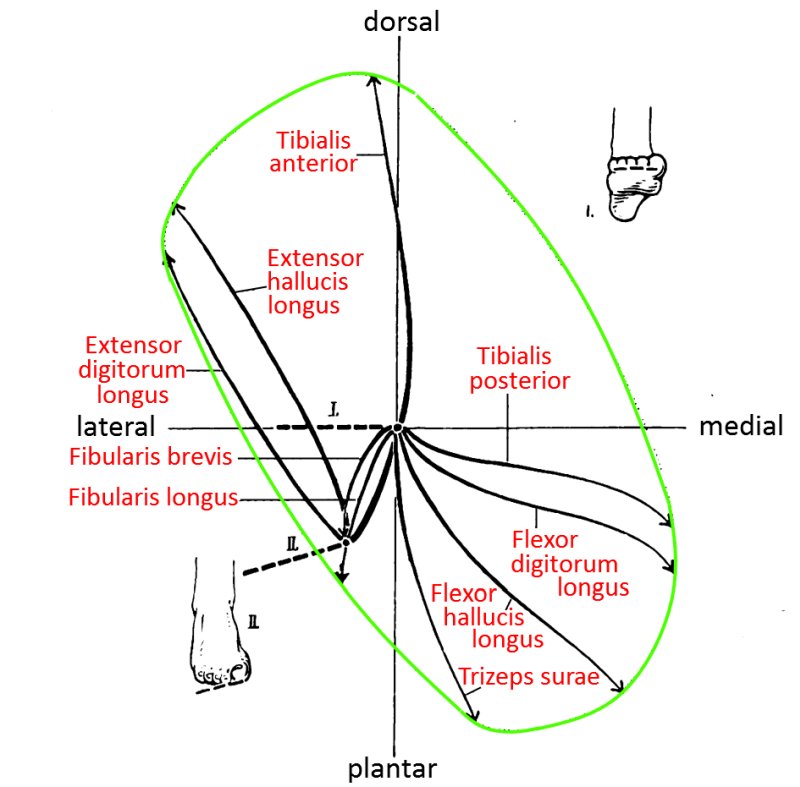
- 10.11 Directions of the muscles moving in the ankle joint (image links to linkmap)
Image: Views of the foot (4 Linkmaps)
(see also images below)
The ankle
The foot is a collection of many bones with different relative movements, from which the known movements arise in total. In contrast, the hand is much more „two-dimensional“. The difference arises from the fact that the talus and calcaneus are arranged one above the other and their joints allow different movements in relation to each other and to the neighboring bones.
Movements of the foot in itself
Apart from the movements of the foot in the OSG and posterior USG (see below), flexion and extension movements take place in the Chopart joint line and Lisfranc joint line in particular, but also torsions, so that the foot can adapt to the surface. This is particularly necessary when wearing lighter footwear or walking barefoot on uneven surfaces. Heavy, sturdy footwear such as hiking boots prevents most movements of the foot and mainly allows a certain degree of plantarflexion/dorsiflexion in the ankle joint. There is hardly any pronation and supination in the subtalar joint. Movements of the foot in the sense of flexion/extension in the Chopartor Lisfranc line or torsion of the foot (also in these two lines) practically do not take place.
The extension and flexion movements in the two joint lines Chopart and Lisfranc are an important part of the natural kinetics of the foot, which under load (as a standing foot) gives way in the longitudinal arch. When unloaded, it restores itself under the tension of various muscles and the tension of the plantar fascia and the plantar long ligament. Torsion of the foot occurs when adapting to uneven ground when the foot is loaded and is restored after the load is removed. In the case of foot valgus or hindfoot varus, torsion also occurs under load and is restored when the load is removed. Some foot deformities, on the other hand, are characterized by contracted torsion. Contractual torsions generally elude conservative therapy.
Bones of the foot
Like the bones of the hand, the bones of the foot are divided into two rows of tarsal bones (tarsus),metatarsal bones(metatarsalia) and bones of the phalanges (phalanges) of the digiti (toes).
The different joints
Chopart joint line
The bent curve between proximal talus and calcaneus and distal cuboid and scaphoid is called the Chopart joint line, so it consists of Art. talonavicularis and Art. calcaneocuboidea. For more details see below.
The scaphoid articulates distally with the three cuneiform bones (ossa cuneiforme), to which the five metatarsals are connected.
Lisfranc joint line
Together with the slightly longer cuboid bone, the cuneiform bones form the Lisfranc joint line towards the metatarsal bones. In other words, the Lisfranc line is a line through all tarsometatarsal joints.
Interphalangeal joints
The metatarsophalangeal joint is the joint in which the metatarsals and toes articulate. For more information, see a separate page
The joints located in the toes between the different phalanges are called interphalangeal joints. For more information, see a separate page
„The“ ankle
In common parlance, „the ankle“ is „the“ joint in which the foot moves relative to the lower leg. In reality, there are two or three joints:
- Upper ankle joint / talocrural joint: proximal: tibia (with malleolus medialis, „inner malleolus“), fibula (with malleolus lateralis, „outer malleolus“); distal: talus (with trochlea tali „ankle bone roller“). The OSG is functionally not an exact hinge joint, not least due to the fact that the talus is narrower at the back than at the front, which allows further dimensions of movement to a lesser extent.
In addition to the movement described, the ankle joint during dorsiflexion causes external rotation of the talus relative to the tibia. This is in the opposite direction to the adduction of the tibia during extension of the knee joint. - lower anterior ankle joint (talocalcaneonavicularis, Chopart joint), talus and calcaneus dorsally and scaphoid and cuboid bone ventrally. The Chopart joint is a ball-and-socket joint in the talonavicular joint (TN) that allows inversion/eversion and a modified saddle joint in the calcaneocuboid joint (CC) that allows a little flexion/extension and a little forefoot rotation.
- lower posterior (talocalcaneal) ankle joint (subtalar joint, talocalcanearis), a pivot joint between the talus and calcaneus
The linguistic usage is not uniform, often only the OSG or the OSG together with the hUSG is referred to as the „ankle joint“. In addition to the extension(plantar flexion) and flexion of the foot/ankle(dorsiflexion), the movement is supination and pronation, i.e. tilting sideways around its longitudinal axis.
The movements in detail:
- talocrural joint: dorsiflexion up to approx. 20°, plantar flexion up to approx. 30°. In addition, movements of the other two movement dimensions abduction/adduction (or rotation in the transverse plane) and pronation/supination only occur to a very limited extent. Abduction/adduction is hardly possible with a dorsiflexed foot, the range of motion increases in the direction of plantar flexion.
- subtalar: supination up to approx. 50°, pronation up to approx. 30°, inversion (supination + plantar flexion + adduction), eversion(pronation + dorsiflexion + abduction)
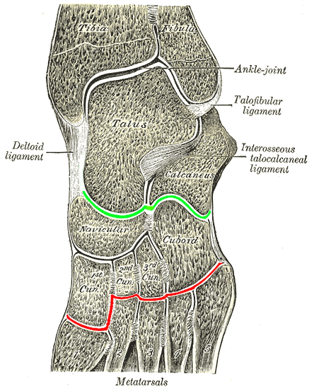
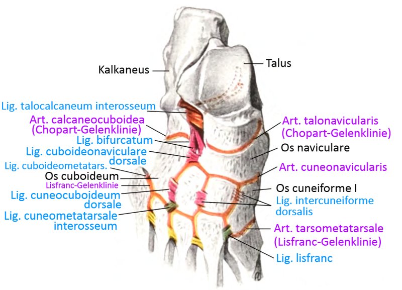
- Chopart joint: supination, pronation, plus a little flexion and extension of the foot as changes in the longitudinal arch (the Lisfranc joint provides further flexion and extension). The following image shows the Lisfranc joint line in front of the metatarsals in red and the Chopart joint line in front of the calcaneus in green.
Talocrural joint
The talocrural joint (also: tibiotalar joint), consisting of the distal tibia, the medialmalleolus of the tibia, the lateralmalleolus of the fibula and the trochlea of the talus, is referred to as the upper ankle joint (talocrural joint). The joint is stabilized by various ligaments:
- the deltoid ligament as a medial collateral ligament
- the lateral collateral ligament(anterior fibulotalar ligament, fibulocalcaneal ligament and posterior fibulotalar ligament)
- the anterior inferior tibiofibular ligament
- the posterior tibiofibular ligament
- the transverse tibiofibular ligament Lig. tibiofibulare transversum
- the Interosseus Band
The deltoid ligament mainly restricts talar abduction. The tibiocalcaneal ligament, the strongest superficial ligament, only restricts abduction of the talus; its deeper parts restrict exorotation. Dorsiflexion in the OSG is mainly limited by the posterior fibulotalar ligament. Plantar flexion is hardly limited by the anterior fibulotalar ligament, as its origin is not above the center of rotation. The limit of movement here is soft-elastic (muscular) or hard-elastic (bony).
Posterior lower ankle joint / subtalar joint
Art. subtalaris: The posterior USG or subtalar, talocalcaneal joint, ST, is the joint between the talus and calcaneus, which has three articular surfaces: the posterior, medial and anterior articular surfaces. The posterior articular surface is the largest of the three. The tarsal tunnel or tarsal canal is located medially between the three articular surfaces. The USG is secured by intrinsic and extrinsic ligaments, the intrinsic ones being the interosseous talocalcaneal ligament ITCL and the two-headed cervical ligament. Both can be affected by a sprain or arthritis, which can lead to instability of the joint. Extrinsic ligaments include the ligamentum fibulocalcaneare LFC, the lateral talocalcaneal ligament, the tibiocalcaneal ligament, the medial talocalcaneal ligament and the posterior talocalcaneal ligament.
Anterior lower ankle joint / Chopart joint
Art. transversa tarsi: The anterior ankle joint or Chopart joint is the transverse joint running proximally/ventrally from the calcaneus and talus to the scaphoid and cuboid bone.
This joint consists of three partial joints:
- Talonavicular joint (Art. talonaviculare, TN)
- Calcaneocuboid joint (Art. calcaneocuboidea, CC)
- Articulatio talocalcaneonavicularis
Important ligaments of the Chopart joint are the
- The plantar calcaneonavicular ligament, also known as the „spring ligament“: Plantar calcaneonavicular ligament
- the calcaneonavicular ligament: dorsal calcaneonavicular ligament
- the dorsal talonavicular ligament: Lig. talonaviculare (dorsal)
- the dorsal calcaneocuboid ligament: dorsal calcaneocuboid ligament
- the plantar calcaneocuboid ligament: Lig. calcaneocuboideum plantare
Tarsometatarsal joint (Chopart joint, TMT)
The more distal/ventral joint between the dorsal cuneiform and cuboid bones and ventral to the base of the 5 metatarsals is known as the Lisfranc joint or tarsometatarsal joint (TMT). The middle cuneiform bone is connected to the second and longest metatarsal bone with the stable so-called Lisfranc ligament, which has two strands in 20% of cases. The base of the first two metatarsals is not connected by a ligament, which signifies relative weakness. The plantar aponeurosis or plantar fascia is a strong fibrous tissue that can be divided into three components: central, medial and lateral. It extends from the tuber calcanei to the heads of the metatarsals and plantar surfaces of the toes. The TMT allows some flexion/extension.
Metatarsophalangeal joint (MTP, metatarsophalangeal joint)
The metatarsophalangeal joint, metatarsophalangeal joint or MTP joint also no longer belongs to the ankle joints.
The metatarsophalangeal joint is the joint in which the metatarsal bones and toes articulate. The bent curve through the metatarsophalangeal joints is the Lisfranc joint line. For more information, see a separate page.
In the case of the hallux, two sesamoid bones lie under the metatarsal head. They are integrated into the two tendons of the flexor hallucis brevis. The first MTP joint, i.e. that of the hallux, is stabilized plantar by medial and lateral fan-shaped collateral ligaments that act sesamoidally. The MTP allows pronounced flexion/extension and a little abduction/adduction to spread the toes. During the rolling movement towards the end of the stance leg phase, there is a clear extension. In this respect, the MTP differs from the MCP, in which only slight hyperextension is physiological.
Achilles tendon
The Achilles tendon is the attachment tendon of the mainly plantar-flexing triceps surae from the gastrocnemius and soleus.
The movements and the muscles involved
Movements in the ankle joint (tibiotaler joint, talocrural joint)
Dorsalflexion: M. tibialis anterior, M. extensor digitorum longus, M. extensor hallucis longus, Fibularis tertius
Plantarflexion: M. triceps surae, M. fibularis (peronaeus) longus, M. fibularis (peronaeus) brevis, M. flexor digitorum longus,
M. tibialis posterior, Plantaris, M. flexor hallucis longus
Movements in the subtalar joint
Pronation: M. fibularis (peronaeus) longus, M. fibularis (peronaeus) brevis, M. fibularis (peronaeus) tertius, M. extensor digitorum longus
Supination: M. triceps surae, M. tibialis anterior, M. tibialis posterior,
M. flexor digitorum longus, M. flexor hallucis longus, M. tibialis posterior
Bursae
Burses of the OSG region
Bursa subcutanea calcanea (Bursa praeachillaea)
The bursa subcutanea calcanea is a bursa located laterally on the calcaneus between the Achilles tendon and the skin and is larger than the bursa tenidis calcanei.
Images: (still without)
Bursa subcutanea malleoli fibularis
Subcutaneous bursa over the lateralmalleolus
Bursa subcutanea malleoli tibialis
Subcutaneous bursa over the medial malleolus
Bursa subtendinea tendinis calcanei (Bursa subachillea, Bursa tendinis calcanei)
The calcaneal tendon bursa is located between the Achilles tendon and the proximal surface of the calcaneal tuberosity of the calcaneus. Each dorsiflexion of the ankle causes an increase in pressure in this bursa, which is filled with approx. 1 – 1.5 ml of synovial fluid. Foot misalignments and irritation of the Achilles tendon favor the development of bursitis
Pictures: (not yet available)
Bursa subtendinea tibialis anterioris
The subtending tibialis anterior bursa lies between the tendon sheath of the tibialis anterior, the medial surface of the medial sphenoid bone and the first metatarsal bone. It communicates with the adjacent medial tarsometatarsal joint.
Images: (still without)
Bursing the foot
Bursae intermetatarsophalangeae
Bursae located dorsally between the metatarsal heads and the bases of the proximal phalanges, which sometimes communicate with other regional bursae from about the 15th ly.
Bursa interossei
Subtendinous bursae between the attachment tendons of the interossei and the joint capsules. These occasionally communicate with each other.
Bursa lumbricalis
Bursae that surround the lumbricales in a sheath and are sometimes surrounded by accessory bursae.
Bursa sinus tarsi
Inconstant, but usually present subtendinous bursa between the loop of the extensor digitorum longus tendon insertion and the collum tali.
Bursa subcalcanea
Subcutaneous bursa over the plantar surface of the calcaneus
Bursa subcutanea basis ossis metatarsalis 5 dorsalis
Inconstant subcutaneous bursa dorsally over the base of the 5th metatarsal bone.
Bursa subcutanea basis ossis metatarsalis 5 plantaris
Inconstant subcutaneous plantar bursa under the base of the metatarsal bone 5.
Bursa subcutanea capitis ossis metatarsalis 1 dorsalis
Inconstant subcutaneous bursa dorsally over the caput of the metatarsal bone 1. This bursa occurs more frequently in the presence of hallux valgus.
Bursa subcutanea capitis ossis metatarsalis 5 dorsalis
Inconstant subcutaneous bursa dorsally over the caput of the metatarsal bone 5.
Bursa subcutanea capitis phalangis
Subcutaneous dorsal bursae over the interphalangeal joints of toes 2-4, sometimes communicating with the joint cavity.
Bursa subcutanea capitis tali medialis
Subcutaneous medial bursa over the caput tali, which is mainly found in the presence of a pes planus(flat foot).
Bursa subcutanea capitis tali lateralis
Inconstant subcutaneous lateral bursa over the caput tali, which is mainly found in the presence of a pes varus (tilt foot, often part of the clubfoot).
Bursa subcutanea ossis cuneiformis medialis
Inconstant, rarely occurring subcutaneous bursa over the medial cuneiform bone
Bursa subcutanea plantaris capitis ossis metatarsalis 5
Mostly accessory inconstant subcutaneous plantar bursa over the caput of the metatarsal bone 1
Inconstant subcutaneous bursa over the tuber ossis navicularis, which is sometimes found in the presence of a pes planus(flat foot).
Bursa subcutanea tuberositas ossis metatarsalis 5
Inconstant subcutaneous bursa over the laterally protruding base of the 5th metatarsal bone. This bursa is often found in the presence of a pes planus(flat foot), sometimes also accessory.
Bursa subtendinea extensoris hallucis longus
Rare inconstant subtendinous bursa between the insertion tendon of the extensor hallucis longus and the caput os metatarsale 1
Bursa subtendinea fibularis brevis
Subtendinous bursa between the insertion tendon of the fibularis brevis and the base of the metatarsal bone V
Bursa subtendinea tibialis anterioris
Subtendinous bursa between the tendon of the tibialis anterior and that of the medial side of the medial os cuneiforme
Bursa subtendinea tibialis posterioris
Subtendinous plantar bursa between the tendon of the posterior tibialis and the plantar calcaneonavicular ligament
Bursa synovialis subretinacularis
Subligamentary bursa between the profound part of the extensorretinaculum inferius and the talus.
Bursa tendinea extensoris hallucis longus
Synonym zu Bursa subtendinea extensoris hallucis longus
Bursa tendinis fibularis tertius
Subtendinous bursa between the attachment tendon of the fibularis tertius and the base of the metatarsal bone V
Ligaments, overview
alphabetisch sortiert
- distal tibiofibular syndesmosise:
– Lig. tibiofibulare interosseum
– Lig. tibiofibulare anterius
– Lig. tibiofibulare posterius
– Lig. tibiofibulare transversum - Lig. bifurcatum:
– Lig. calcaneonaviculare dorsale
– Lig. calcaneocuboideum dorsale - Lig. calcaneocuboideum plantare
- Lig. calcaneocuboideum (dorsale)
- Lig. calcaneocuboideum mediale
- Lig. calcaneofibulare ((CFL), part of Lig. collaterale laterale)
- Lig. calcaneonaviculare plantare
- Lig. calcaneonaviculare dorsale
- Lig. calcaneonaviculare superius (SMCNL)
- Lig. calcaneonaviculare inferius (ICNL)
- Lig. collaterale laterale:
– Lig. calcaneofibulare
– Lig. talofibulare anterius
– Lig. talofibulare posterius
with common origin at the malleolus lateralis, and depending on the literature also:
– Lig. talonaviculare
– Lig. tibiofibulare posterius - Lig. collaterale mediale
- Lig. cuboideonaviculare dorsale
- Lig. cuboideonaviculare plantare
- Lig. cuneocuboideum dorsale
- Lig. cuneocuboideum interosseum
- Lig. cuneocuboideum plantare
- Ligg. cuneometatarsalia dorsalia
- Ligg. cuneometatarsalia interossea
- Ligg. cuneometatarsalia plantaria
- Ligg. cuneonavicularia dorsalia
- Ligg. cuneonavicularia plantaria
- Lig. deltoideum (Lig. collaterale mediale, medial collateral ligament)
- Ligg. intercuneiformia dorsalia
- Ligg. intercuneiformia interossea
- Ligg. intercuneiformia plantaria
- Ligg. intercuneometatarsalia interossea
- Lig metatarsale transversum profundum
- Lig metatarsale transversum superficiale
- Ligg. metatarsalia dorsalia
- Ligg. metatarsalia plantaria
- Ligg. metatarsalia interossea dorsalia
- Ligg. metatarsalia interossea plantaria
- Lig. plantare longum (LPL)
- Lig. talocalcaneum interosseum
- Lig. talocalcaneum laterale
- Lig. talocalcaneum mediale
- Lig. talocalcaneum posterius
- Lig. talofibulare anterius (part of Lig. collaterale laterale)
- Lig. talofibulare posterius (part of Lig. collaterale laterale)
- Lig. talonaviculare (part of Lig. collaterale laterale)
- Ligg. tarsi dorsalia
- Ligg. tarsi interossea
- Ligg. tarsi plantaria
- Lig. tibiocalcaneare (TCL), superficial
- Lig. tibiofibulare anterius (anterior syndesmosis ligament, Band der Malleolengabel)
- Lig. tibiofibulare posterius (posterior syndesmosis ligament, Band der Malleolengabel, part of Lig. collaterale laterale)
- Lig. tibiofibulare anterius inferior
- Lig. tibiofibulare transversum
- Lig. tibionaviculare (TNL), superficial
- Lig. tibiotalare anterius (ATTL), profound
- Lig. tibiotalare posterius (PTTL), profound
- Lig. tibiotalare superficiale (posterius) (STTL), superficial
- Tibiospring-ligament (TSL)
- Ligg. tarsometatarsalia dorsalia
- Ligg. tarsometatarsalia plantaria
- Retinaculum (musculorum) extensorum inferius
- Retinaculum (musculorum) extensorum superius
- Retinaculum (musculorum) flexorum inferius
- Retinaculum (musculorum) flexorum superius
sorted by location
- Lig. calcaneonaviculare plantare
- Lig. calcaneonaviculare dorsale
- Ligg. metatarsalia dorsalia
- Ligg. metatarsalia plantaria
distal tibiofibular syndesmosis
The distal tibiofibular syndesmosis is a ligamentous structure that secures the ankle joint. Some authors refer to the ligamentous area consisting of fibers from the interosseous ligament as the transverse tibiofibular ligament.
The distal tibiofibular syndesmosis consists of :
- Lig. tibiofibulare interosseum
- Lig. tibiofibulare anterius
- Posterior tibiofibular ligament
- Transverse tibiofibular ligament
dorsal ligaments that connect the tibia and fibula to the tarsus
- Lig. collaterale mediale (mediales Kollateralband, Lig. deltoideum)
- Lig. deltoideum (mediales Kolalteralband, Lig. collaterale mediale)
- Lig. collaterale laterale
– Lig. talofibulare anterius
– Lig. talofibulare posterius
– Lig. calcaneofibulare
– Lig. tibiofibulare anterius
– Lig. tibiofibulare posterius - Lig. tibiofibulare anterius inferior
dorsal ligaments that connect the talus to the rest of the tarsus
- Lig. talonaviculare
- Lig. talocalcaneum interosseum
- Lig. talocalcaneum laterale
- Lig. talocalcaneum mediale
- Lig. talocalcaneum posterius
Other tarsal ligaments
Other dorsal ligaments
- Lig. bifurcatum
– Lig. calcaneonaviculare
– Lig. calcaneocuboideum - Ligg. intercuneiformia dorsalia
- Lig. cuneocuboideum dorsale
- Lig. cuboideonaviculare dorsale
- Ligg. cuneonavicularia dorsalia
- Ligg. calcaneocuboidea dorsalia
plantar ligaments, general
plantar ligaments of the tarsus
- Lig. calcaneocuboideum plantare
- Ligg. cuneonavicularia plantaria
- Ligg. intercuneiformia plantaria
- Lig. cuneocuboideum plantare
- Lig. cuneocuboideum interosseum
- Ligg. intercuneiformia interossea
- Ligg. tarsometatarsalia dorsalia
- Ligg. tarsometatarsalia plantaria
- Ligg. intercuneometatarsalia interossea
Ligaments between the metacarpals
Lig. bifurcatum (Chopart ligament, bifurcate ligament)
The bifurcate ligament consists of two individual ligaments, the medial dorsal calcaneonavicular ligament and the lateral dorsal calcaneocuboid ligament, and connects the dorsal ventral calcaneus with the neighboring cuboid and navicular bones. This makes the bifurcate ligament the dorsal part of the ligamentous protection of the Chopart joint line, in which a limited degree of extension and flexion of the foot takes place. While the plantar ligament and calcaneonavicular plantar ligament with the plantar fascia and intrinsic foot muscles limit extension, the bifurcate ligament sets a limit in the direction of flexion of the foot. Inversion trauma (see supination trauma) frequently causes injuries to this ligament, including avulsion fractures of the anterolateral process of the calcaneus.
Images:
Linkmap: Ankle joint, ligaments, lateral
Linkmap: Dorsal view of the ankle, ligaments
Linkmap: Lateral ankle, ligaments
Linkmap: Ankle from medial, ligaments
Linkmap: Tarsus, internal ligaments
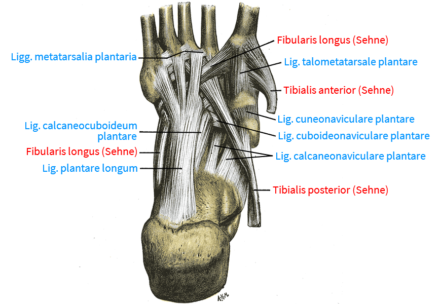
Lig. calcaneocuboideum plantare (Ligamentum plantare breve)
Fiber tract considered by many authors to be a separate part of the plantar long ligament. It runs from the calcaneal tuber to the plantar surface of the cuboid bone (cuboid bone)
Images:
Linkmap: Foot, ligaments, medial
Linkmap: Foot, ligaments, plantar
Ligg. calcaneocuboidea
The plural is used less frequently in the literature, although „the calcanocuboid ligament“ consists of four ligaments, see here.
Lig. calcaneocuboideum
The calcaneocuboid ligament supports the pronounced medial and the weak lateral arch of the foot. It consists of four ligaments:
- Dorsal calcaneocuboid ligament
- Medial calcaneocuboid ligament
- Lig. calcaneocuboideum plantare
- Lig. plantare longum (LPL )
The ligaments can be damaged by inversion trauma(supination trauma).
Images:
Linkmap: Foot, ligaments, medial
Dorsal calcaneocuboid ligament
The calcaneocuboid ligament is a dorsal ligament of the foot and part of the bifucate ligament (together with the calcaneonavicular ligament). It contributes to the stability of the calcaneocuboid joint as part of the Chopart joint.
Images:
Linkmap: Foot, ligaments, dorsal
Linkmap: Foot, ligaments, medial
Medial calcaneocuboid ligament
represents a part of the bifurcate ligament.
Pictures: (still without)
Lig. calcaneocuboideum plantare
Ligament from the calcaneal tuber to the plantar surface of the cuboid bone, considered by some authors to be part of the plantar ligament.
Images:
Linkmap: Ankle joint, ligaments, medial
Linkmap: Foot, ligaments, plantar
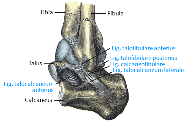
Calcaneofibular ligament (CFL)
The calcaneofibular ligament, together with the anterior talofibular ligament and the posterior talofibular ligament, is part of the lateral collateral ligament complex. It runs as a narrow, strong ligament of the ankle joint from the apex of the lateral venrtolateral malleolus backwards and downwards to a tubercle on the posterolateral calcaneus, thus covering the talocrural joint(OSG) and the subtalar joint. It limits supination and inversion of the foot in the subtalar joint. It is covered by the tendons of the fibularis longus and fibularis brevis. It is frequently affected in classic supination trauma in the direction of inversion, as is the anterior talofibular ligament.
Images:
Linkmap: Foot, ligaments, lateral
Linkmap: Ankle joint, ligaments, lateral
Linkmap: Ankle joint, ligaments, capsule, lateral
Linkmap: Ankle joint, ligaments, lateral
Linkmap: Ankle joint, ligaments, medial
Often used as a synonym for the dorsal calcaneonavicular ligament, see there.
Together with the calcaneocuboid ligament, the dorsal calcaneonavicular ligament forms the bifurcate ligament. It stabilizes the talocalcaneonavicular joint and is part of the ligamentous protection of the Chopart joint line.
Images:
Linkmap: Ankle joint, ligaments, lateral
Linkmap: Ankle joint, ligaments, medial
Linkmap: Ankle joint, ligaments, dorsal
The conical ligament, also known as the spring ligament (SL), runs from the sustentaculum tali of the calcaneus to the plantar side of the navicular bone. It belongs to the subtalar joint and is covered with cartilage on its dorsal side, as it articulates with the articular surface of the caput of the talus. The tendon of the posterior tibialis runs plantar to this ligament. As this ligament is involved in maintaining the longitudinal arch of the foot in the talocalcaneal joint alongside the long plantar ligament as part of the passive tension girdle, its insufficiency contributes to the formation of a flat foot, which explains its name. It limits the medial rotation and plantar flexion of the talus as well as the dosal flexion, eversion and abduction of the navicular bone. Anterolaterally, the ligament is protected from friction by a corpus adiposum.
Images:
Linkmap: Calcaneus
Linkmap: Ankle joint, ligaments, plantar
Linkmap: Ankle joint, ligaments, medial
Linkmap: Foot, ligaments, plantar
Reinforcing ligament of the talocalcaneonavicularis species, part of the bifurcate ligament.
Pictures: (still without)
The superior calcaneonavicular ligament runs from the sustentaculum tali of the talus to the navicular bone and forms a loop-like enclosure of the head of the talus together with the TSL. In the contact zone with the talus, the ligament is covered with fibrocartilage.
Images: (still without)
Lateral collateral ligament („outer ligament“ or „outer ligament complex“)
The outer ligament complex consists of the ligaments
- Lig. talofibulare anterius (ATFL)
- Lig. talofibulare posterius (PTFL)
- Lig. calcaneofibulare (CFL)
Medial connecting fibers create a lateral fibulotalocalcaneal ligament complex, the “ lateral collateral ligament“, from these ligaments, both anatomically and functionally.
Images: (still without)
Medial collateral lig.
Siehe Lig. deltoideum
The cuboideonavicular dorsal ligament is a dorsal ligament between the cuboid bone and the navicular bone which, together with the transverse metatarsal ligament, braces the posterior transverse arch of the foot.
Images:
Linkmap: Ankle joint, ligaments, lateral
Linkmap: Lateral view of the ankle, ligaments
Linkmap: Tarsus, internal ligaments
The cuboideonavicular plantar ligament connects the plantar cuboid and scaphoid bones.
Images:
Linkmap: Foot, ligaments, plantar
Ligg. cuneocuboideae
the three ligaments between the cuboid bone and the lateral sphenoid bone: Lig. cuneocuboideum dorsale, Lig. cuneocuboideum interosseum, Lig. cuneocuboideum plantare
Images:
Dorsal cuneocuboid ligament
Dorsal ligament connection of the cuboid bone with dorsolateral areas of the 3 sphenoid bones, widening towards the cuboid.
Images:
Linkmap: Ankle joint, ligaments, lateral
Linkmap: Dorsal view of theankle, ligaments
Linkmap:Ankle from lateral, ligaments
Linkmap: Tarsus, internal ligaments
Lig. cuneocuboideum interosseum
Ligament attached distal to the articular facet of the cuboid and extending to the medial side of the cuboid, which serves to tighten the bone fiber structure.
Images: (still without)
Lig. cuneocuboideum plantare
Short plantar ligament connection of the cuboid bone with the three cuneiform bones, made of capsular ligament.
Pictures: (still without)
Ligg. cuneometatarsalia interossea
Ligaments that connect the cuneiform bones with the opposing metatarsals. See also lisfranc ligament
Images:
Linkmap: Tarsus, internal ligaments
Strong dorsal ligaments connecting the scaphoid to the three cuneiform bones.
Images:
Linkmap: Ankle joint, ligaments, lateral
Linkmap: Ankle joint, ligaments, medial
Linkmap: Ankle from dorsal, ligaments
Linkmap: Lateral ankle, ligaments
Linkmap: Ankle from medial, ligaments
Strong plantar ligaments connecting the scaphoid to the three cuneiform bones.
Images:
Linkmap: Foot, ligaments, plantar
Lig. deltoideum (Lig. collaterale mediale, medial collateral ligament)
The deltoid ligament is a grouping of distinct ligaments that originate from the medialmalleolus and extend to the respective bones. A distinction is made between
superficial ligaments:
- Superficial tibiotalar ligament (posterius) (STTL), superficial
- Tibionavicular ligament (TNL), superficial
- Tibiocalcaneal ligament (TCL), superficial
- Tibiospring Band (TSL)
profound ribbons:
- Anterior tibiotalar ligament (ATTL), profound
- Posterior tibiotalarligament (PTTL), profound
The deltoid ligament generally stabilizes the medial OSG against lateral, ventral and dorsal translation of the talus during extension and flexion. The posterior and medial part is covered by the sheath of the posterior tibialis.
Images:
Linkmap: Ankle joint, ligaments, medial
Linkmap: Medial ankle joint, ligaments
Anterior fibulotalar ligament (ATFL)
other name for anterior talofibular ligament.
Posterior fibulotalar ligament
other name for posterior talofibular ligament.
Ligg. intercuneiformia dorsalia
These ligaments connect the dorsal intercuneiform ossa with each other.
Images:
Linkmap: Ankle joint, ligaments, lateral
Linkmap: Dorsal view of the ankle, ligaments
Linkmap: Lateral ankle, ligaments
Linkmap: Lateral ankle, ligaments
Linkmap: Tarsus: internal ligaments
Ligg. intercuneiformia interosseosae
Ligamentous cross-connection of the sphenoid bones, which reinforces the effect of the Ligg. intercuneiformia dorsalia and Ligg. intercuneiformia plantaria.
Pictures: (still without)
Ligg. intercuneiformia plantaria
These ligaments connect the ossa intercuneiformia plantar with each other.
Images: (still without)
Laciniatum ligament
Synonym for Retinaculum flexorum
Lig. lisfranc
The lisfranc ligament runs from the lateral side of the medial cuneiform bone to the medial side of the base of the second metatarsal bone.
Images:
Linkmap: Tarsus, internal ligaments
Lig. malleoli lateralis anterius
Old name for the anterior tibiofibular ligament (anterior syndesmosis ligament), see there.
Images:
Linkmap: Lateral view of the ankle, ligaments
Linkmap: Ankle joint, ligaments, lateral
Linkmap: Ankle joint, ligaments, lateral
Lig. malleoli lateralis posterius
Old name for the posterior tibiofibular ligament (anterior syndesmosis ligament), see there.
Images:
Linkmap: Lateral view of the ankle, ligaments
Linkmap: Medial view of the ankle, ligaments
Linkmap: Ankle joint, ligaments, lateral
Linkmap: Ankle joint, ligaments, medial
Ligg. metatarsalia dorsalia
Dorsal, transverse, dense ligaments that connect the dorsal bases of the 2nd to 5th metatarsal bones. They are involved in maintaining the transverse arch of the foot.
Images: (still without)
Ligg. metatarsalia interossea dorsalia
These ligaments dorsally connect the opposing bone surfaces of the bases of the 2nd to 5th metatarsal bone with each other in a transverse direction.
Images: (still without)
Ligg. metatarsalia interossea plantaria
These plantar ligaments connect the opposite bone surfaces of the bases of the 2nd to 5th metatarsal bones with each other in a transverse direction.
Images: (still without)
Ligg. metatarsalia plantaria
plantar, transverse dense ligaments that connect the plantar bases of the 2nd to 5th metatarsal bones. These ligaments are involved in maintaining the transverse arch of the foot. Distally, these ligaments merge into those of the metatarsophalangeal joints. At the same time, the transverse caput of the adductor hallucis muscle originates here.
Images:
Linkmap: Foot, ligaments, plantar
Lig. metatarsale transversum profundum
This ligament is the plantar connection of the metatarsal heads. These ligaments are involved in maintaining the transverse arch of the foot.
Images: (still without)
Lig. metatarsale transversum superficiale
This ligament consists of fibrous bands that reinforce the plantar aponeurosis at the level of the metatarsal heads. These ligaments are involved in maintaining the transverse arch of the foot.
Images: (still without)
old name for the Ligg. cuneonavicularia dorsalia or Ligg. cuneonavicularia plantaria.
Pictures: (still without)
Lig. plantare longum
The plantar long ligament, also known as the long plantar ligament, is the strongest ligament on the sole of the foot. It runs from the calcaneus to the bases of the metatarsals. Deep fibers run from the calcaneus to the cuboid (cuboid bone) as the plantar calcaneocuboid ligament or plantar ligament breve.
Images:
Linkmap: Ankle joint, ligaments, lateral
Linkmap: Ankle joint, ligaments, medial
Linkmap: Lateral ankle, ligaments
Linkmap: Medial ankle, ligaments
Linkmap: Foot, ligaments, plantar
Linkmap: Ankle joint, ligaments, medial
Linkmap: Ankle, ligaments, lateral
Lig. talocalcaneum anterius
The anterior talocalcaneal ligament runs from the ventrolateral surface of the neck of the talus to the tarsal sinus of the calcaneus.
Images:
Linkmap: Ankle joint, ligaments, lateral
Linkmap: Lateral view of ankle joint, ligaments, capsule
Linkmap: Lateral ankle joint, ligaments
Linkmap: Ankle from dorsal, ligaments
Linkmap: Calcaneus
Lig. talocalcaneum interosseum
The interosseous talocalcaneal ligament is the strongest ligament of the subtalar joint, which is why it is sometimes referred to as the cruciate ligament of the foot. It consists of two parts, each of which has its own name:
- Lig. canalis tarsi: flat ligament com sulcus calcanei ventral to the capsule of the posterior chamber to the medial section of the sulcus tali. This part mainly limits eversion.
- Lig. colli: strong ligament that runs from the anteromedial sinus tarsi (tuberosity) to the cranio-ventro-medial to the cervical tuberosity of the talus. This part mainly limits the inversion.
Images:
Linkmap: Tarsus, internal bands
Linkmap: Ankle joint, ligaments, lateral
Linkmap: Ankle joint, lateral, ligaments
Linkmap: Ankle joint, lateral ligaments
Linkmap: Ankle joint, dorsal view, ligaments
Linkmap: Calcaneus
Lateral talocalcaneal ligament
The lateral talocalcaneal ligament runs from the lateral talar process to the lateral side of the calcaneus, almost parallel to the calcaneofibular ligament. It limits the lateral opening (gapping) of the subtalar joint.
Images:
Linkmap: Ankle joint, ligaments, medial
Linkmap: Ankle joint, ligaments, lateral
Linkmap: Ankle joint, ligaments, lateral capsule
Linkmap: Lateral ankle joint, ligaments
Linkmap: Ankle from dorsal, ligaments
Linkmap: Calcaneus
Medial talocalcaneal ligament
The medial talocalcaneal ligament is a short, strong ligament from the medial tubercle of the posterior talar process to the dorsal edge of the talar sustentaculum. It limits the medial opening (gapping) of the subtalar joint.
Images:
Linkmap: Dorsal view of the ankle joint, ligaments
Linkmap: Ankle joint, ligaments, medial
Lig. talocalcaneum posterius
The posterior talocalcaneal ligament is a short, falciform ligament from the medial tubercle of the posterior talar process to the craniomedial surface of the calcaneal tuber to limit dorsiflexion.
Images:
Linkmap: Ankle joint, ligaments, medial
Linkmap: Ankle joint, ligaments, lateral
Linkmap: Ankle joint, medial, ligaments
Linkmap: Lateral ankle, ligaments
Linkmap: Ankle from dorsal, ligaments
Linkmap: Ankle joint, ligaments, medial
Lig. talofibulare anterius
The anterior talofibular ligament is part of the lateral collateral ligament. It runs from the anterior edge of the lateral malleolus approximately horizontally to the talus. It is the weakest part of the lateral collateral ligament and limits plantar flexion and ventral translation of the talus.
Images:
Linkmap: Ankle joint, ligaments, lateral
Linkmap: Lateral view of the ankle, ligaments and capsule
Linkmap: Ankle joint, ligaments from lateral
Linkmap: Ankle joint, ligaments, lateral
Posterior talofibular ligament
The posterior talofibular ligament is part of the lateral collateral ligament. It runs from the malleolar fossa of the lateralmalleolus of the fibula to the posterolateral talus. It runs almost horizontally and only stretches during dorsiflexion, which limits it. It is relaxed when the foot is in neutral-zero position and in plantar flexion.
Images:
Linkmap: Ankle joint, ligaments, medial
Linkmap: Ankle joint, ligaments, lateral
Linkmap: Ankle joint, ligaments, lateral capsule
Linkmap: Ankle joint, lateral, ligaments
Linkmap: Ankle joint, ligaments, lateral
Plantar talometatarsal ligament
The plantar talometatarsal ligament is the plantar talus-metatarsal ligament.
Images: (still without)
The talonavicular ligament is a ligament located on the back of the foot that connects the talar neck (collum) to the navicular bone and reinforces the capsule dorsally. Together with the bifurcate ligament, it reinforces the anterior joint capsule of the lower ankle joint. It presumably plays a role in the windlass mechanism, which is important for power transmission when walking. It is part of the lateral collateral ligament and is covered by tendons of the extensors. This ligament is more frequently injured in plantar flexion/inversion trauma (see supination trauma) and sometimes tears a piece of bone from the navicular bone(avulsion).
Images:
Linkmap: Ankle joint, ligaments, medial
Linkmap: Ankle joint, ligaments, lateral
Linkmap: Ankle joint, ligaments, medial
Linkmap: Ankle joint, lateral, ligaments
Linkmap: Ankle joint, dorsal, ligaments
Linkmap: Ankle joint, medial ligaments
Linkmap: Ankle joint, ligaments, lateral
Synonym for Lig Lig. talonaviculare
Anterior talotibial ligament
Old name for anterior tibiotalar ligament
Posterior talotibial ligament
Old term for posterior talotibial ligament.
Dorsal tarsal ligament
The dorsal tarsal ligaments are the ligaments that hold the bones of the tarsus together dorsally and limit the relative movements of the bones to each other:
- Lig. bifurcatum
- Talonavicular ligament
- Dorsal calcaneocuboid ligament
- Dorsal cuboideonavicular ligament
- Dorsal cuneocuboid ligament
- Ligg. cuneonavicularia dorsalia
- Ligg. intercuneiformia dorsalia
- Dorsal cuneocuboid ligament
- Talonavicular ligament
Ligg. tarsi interossea
The tarsal plantar ligaments are the ligaments that hold the bones of the tarsus together in depth, between the bones
Ligg. tarsi plantaria
The ligg. tarsi plantaria are the ligaments that hold the bones of the tarsus plantar together and limit the relative movements of the bones to each other:
- Lig. plantare longum
- Lig. calcaneocuboideum plantare
- Plantar calcaneonavicular ligament
- Ligg. cuneonavicularia plantaria
- Lig. cuboideonavicular plantar
- Ligg. intercuneiformia plantaria
- Lig. cuneocuboideum plantare
Ligg. tarsometatarsalia dorsalia
Ligaments that dorsally connect the tarsal bones with the adjacent metatarsal bones, i.e. stabilize the Lisfranc joint line dorsally.
Images:
Linkmap: Ankle joint, ligaments, medial
Linkmap: Ankle joint, ligaments, lateral
Ligg. tarsometatarsalia plantaria
Ligaments that connect the plantar tarsal bones to the adjacent metatarsal bones, i.e. stabilize the Lisfranc joint line plantar.
Images:
Linkmap: Ankle joint, ligaments, medial
Tibiocalcaneal ligament (TCL)
The medial tibiocalcaneal ligament runs from the medialmalleolus approximately vertically down to the sustenaculum tali of the medial calcaneus.
Images:
Linkmap: Ankle joint, ligaments, medial
Recent anatomical studies suggest that the glenoid ligament SL and the deltoid ligament Lig. deltoideum should be regarded as a ligament complex.
Images: (still without)
Ligg. tibiofibularia (tibiofibular syndsmosis)
The tibiofibular syndesmosis stabilizes the distal connection between the tibia and fibula in the region of the ankle joint. The ligamentous attachment enables internal rotation of the fibula during maximum dorsiflexion of the foot and inversion of the fibula during plantar flexion of the foot.
It consists of four tibiofibular ligaments:
- Lig. tibiofibulare anterius (inferius)
- Lig. tibiofibulare interosseum
- Posterior tibiofibular ligament
- Transverse tibiofibular ligament
Lig. tibiofibulare anterius (inferius) (anterior syndesmosis ligament, AITFL)
The anterior tibiofibular ligament is a flat, triangular ligament and runs obliquely from the anterior tuberosity of the lateral distal tibia to the anterior tuberosity of the fibula. It is wider distally than proximally. At around 35%, it is the ligament that contributes most to the stability of the syndesmosis. The anterior, posterior part of the syndesmosis is the posterior tibiofibular ligament.
Lig. malleoli lateralis anterius is synonymous with lig. tibiofibuilare anterius.
Images:
Linkmap: Ankle joint, ligaments, lateral
Lig. tibiofibulare interosseum
The interosseous tibiofibular ligament is the distal part of the interosseous membrane, which can be delimited and understood as a separate 2-3 cm wide ligament that ends about 1 cm above the joint space. The fibers run from the tibia in alateral-distal-anterior direction to the fibula. The ligament accounts for around 22% of the stability of the syndesmosis of the ankle joint.
Images: (not yet available)
Posterior tibiofibular ligament (posterior syndesmosis ligament)
The posterior tibiofibular ligament runs from the posterior edge of the lateralmalleolus to the posterior tuberosity of the lateral posterior tibia.
It is part of the lateral collateral ligament and, together with the anterior tibiofibular ligament, the syndesmosis. It runs much flatter (closer to horizontal alignment) than the anterior tibiofibular ligament. It contributes about 9% to the stability of the syndesmosis. Old name: Lig. malleoli lateralis posterius.
Images:
Linkmap: Ankle joint, ligaments, lateral
Lig. tibiofibulare transversum (part of the syndesmosis ligament)
Depending on the author, the transverse tibiofibular ligament is regarded as a separate ligament or as a profound part of the posterior syndesmosis ligament, the posterior tibiofibular ligament. It runs from the proximal malleolar fossa of the fibula to the posterior edge of the tibia and prevents dorsal translation of the talus. It therefore contributes one third of the stability of the upper ankle joint (OSG), i.e. slightly less than the anterior syndesmosis ligament.
Images: (not yet available)
The tibionavicular ligament runs from the ventral edge of the medialmalleolus to the dorsomedial surface of the navicular bone.
Images:
Linkmap: Ankle joint, ligaments, lateral
Linkmap: Ankle joint, ligaments, medial
Linkmap: Ankle joint, dorsal, ligaments
Tibiospring (TSL)
The tibiospring ligament runs from the anterior colliculus of the medialmalleolus to the distal fibers of the calcaneonavicular ligament. The tibiospring ligament is the only ligament of the deltoid ligament complex without an osseous distal insertion.
Images: (still without)
Anterior tibiotalar ligament (ATTL)
The anterior tibiotalar ligament runs from the ventral medialmalleolus approximately horizontally to the collum of the talus and with deep fibers to the joint capsule.
Images: (still without)
Posterior tibiotalar ligament (PTTL)
The posterior tibiotalar ligament is a dorsomedial ligament between the dorsal medialmalleolus of the tibia and the medial tubercle of the posterior process of the medial talus. It is the strongest ligament of the medial collateral ligament.
Images:
Linkmap: Ankle joint, ligaments, medial
Posterior superficial tibiotalar ligament (STTL)
Superficial part of the posterior tibiotalar ligament
Images: (still without)
Retinaculi
Retinaculum (musculorum) extensorum inferius
The two-part lower restraining ligament that holds the tendons of the dorsiflexors to the lower leg.
Images:
Linkmap: Ankle joint, medial, tendons
Linkmap: Ankle joint, lateral, tendons
Retinaculum (musculorum) extensorum superius
The one-piece upper restraining ligament that holds the tendons of the dorsiflexors to the lower leg.
Images:
Linkmap: Ankle joint, medial, tendons
Linkmap: Ankle joint, lateral, tendons
Retinaculum (musculorum) fibularium inferius
Also known as the retinaculum (musculorum) peroneorum inferius, the lateral continuation of the Y-shaped retinaculum extensorum inferius, which extends caudally-inferiorly and attaches to the trochlea fibularis of the calcaneus. It covers the tendons of the fibularis longus and fibularis brevis.
Images:
Linkmap: Ankle joint, lateral, ligaments
Linkmap: Ankle joint, dorsal, ligaments
Retinaculum (musculorum) fibularium superius
Also known as retinaculum (musculorum) peroneorum superius, the ligament from the lateralmalleolus to the caudal-inferior calcaneus. It covers the tendons of the fibularis longus and fibularis brevis.
Images: (still without)
Retinaculum flexorum
The ligament, also known as the laciniatum ligament, which holds the ankle plantar flexors and their tendons in position, runs from the medialmalleolus to the calcaneus. The retinaculum flexorum has its lateral counterpart in the retinaculum fibularium superius and retinaculum fibularium inferius, which extend from the lateral malleolus.
Spring Ligament (SL)
A name often used today for the plantar calcaneonavicular ligament
Pathology
80% of the population suffer from foot problems. Consider the foot as part of the lower limb system, its pathology can affect the other joints of the lower limb. Assess it in standard anatomical position and unloaded! The wear pattern of the shoes can also be informative. Metatarsalgia is the most common picture – although not a disease in its own right – and splayfoot is its most common cause. Claw toes and hammer toes as well as hallux valgus are often the result. Forefoot pain results, for example, from arthroses such as hallux rigidus, neuromas(Morton’s neuralgia), diseases of the sesamoid bones, plantar warts, nerve compression syndromes(tarsal tunnel syndrome). Systemic diseases cause secondary disorders, e.g. diabetes mellitus, gout, paVK, psoriasis, collagen diseases, RA. Common conditions of the ankle joint: fracture, dislocation, supination trauma. Impingements are also possible, more frequently anterolateral (especially soccer players‘ ankles and dancers), less frequently dorsal. The ankle joints are affected in 20% of all sports injuries.
In a study of over 12,000 sports injuries, the foot and ankle accounted for a quarter. Depending on the type of sport, these are more likely to be sprains, accidents or overuse. Some sports such as soccer, basketball and snowboarding are particularly risky, while others such as rowing, cycling, horse riding, bowling and golf are low-risk. Teammates, opponents, obstacles, speed, training methods, equipment and the way in which the direction is changed play a significant role in the type and frequency of injuries and damage. Idiopathic physical and intrinsic psychological factors also play a role. The amount of strength, flexibility, proprioception, power, muscular imbalances, inadequate footwear and deviations from the norm of the axes or the foot also play a significant role. It is known, for example, that hyperpronation predisposes to kneeand ankle injuries. The exact condition of the foot can also have an influence on the development of stress fractures. Initial hyperpronation during running has not yet been confirmed as a risk factor. However, there is evidence of a higher risk of foot and ankle injuries with a hindfoot valgus. The preventive effect of foot orthoses in this case has also been proven. Restricted dorsiflexion of the OSG as a result of a shortened Achilles tendon or a shortened triceps surae is known to be a predisposing factor for the formation of anterior tibial osteophytes. Whether these are a consequence of repeated plantar hyperflexion or rather repeated impact trauma from a ball in soccer has not yet been conclusively clarified. Some other disorders are due to the risk factor of insufficient dorsiflexion:
- A tear of the plantar plate of the metatarsophalangeal joint of the big toe is known as a turf toe
- Hallux valgus
- Metatarsalstrains
- Plantar fasciitis and OSG distortions
- Achillodynia
- Calf strains
- Hyperpronation or kinked foot
In these cases, there is a strong initial suspicion, but there are not yet sufficient studies. It is discussed that old turf toe injuries are a risk factor for a lack of dorsiflexion in the first MTP. The relationship between flexibility deficiencies in interphalangeal joints and the deformities claw toes and hammer toes has not been sufficiently clarified etiologically. It is also not sufficiently clear whether a high degree of flexibility, for example in ballet, gymnastics and high diving, predisposes to foot damage or whether the way the foot is used in the direction of end-degree positions. Extreme plantar flexion can lead to posterior impingement of the ankle joint under load. Sports such as soccer, basketball and orienteering predispose to the development of instability in the ankle joint. It can be categorized as acute or chronic and according to functionality as lateral, medial or rotatory. It is now a significant disorder. Not least because it represents a risk for post-traumatic arthrosis of the ankle joint. Supination trauma, also known as inversion trauma or „sprained ankle“, is one of the most common injuries in sport. The daily incidence in the USA is 23,000. In most cases, a conservative approach is sufficient, but 20% to 40% lead to chronic instability with the risk of subsequent disability. A sustained supination trauma predisposes to further supination traumas, although it is unclear whether this is due more to behavior, i.e. the use of the musculoskeletal system, or to the damage caused by the first trauma in the form of instability. There is evidence that chronic ankle instability leads to deficits in proprioception, nerve conduction velocity and neuromuscular reaction time as well as postural control and strength. This appears to be due to an altered activity of the muscle spindles rather than a change in the mechanoreceptors of the joint. Ankle instability predisposes to loss of strength in the direction of inversion and eversion. There is also evidence of a connection between muscular weakness and the risk of injury. Ankle injuries are often observed in taller, less muscularly equipped athletes. side discrepancies in available strength also increase the risk of injury. In general, a protective effect of muscle-building training on the risk of injury can be demonstrated. In various sports, material properties play a major role in the risk of injury. Individually adapted running shoes, possibly with insoles, can significantly reduce the risk of overuse. A further protective effect can be found in sufficient warming up as well as improving the stretching condition and supervision by a physiotherapist or sports doctor. Adapted, sufficiently flexible footwear also helps.
In ankle injuries such as supination trauma, 1/6 of cases also involve ligaments of the distal syndesmosis.
Some of the most common disorders:
- Supination trauma
- Rare fractures of the lateral or medial malleolus
- Less common: rupture of the ligament connecting the tibia and fibula
- Ankle arthrosis. This is usually the result of an injury to the ankle joints with subsequent instability. Without injury, these joints are rarely prone to osteoarthritis.
- Tarsal tunnel syndrome
- Torn and overstretched ligaments
Tests
Tests of the ankle joint
Hindfoot valgus
Splayfoot
Morton-neuralgia
Hallux rigidus
Metatarsopahlangeal joints (damage)
Talipes valgus and posterior tibial muscle/tendon insifficiency (PTTD):
baby’s talipes varus (pigeon toe)
Lateral ligaments
- Talar-Tilt-Test 1 (Inversion or varus stress test)
- Talar-Tilt-Test 2 (Eversion or valgus stress test)
- Anterior and posterior drawer test
Achilles tendon
Tibiofibular syndesmosis
Impingement
Tarsal tunnel syndrome
Plantarfasciitis
Hyperpronation
Foot torsion
Calcaneus Stress fracture
Shin splint
Tests of the movement directions
Supination: definite yoga ankle supination test in warrior 2 pose
Pronation: definite yoga Pronationsfähigkeitstest Fußgelenk, definite yoga pronation test of the ankle in malasana
Plantar flexion: definite yoga plantar flexion test in baddha padasana, definite yoga plantar flexion test in hip opener 5
Tests of the spanning muscles
Triceps surae
- Silfverskjöld-Test (shortening)
Images
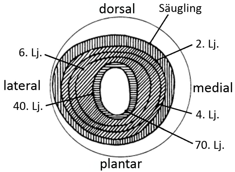
Increase in mobility restrictions with age
The following image illustrates the average progressive restriction of mobility with age.
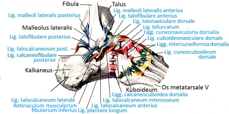
Lateral ligaments (image links to linkmap)
Plantar ligaments (image links to linkmap)
Bones from plantar (image links to linkmap)
Bones from dorsal (image links to linkmap)
Lateral ligaments (image links to linkmap)
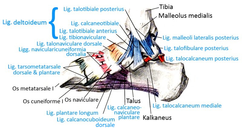
Ligaments from medial (image links to linkmap)
Ligaments from dorsal (image links to linkmap)
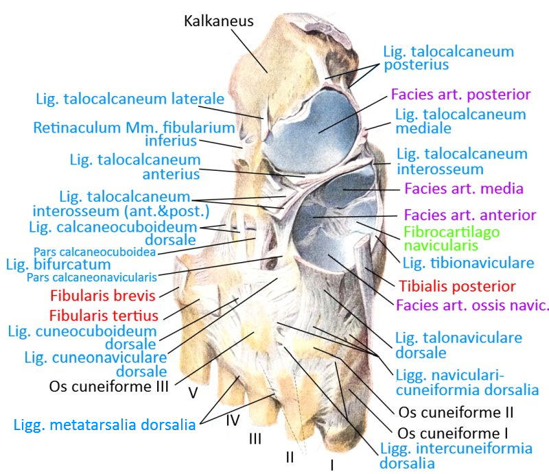
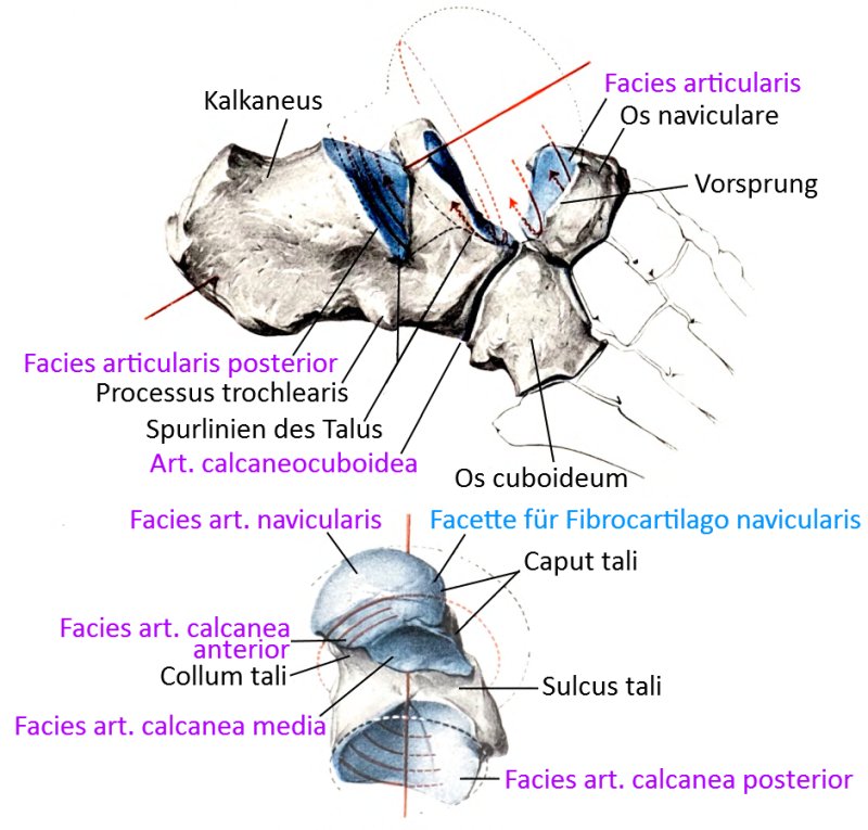
Talotarsal joint
Internal ligaments of the tarsus (image links to linkmap)
Directions of the muscles moving in the ankle joint (image links to linkmap)