Contents
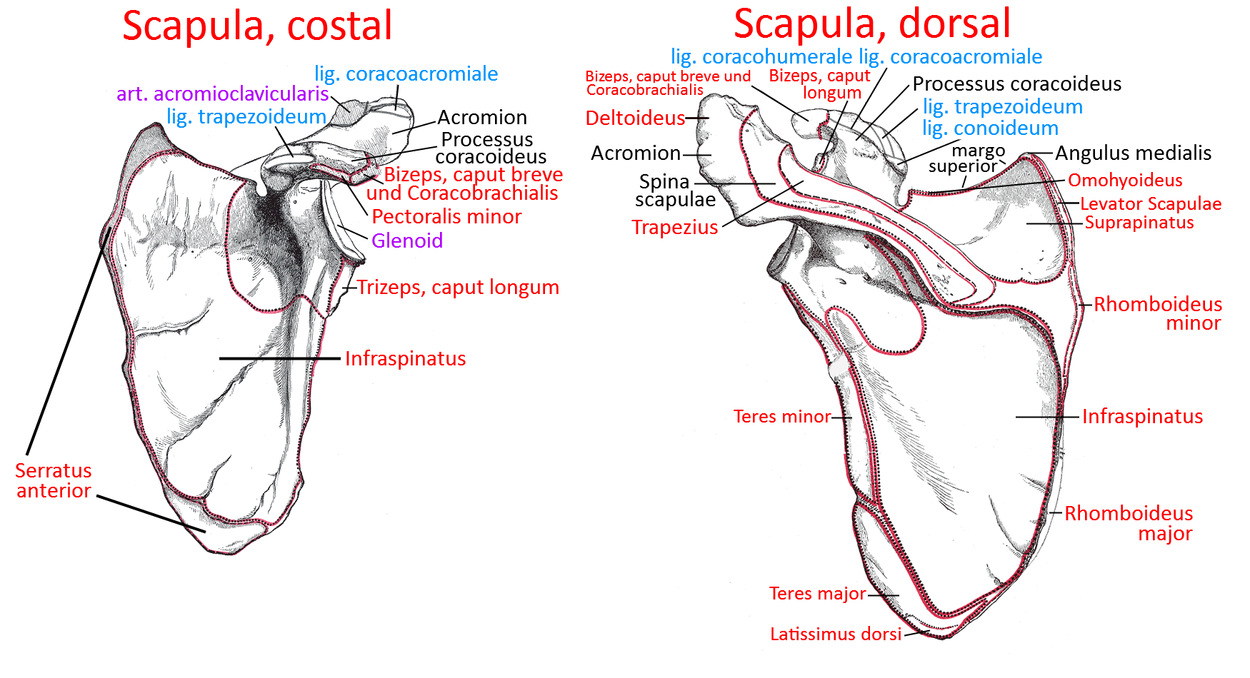
Image: Scapula costal and dorsal (link to two linkmaps)
Scapula
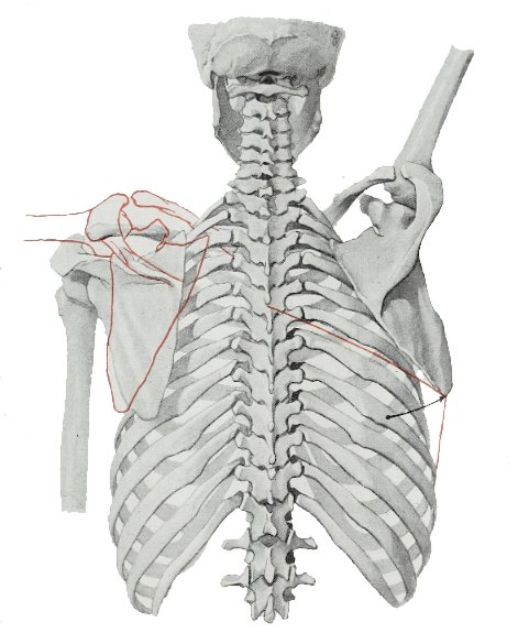
The shoulder blade is a mainly flat bone that is largely covered by muscles and lies on the dorsal side of the torso, connecting the arm to the torso. The connection between the scapula and the rib cage is not a true joint, especially as the bones do not articulate directly and therefore lack cartilage coverings. It is therefore referred to as the scapulothoracic gliding joint, as the scapula glides on the two serratus anterior(ventral, i.e. more profound, on the rib side) and subscapularis (dorsal, i.e. more superficial, originating on the costal side of the scapula ) muscles, which lie in front of the ribs on their ventral/costal side. The scapula articulates with the humerus of the upper arm in its articular surface called the glenoid. The scapula can move freely in three dimensions on the back.
Surfaces
Facies dorsalis
The spina scapula divides the dorsal surface of the scapula into a small superior part with the supraspinous fossa, in which the supraspinatus originates, and a large inferior part, which largely serves as the origin of the infraspinatus. Laterally, diagonally between the lateral and inferior angles, there is a bony ridge that separates the large medial origin area of the infraspinatus from that of the greater(inferior) teres and above that of the lesser teres.
Supraspinous fossa
The supraspinous fossa is the fossa above the spina scapulae and in front of the superior margo of the scapula, in which the suprapinatus, which extends laterally over the humeral head, originates.
Facies costalis / ventralis
The facies costalis contains an extensive fossa, the subscapularis fossa, in the superior two-thirds of which the subscapularis originates. The serratus anterior inserts in triangular areas in front of the angulus inferior and angulus superior.
Fossa subscapularis
Extensive pit in the facies costalis, in the superior two-thirds of which the subscapularis originates.
Margins
Margo superior
Between the superior angulus and the base of the coracoid process lies the superior margo, in which the incisura scapulae covered by the lig, transversum scapulae superius lies. The base of the omohyoid muscle borders the incisure.
Margo lateralis / axillaris
The lateral margo lies between the glenoid cavity and the inferior angulus. Below the glenoid lies the infraglenoid tubercle, from which the caput longum of the triceps originates.
Margo medialis / vertebralis
The margo medialis, to which several muscles attach, lies between the superior angulus and the inferior angulus:
- Rhomboideus major
- Rhomboideus minor
- Levator scapulae
- Pars inferior of the serratus anterior
The first of these three pull medially towards the margo medialis of the scapula and thus cancel out external rotation, at the same time pulling it cranially and thus elevating it. The serratus anterior coming from the lateral, on the other hand, is part of the scapulothoracic gliding bearing and pulls the scapula into protraction.
Angles / corners
Angulus inferior (shoulder blade)
The margo medialis and margo lateralis meet at the lower tip of the shoulder blade, the angulus inferior. During external rotation it moves recognizably and palpably laterally and during elevation recognizably and palpably cranially and therefore represents a good point of orientation. The teres major and some fiber tracts of the latissimus dorsi are located on the dorsal side.
Angulus superior
Margo medialis and margo superior meet at the angulus superior. Fibers of the levator scapulae insert here.
Angulus lateralis
A cavitas glenoidalis, a flat cartilage socket in which the humerus articulates, lies far superior to the more massive angulus lateralis, the junction of margo lateralis and margo superior. Above this lies the supraglenoid tuberosity and below it the infraglenoid tuberosity.
Distinctive structures
Spina
The spina of the scapula is the far superior, largely transverse and medially caudally running scapular ridge, above which the supraspinatus arises (giving it its name) in the supraspinatus fossa, and below which the infraspinatus arises over a large area.
Glenoid / glenoid cavity
The glenoid is the articular surface of the shoulder blade in which the humerus(upper arm bone) articulates. The vertical diameter is larger than the horizontal diameter. As the articular surface is very small and relatively flat in order to achieve the greatest possible range of movement, it is surrounded by a labrum (a cartilage lip), to the upper part of which fibers of the tendon of origin of the caput longum of the biceps attach. The main part of the tendon attaches directly above to the supraglenoid tubercle. Because of this connection, the glenoidlabrum is often also affected when the tendon of origin is torn, which is referred to as a SLAP lesion. It is caused, for example, by injuries caused by extreme loads, such as those that can occur when catching heavy objects, through trauma or in opponent contact situations in sport, when the biceps are jerkily loaded in a powerful eccentric contraction. A SLAP lesion can also occur as a result of repeated microtrauma in the sense of overuse.
The shoulder joint can dislocate more easily than other joints due to the extreme ratio of the small, fairly flat articular surface of the glenoid and the large humeral head:
- in 95-97% anteriorly (arm extended dorsally during a fall dislocates anteriorly)
- in 2-4% after posterior(cramp, electrical accident, which is often overlooked as a cause)
- in 0.5% to inferior (fall on the frontally abducted arm)
- A superior dislocation is not possible because the acromium stops the movement of the humerus.
Below the glenoid lies the infraglenoid tubercle, from which the caput longum of the triceps originates.
Glenoid labrum
The glenoidlabrum is the cartilaginous lip that surrounds the glenoid at its edge and gives the shoulder joint additional stability. It is designed primarily for three-dimensional flexibility with a large ROM, which makes a small and flat glenoid necessary, but which must be given additional stability through additional measures (such as muscular support from the rotator cuff) and the labrum glenoidale cartilage lip. A fundamentally similar construction in the sense of a ball-and-socket joint at the end of the trunk at the base of the limb is found in the hip joint, which is also designed for a rather high degree of three-dimensional flexibility, but in addition to a cartilage lip (labrum acetabuli) has a robust ligamentous apparatus due to the large partial body weight to be supported and whose mobility is reduced in favor of stability; it is also referred to as a nut joint due to the more than full coverage of the caput femoris by the acetabulum. Connected to the glenoidlabrum are fibers of the tendon of origin of the long head of the biceps brachii. In overload situations, not only an avulsion is therefore possible, but often also a tear of the biceps anchor at the cartilage lip.
Biceps anchor
The origin of the tendon of the long head of the biceps at the superior cartilage lip of the glenoid or the supraglenoidal tubercle above the glenoid is referred to as the biceps anchor.
According to studies, only the posterosuperior labrum is the origin of the biceps in 50% of cases, only the supraglenoid tubercle in 20% and both in 30%. The origin on a cartilage is naturally less resilient than a bony origin. Extraordinary tensile stress in particular can therefore cause this area of the cartilage to tear especially in the first case, which is referred to as a SLAP lesion.
There are already three variants for the superior labrum; the anchoring of the tendon to it shows another four types, depending on how the fibres are distributed between the anterior and posterior labrum
Processus coracoideus (coracoid process)
the ventrally protruding process on the outer upper scapula, slightly medial and caudal to the acromion, from which the pectoralis minor and the caput breve of the biceps brachii and the coracobrachialis originate in a common tendon. The main function of the muscles that originate there is to abduct the arm frontally and depress the scapula. The coracoacromial ligament and the coracoclavicular ligament also insert at the coracoid process.
Acromion
The acromion is the shoulder level, the outermost area in the upper shoulder blade, still above the glenoid. The deltoid pars acromialis arises laterally from the superior surface of the acromion, and the joint with the clavicle, the acromioclavicular joint (ACJ ), is located far medially.
Infraglenoid tubercle
Protrusion located below the glenoid, where the caput longum of the triceps originates.
Supraglenoid tubercle
Protrusion located above the glenoid, from which the caput longum of the biceps originates.
Movements
Protraction / retraction: the movement away from the spine to the side-front (protraction) or to the back-inside(retraction) of the spine
Elevation / depression: the cranial lifting(elevation) or caudal lowering (depression) of the shoulder blade.
Rotation: with its lower tip(angulus inferior) pointing outwards and upwards (external rotation) or back again (internal rotation). To raise the arm above 90°, the scapula must externally rotate; when the arm is lowered, the external rotation is reversed.
This means that the scapula has three main dimensions of movement. Pathologically, especially in the case of weakness of the retractors such as the rhomboids, and also in the case of weakness of the protractors such as the serratus anterior, which together press the scapula onto the thorax, it can perform a tilting movement that lifts it away from the back, which can be clearly seen in the various forms of scapula alata (see Wikipedia).
Joints
Acromioclavicular joint
The acromioclavicular joint is the joint in which the scapula articulates with the clavicle, see acromioclavicular joint.
Glenohumeral joint
The glenohumeral joint is the joint in which the scapula articulates with the humerus, the shoulder joint, see under shoulder joint.
Scapulothoracic bearing
The serratus anterior( lyingventrally, i.e. more profound, on the rib side) attaches to the ventral inner edge(margo medialis) of the scapula. The subscapularis originates on the costal side of the scapula. These muscles therefore lie directly on top of each other and at the same time between the thorax and scapula. They form the scapulothoracic gliding bed in which the scapula glides on the fasciae of the muscles and the thorax.
Pictures
Shoulder girdle from cranial
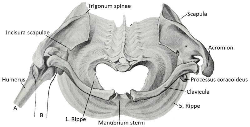
Scapula in transverse adduction with movement in the sternoclavicular joint
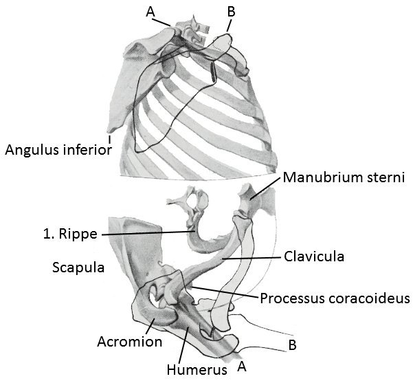
Scapula at 30° transverse adduction without movement in the sternoclavicular joint
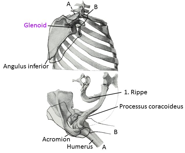
Shoulder blade with lateral abduction
