yogabook / ligaments
Contents
- 1 Ligaments, sorted by associated joint
- 2 Finger joints
- 3 Hand/wrist
- 3.1 Lig. capitohamatum
- 3.2 Lig. carpi arcuatum
- 3.3 Lig. carpi palmare
- 3.4 Lig. carpi radiatum (Lig. deltoideum)
- 3.5 Lig. carpi transversum
- 3.6 Ligg. carpometacarpalia palmaria
- 3.7 Dorsal carpometacarpal ligament
- 3.8 Lig. collaterale carpi radiale
- 3.9 Lig. collaterale carpi ulnare
- 3.10 Ligg. intercarpalia dorsalia
- 3.11 Ligg. intercarpalia interossea
- 3.12 Ligg. intercarpalia palmaria
- 3.13 Ligg. metacarpalia dorsalia
- 3.14 Ligg. metacarpalia interossea
- 3.15 Ligg. metacarpalia palmaria
- 3.16 Lig. metacarpale transversum superficiale
- 3.17 Lig. metacarpale transversum profundum
- 3.18 Ligg. palmaria
- 3.19 Lig. pisohamatum
- 3.20 Pisometacarpal ligament
- 3.21 Dorsal radiocarpal ligament
- 3.22 Lig. radiocarpale palmare
- 3.23 Dorsal radiotriquetral ligament
- 3.24 Dorsal radioulnar ligament
- 3.25 Lig. radioulnare palmare
- 3.26 Retinaculum extensorum
- 3.27 Retinaculum flexorum
- 3.28 Lig. trapezoideocapitatum
- 3.29 Lig. ulnocapitatum
- 3.30 Ulnocarpal ligament
- 3.31 Dorsal ulnocarpal ligament
- 3.32 Lig. ulnolunatum
- 3.33 Lig. ulnotriquetrum
- 3.34 Vaginal ligament
- 3.35 Triangular fibrocartilaginous complex (TFCC, ulnocarpal complex)
- 4 Elbow joint
- 4.1 Lig. anulare radii
- 4.2 Outer ligament complex (LCLC)
- 4.3 Chorda obliqua
- 4.4 Lateral collateral accessorius ligament
- 4.5 Lig. collaterale radiale (RCL: radial collateral ligament, LCL: lateral collateral ligament)
- 4.6 Lig. collaterale ulnare (medial collateral ligament, MCL: medial collateral ligament, UCL: ulnar collateral ligament)
- 4.7 Lateral ulnar collateral ligament (LUCL)
- 4.8 Lig. collaterale ulnare mediale (MUCL: medial ulnar collateral ligament)
- 4.9 Lig. quadratum
- 5 Shoulder joint
- 5.1 Acromioclavicular ligament
- 5.2 Inferior acromioclavicular ligament
- 5.3 Lig. acromioclaviculare superius
- 5.4 Lig. conoideum
- 5.5 Coracoacromial ligament
- 5.6 Coracoclavicular ligament
- 5.7 Coracoglenoid ligament
- 5.8 Coracohumeral ligament
- 5.9 Ligg. glenohumeralia
- 5.10 Inferior glenohumeral ligament
- 5.11 Medial glenohumeral ligament
- 5.12 Lig. glenohumerale superius
- 5.13 Lig. transversum humeri
- 5.14 Lig. transversum scapulae inferius
- 5.15 Lig. transversum scapulae superius
- 5.16 Lig. trapezoideum
- 6 Acromioclavicular joint (see also: Shoulder joint)
- 7 Sternoclavicular joint
- 8 Sternocostal joint
- 9 SIJ (sacroiliac joint)
- 9.1 Lig. ileopectineum (Lig. cooperi)
- 9.2 iliolumbar ligament
- 9.3 Lig. inguinale (inguinal ligament, Vesalius ligament)
- 9.4 Lig. lacunare (Lig. gimbernati)
- 9.5 Lumbosacral ligament
- 9.6 Pubic ligament
- 9.7 Lig. pubicum superius
- 9.8 Lig. pubicum inferius (Lig. arcuatum pubis)
- 9.9 Lig. reflexum
- 9.10 Ligg. sacrococcygea
- 9.10.1 Lig. sacrococcygeum posterius profundum (dorsal profundum)
- 9.10.2 Lig. sacrococcygeum posterius superficiale (dorsal superficiale)Images:
- 9.10.3 Lig. sacrococcygeum anterius
- 9.10.4 Lig. sacrococcygeum interosseum (interosseum axiale)
- 9.10.5 Lateral sacrococcygeal ligament
- 9.10.6 Sacrococcygeal articular ligament (intraalticular)
- 9.11 Sacroiliac ligament
- 9.12 Sacrospinous ligament
- 9.13 Sacrotuberous ligament
- 10 Hip joint
- 10.1 Lig. capitis femoris
- 10.2 Lig. cooperi
- 10.3 Lig. ischiocapsulare
- 10.4 Iliofemoral ligament
- 10.5 Lig. ischiofemoral
- 10.6 Lacunar ligament
- 10.7 Lig. orbicularis
- 10.8 Lig. pectineum
- 10.9 Pubocapsular ligament
- 10.10 Pubofemoral ligament
- 10.11 Lig. transversum acetabuli
- 10.12 Membrana obturatoria
- 10.13 Lig. teres femoris
- 11 Knee joint
- 11.1 Patellar ligament
- 11.2 Collateral ligaments Ligg. collateralia
- 11.3 Cruciate ligaments Ligg. cruciata
- 11.4 Meniscofemoral ligaments Ligg. meniscofemoralia
- 11.5 Lig. popliteofibulare (LPF)
- 11.6 Lig. popliteum arcuatum
- 11.7 Lig. popliteum obliquum (lig. bourgery)
- 11.8 Lig. collaterale mediale posterius (posterior oblique ligament, POL, posterior inner ligament, medial capsular ligament)
- 11.9 Lig. capitis fibulae anterius
- 11.10 Lig. capitis fibulae posterius
- 11.11 Lig. transversum genus
- 11.12 Retinaculum patellae
- 11.13 Medial capsular ligament
- 12 Foot/ankle
- 12.1 Lig. bifurcatum (Chopart ligament, bifurcate ligament)
- 12.2 Lig. calcaneocuboideum plantare (Ligamentum plantare breve)
- 12.3 Ligg. calcaneocuboidea
- 12.4 Lig. calcaneocuboideum
- 12.5 Dorsal calcaneocuboid ligament
- 12.6 Medial calcaneocuboid ligament
- 12.7 Lig. calcaneocuboideum plantare
- 12.8 Calcaneofibular ligament (CFL)
- 12.9 Calcaneonavicular ligament
- 12.10 Dorsal calcaneonavicular ligament
- 12.11 Lig. calcaneonavicular plantar (SL, spring ligament, glenoid ligament)
- 12.12 Inferior calcaneonavicular ligament (ICNL)
- 12.13 Lig. calcaneonavicular superius (SMCNL, superiomedial calcaneo-navicular ligament)
- 12.14 Lateral collateral ligament („outer ligament“ or „outer ligament complex“)
- 12.15 Lig. collaterale mediale
- 12.16 Dorsal cuboideonavicular ligament
- 12.17 Lig. cuboideonaviculare plantare
- 12.18 Ligg. cuneocuboideae
- 12.19 Ligg. cuneometatarsalia interossea
- 12.20 Ligg. cuneonavicularia dorsalia
- 12.21 Ligg. cuneonavicularia plantaria
- 12.22 Lig. deltoideum (Lig. collaterale mediale, medial collateral ligament/collateral ligament)
- 12.23 Anterior fibulotalar ligament (ATFL)
- 12.24 Posterior fibulotalar ligament
- 12.25 Ligg. intercuneiformia dorsalia
- 12.26 Ligg. intercuneiformia interossea
- 12.27 Ligg. intercuneiformia plantaria
- 12.28 Laciniatum ligament
- 12.29 Lig. lisfranc
- 12.30 Lig. malleoli lateralis anterius
- 12.31 Lig. malleoli lateralis posterius
- 12.32 Ligg. metatarsalia dorsalia
- 12.33 Ligg. metatarsalia interossea dorsalia
- 12.34 Ligg. metatarsalia interossea plantaria
- 12.35 Ligg. metatarsalia plantaria
- 12.36 Lig. metatarsale transversum profundum
- 12.37 Lig. metatarsale transversum superficiale
- 12.38 Ligg. naviculocuneiforme dorsalia / plantaria
- 12.39 Lig. plantare longum
- 12.40 Lig. talocalcaneum anterius
- 12.41 Lig. talocalcaneum interosseum
- 12.42 Lateral talocalcaneal ligament
- 12.43 Medial talocalcaneal ligament
- 12.44 Posterior talocalcaneal ligament
- 12.45 Lig. talofibulare anterius
- 12.46 Posterior talofibular ligament
- 12.47 Plantar talometatarsal ligament
- 12.48 Lig. talonaviculare (also: talonaviculare dorsale)
- 12.49 Dorsal talonavicular ligament
- 12.50 Anterior talotibial ligament
- 12.51 Posterior talotibial ligament
- 12.52 Dorsal tarsal ligament
- 12.53 Ligg. tarsi interossea
- 12.54 Ligg. tarsi plantaria
- 12.55 Ligg. tarsometatarsalia dorsalia
- 12.56 Ligg. tarsometatarsalia plantaria
- 12.57 Tibiocalcaneal ligament (TCL)
- 12.58 Tibiocalcaneonavicular ligament (TCNL)
- 12.59 Ligg. tibiofibularia (tibiofibular syndsmosis)
- 12.60 Lig. tibiofibulare anterius (inferius) (anterior syndesmosis ligament, AITFL)
- 12.61 Lig. tibiofibulare interosseum
- 12.62 Posterior tibiofibular ligament (posterior syndesmosis ligament)
- 12.63 Lig. tibiofibulare transversum (part of the syndesmosis ligament)
- 12.64 Tibionavicular ligament (TNL)
- 12.65 Tibiospring (TSL)
- 12.66 Anterior tibiotalar ligament (ATTL)
- 12.67 Posterior tibiotalar ligament (PTTL)
- 12.68 Posterior superficial tibiotalar ligament (STTL)
- 12.69 Retinaculi
- 13 Retinaculum flexorum
- 14 Spring Ligament (SL)
- 15 Toes
- 16 Spine
- 16.1 Fasciculi longitudinales
- 16.2 Lig. alare
- 16.3 Lig. apicis dentis
- 16.4 Atlantoaxial ligament
- 16.5 Lig. atlantoaxiale anterius
- 16.6 Posterior atlantoaxial ligament
- 16.7 Lig. atlantooccipitale
- 16.8 Lig. atlantooccipitale anterius
- 16.9 Lateral atlantooccipital ligament
- 16.10 Lig. atlantooccipitale posterius
- 16.11 Lig. capitis costae intraarticulare
- 16.12 Lig. capitis costae radiatum
- 16.13 Lig. costotransversarium
- 16.14 Lig. cruciatum atlantis
- 16.15 Lig. flavum (interosseous ligament)
- 16.16 Interspinous ligament (interspinosum)
- 16.17 Intertransverse ligament
- 16.18 Lig. longitudinale anterius (anterior longitudinal ligament)
- 16.19 Lig. longitudinale posterius (posterior longitudinal ligament)
- 16.20 Lumbocostal ligament
- 16.21 Lig. nuchae (nuchal ligament)
- 16.22 Supraspinous ligament
- 16.23 Lig. transversum atlantis (transverse ligament of the first cervical vertebra)
- 16.24 Membrana atlantoaxialis anterior (Lig. atlantoaxiale)
- 16.25 Membrana atlantoaxialis posterior (Lig. atlantoaxiale)
- 16.26 Membrana atlantooccipitalis anterior (Lig. atlantooccipitale)
- 16.27 Membrana atlantooccipitalis posterior (Lig. atlantooccipitale posterior)
- 16.28 Membrana tectoria
- 16.29 Costovertebral joint
Ligaments, sorted by associated joint
Finger joints
Ligg. anularia
The anular ligaments are attached palmarly at the level of the respective interphalangeal joints with fibre tracts to the shafts of the proximal and medial phalanges. Their main purpose is to attach the tendon sheaths so that they do not become detached.
Images: (still without)
Ligg. collateralia
As the collateral ligaments of fingers 2-5 run from proximal/dorsal to distal/palmar, they are tense when the metacarpophalangeal joint is flexed and prevent adduction and abduction. With increasing extension of the metacarpophalangeal joints, the range of motion in this dimension increases.
In the case of the MCP (metacarpophalangeal joint), the collateral l igaments extend from the dorsolateral side of the metacarpals diagonally palmarwards to the bases of the proximal phalanges. The collateral ligaments of the interphalangeal joints run on the medial and lateral sides of the joints from the head of one phalanx to the base of the next.
Ligg. collateralia accessoria
The collateral ligaments are additional collateral ligaments that are also attached to the fibrocartilage plates on the palmar side.
Images: (still without)
Ligg. metacarpalia transvera profunda
The deep transverse ligaments connect the metacarpal heads and extend into the joint capsule and the palmar aponeurosis.
Images: (still without)
Ligg. metacarpalia transvera superficialia
The superficial transverse ligaments connect the metacarpal heads and extend into the joint capsule and the palmar aponeurosis.
Images: (still without)
Ligg. obliqua
The oblique ligaments are „cruciate ligaments“ that run between the anular ligaments and reinforce the tendon sheath.
Images: (still without)
Ligg. palmaria (palmar plate)
The palmar ligaments run from the root of the collateral ligaments to the fibrocartilage plates at the bases of the phalanges.
Images: (still without)
Vaginal ligament
Ligament that fixes the tendon sheath of the finger flexors. Some authors differentiate between the anular ligaments in the vaginal accessory ligament (MCP), caginal 1 ligament (PIP) and vaginal 2 ligament (DIP).
A certain degree of hyperextension (beyond the geometric 180°) is considered physiological and is not uncommon, especially in females. Hyperextensions of 90° at the latest are pathological and (in relation to the little finger) a point in the Beighton Score for the diagnosis of hypermobility syndrome. Noticeable varus mobility or valgus mobility in the interphalangeal joints is pathological and a sign of insufficiency of the collateral ligaments(joint instability).
Images:
Linkmap: Hand, wrist, semi-profound
Linkmap: Hand, palmar, profound
Linkmap: Forearm, palmar, very profound
Hand/wrist
Lig. capitohamatum
Ligament connecting the head bone to the hook bone.
Pictures: (still without)
Lig. carpi arcuatum
The carpi arcuatum ligament runs with proximal fibres from the scaphoid bone to the triquetrum and with distal fibres from the triquetrum to the trapezium (Os trapezium and Os trapetoideum). It stabilises the joint row of the mediocarpal joint in the transverse direction.
Images: (still without)
Lig. carpi palmare
Alongside the flexorretinaculum, the carpi palmare ligament is part of the superficial palmar layer. It connects the tendons of the flexor carpi ulnaris and the palmaris longus, surrounds the tendon of the flexor carpi ulnaris and is connected to the palmar ap oneurosis. It serves to tense the palmar aponeurosis and protect the tendons.
Images:
Linkmap: Hand, palmar, superficial
Linkmap forearm, hand, palmar, superficial
Lig. carpi radiatum (Lig. deltoideum)
The carpi radiatum ligament is a ligament of the middle palmar layer. It is also known as the deltoid ligament because it connects the capitate bone to the hamate bone, the triquetrum, the scaphoid bone, the trapezoid bone, the triapezoideum and, rarely, the lunate bone. Fibre tracts of the flexor carpi radialis radiate into the ligament.
Images:
Linkmap: Wrist, ligaments of the carpus
Linkmap: Ligaments, palmar
Lig. carpi transversum
other name for retinaculum flexorum
Ligg. carpometacarpalia palmaria
The palmar carpometacarpal ligaments are short, tight palmar ligaments that connect the bases of the metacarpal bones Ossa metacarpalia II-V with the distal carpal bones.
Images:
Linkmap: Wrist, ligaments of the carpus
Linkmap: Ligaments, palmar
Dorsal carpometacarpal ligament
The carpometacarpal dorsal ligaments are short, taut dorsal ligaments that connect the bases of the metacarpal bones Ossa metacarpalia II-V with the distal carpal bones.
Images: (still without)
Lig. collaterale carpi radiale
The lig. collaterale carpi radiale runs from the styloid process of the radius to the radial side of the scaphoid bone and its tuberosity. It is connected to the tendon sheath of the flexor carpi radialis and limits the ulnar abduction of the hand.
Images:
Linkmap: Wrist, ligaments of the carpus
Linkmap: Wrist, ligaments, dorsal
Linkmap: Ligaments, palmar
Lig. collaterale carpi ulnare
The carpal collateral ligament runs with dorsal fibres from the styloid process of the ulna and the discus to the triquetrum. A palmar fibre tract runs from the palmar radius to the pisiform pea bone. It is fused with the discus and the meniscus of the TFCC (triangular fibrocartilaginous complex) of the ulnocarpal joint. It limits the radial abduction of the hand.
Images:
Linkmap: Wrist, ligaments of the carpus
Linkmap: Wrist, ligaments, dorsal
Linkmap: Ligaments, palmar
Ligg. intercarpalia dorsalia
The dorsal intercarpal ligaments are profound dorsal ligaments made of strong, firm fibres. They belong to the intrinsic or interosseous ligaments.
Images: (still without)
Ligg. intercarpalia interossea
The intercarpal interosseous ligaments connect the carpal bones profoundly in a transverse direction.
Images: (still without)
Ligg. intercarpalia palmaria
The palmar intercarpal ligaments are deeper interosseous ligaments that connect the carpal bones to each other. They are fused to the joint capsule and ensure good stability against translation of the bones.
Images:
Linkmap: Wrist, ligaments of the carpus
Ligg. metacarpalia dorsalia
Short ligaments that connect the bases of the metacarpals dorsally.
Ligg. metacarpalia interossea
Short ligaments that connect the bases of the metacarpals in depth.
Ligg. metacarpalia palmaria
Short ligaments that connect the palmar bases of the metacarpals.
Lig. metacarpale transversum superficiale
The metacarpal transverse superficial ligament is a fibre tract that runs transversely over the bases of the proximal proximal proximal phalanges and connects to the tendon sheaths of the finger flexors.
Images:
Linkmap: Forearm, hand, palmar, superficial
Lig. metacarpale transversum profundum
The metacarpal transverse ligament is a narrow band that covers the palmar side of metatarsophalangeal joints 2 to 5 (MCP) at the metatarsal bases. The palmar ligaments radiate into the metacarpal transverse ligament.
Images:
Linkmap: Forearm, hand, palmar, profundum
Ligg. palmaria
The palmar ligaments are palmar capsular ligaments of the metacarpophalangeal joints, the proximal interphalangeal joints and the distal interphalangeal joints of fingers II to V. They form a gliding support for the tendons of the finger flexors. They form a sliding support for the tendons of the finger flexors and connect to the transverse metacarpal ligament
Images: (still without)
Lig. pisohamatum
The pisohamate ligament is a short ligament that connects the pisiform bone to the hamulus of the hamate bone. Together with the retinaculum (musculorum) flexorum, it delimits the Guyon’s box dorsally, through which the ulnar artery and ulnar nerve run.
Images:
Linkmap: Forearm, hand, palmar, profound
Linkmap: Hand, palmar, profound
Linkmap: Wrist, ligaments of the carpus
Linkmap: Ligaments, palmar
Pisometacarpal ligament
The pisometacarpal ligament is a palmar ligament between the pisiform bone and the base of the 5th metacarpal bone.
Images:
Linkmap: Forearm, hand, palmar, profound
Linkmap: Wrist, ligaments of the carpus
Linkmap: Ligaments, palmar
Dorsal radiocarpal ligament
The dorsal radiocarpal ligament, along with the arcuate carpal ligament, belongs to the middle dorsal layer. It runs from the radius with three separate fibres to the respective carpal bones of the distal radiocarpal joint and stabilises it.
Images:
Linkmap: Wrist, ligaments, dorsal
Lig. radiocarpale palmare
The radoicarpal ligament is a ligament of the middle palmar layer. It consists of a superficial and a deep layer, both of which are connected to the joint capsule. The superficial layer runs from the styloid process of the radius to the capitate bone and the triquetrum, while the deep layer runs from the radius to the scaphoid bone and the lunate bone.
Images:
Linkmap: Wrist, ligaments of the carpus
Linkmap: Ligaments, palmar
Dorsal radiotriquetral ligament
The dorsal radiotriquetral ligament is an approx. 2 cm long ligament that narrows distally between the dorsal tuberosity of the radius and the dorsal side of the triquetrum. It covers the proximal part of the scaphoid bone (Os scaphoideum) and the lunate bone (Os lunatum). It is part of the TFCC.
Images: (still without)
Dorsal radioulnar ligament
The dorsal radioulnar ligament is an extracapsular ligament of the distal radioulnar joint, articulatio radioulnaris distalis, which connects the radius and ulna dorsally. Together with the ligamentum radioulnare palmare, it forms a ring that holds the bones together and is fused with the discus articularis ulnocarpalis. The ligament stretches during pronation. The dorsal radioulnar ligament is part of the so-called TFCC.
Images:
Linkmap: Wrist, ligaments, dorsal
Lig. radioulnare palmare
The dorsal radioulnar ligament is an extracapsular ligament of the distal radioulnar joint (articulatio radioulnaris distalis), which connects the palmar radius and ulna. Together with the dorsal radioulnar ligament, it forms a ring that holds the bones together and is fused to the ulnocarpal articular disc. The ligament stretches during supination. The dorsal radioulnar ligament is part of the so-called TFCC,
Images: (still without)
Retinaculum extensorum
The extensor retinaculum is the superficial dorsal layer with a total of 6 compartments for one tendon each. It is connected to the radius and ulna via intermediate septa.
Images:
Linkmap: Wrist, dorsal, superficial
Linkmap: Hand, dorsal, tendon sheaths and muscles
Retinaculum flexorum
The flexor retinaculum is sometimes referred to as the transverse carpal ligament. It borders the carpal tunnel palmarwards and is connected to both ulnar and radial carpal bones.
Linkmap: Forearm, hand, palmar, profound
Linkmap: Hand, wrist, palmar, semi-superficial
Linkmap: Hand, palmar, semi-profound
Linkmap: Forearm, palmar, very deep
Linkmap: Wrist, ligaments of the carpus
Linkmap: Hand, section through distal carpus
Linkmap: Hand, dorsal, tendon sheaths and muscles
Lig. trapezoideocapitatum
Ligament connecting the small trapezoid bone to the hook bone.
Images: (still without)
Lig. ulnocapitatum
The ulnocapitate ligament is part of the ulnocapitate palmar ligament and the TFCC and runs from the ulna to the palmar side of the capitate bone.
Images: (still without)
Ulnocarpal ligament
The ulnocarpal ligament is a ligament of the middle palmar layer. It runs from the styloid process of the ulna to the discus and the lunate bone (os lunatum), the triangular bone (os triquetrum) and the capitate bone (os capitatum).
Images: (still without)
Dorsal ulnocarpal ligament
The dorsal ulnocarpal ligament runs from the styloid process of the ulna to the dorsal surfaces of the carpal bones.
Images: (still without)
Lig. ulnolunatum
The ulnolunate ligament is part of the ulnocaraple ligament and the TFCC and runs from the ulna and the radioulnar palmar ligament to the anterior horn of the lunate bone.
Images: (still without)
Lig. ulnotriquetrum
The ulnotriquetrum ligament is part of the ulnocaraple ligament and the TFCC and runs from the ulna and the radioulnar palmar ligament to the palmar side of the triquetrum.
Images: (still without)
Vaginal ligament
Ligament that fixes the tendon sheath of the finger flexors.
Some authors differentiate the anular ligaments into
Lig. vaginale accessorium (MCP), Lig. caginale 1 (PIP) and Lig. vaginale 2 (DIP).
Images:
Linkmap: Hand, palmar, semi-profound
Linkmap: Hand, palmar, profound
Linkmap: Forearm, palmar, very profound
Linkmap: Dorsal hand, tendon sheaths and muscles
Triangular fibrocartilaginous complex (TFCC, ulnocarpal complex)
The triangular fibrocartilaginous complex (TFCC) contains the following structures:
- Lig. radioulnare dorsale
- Lig. radioulnare palmare
- Lig. ulnolunatum
- Lig. ulnotriquetrum
- Lig. ulnocapitatum
- Lig. collaterale carpi ulnare
- Lig. radiotriquetrum dorsale
- Meniscus ulnocarpalis
- Discus ulnocarpalis
Elbow joint
Lig. anulare radii
A capsular ligament extending from both ends of the ulnar radial incisura, which holds the proximal end of the radius (radial head) and is also cartilaginous on the inner side. The two legs of the radial collateral ligament radiate into the radial anular ligament. Limited tensile strength of the elbow joint in children sometimes leads to the radial head becoming trapped in the pronated position.
Images:
Linkmap: Elbow joint from the side with ligaments and supinator
Linkmap: Muscles, supinators and pronators of the forearm
Linkmap: Muscles, forearm and hand, palmar, profound
Linkmap: Muscles, inner elbow, profound
Linkmap: Elbow joint, medial and lateral
Linkmap: Elbow joint, ligaments from flexor side
Linkmap: Elbow joint, ligaments from radial side
Linkmap: Elbow joint with lig. anulare radii and lig. quadratum
Linkmap: Supinator muscle
Outer ligament complex (LCLC)
The following four ligaments are sometimes summarised under the term lateral collateral ligament complex:
- Lig. anulare radii
- Lig. collaterale radiale
- Lig. collaterale ulnare laterale
- Lateral collateral accessorius ligament
Chorda obliqua
The chorda obliqua is a ligament that extends from the ulnar tuberosity distal to the radial tuberosity to limit supination of the forearm. The fibres of the chorda obliqua run in the opposite direction to those of the interosseous membrane of the antebrachial ligament.
Images:
Linkmap: Elbow joint lateral with ligaments and supinator
Linkmap: Elbow joint, ligaments from the flexor side
Linkmap: Elbow joint from medial and lateral side
Lateral collateral accessorius ligament
The lateral collateral accessorius ligament runs from the anular radial ligament via the lateral radial head to its insertion immediately distal to the posterior edge of the radial incisura of the ulna. It stabilises the radial anular ligament.
Images: (still without images)
Lig. collaterale radiale (RCL: radial collateral ligament, LCL: lateral collateral ligament)
This ligament radiates from the lateral humeral epicondyle into the radial anular ligament in an approximately delta-shaped manner via two fibre tracts, one medially and one laterally. Further fibres run to the radial incisura ulnae. It is weaker than its ulnar counterpart and contributes to varus stability.
Images:
Linkmap: Elbow joint lateral with supinator
Linkmap: Muscles, forearm, pronators and supinators
Linkmap: Elbow joint, ligaments from radial
Linkmap: Elbow joint, ligaments from flexor side
Linkmap: Muscles, forearm and hand, palmar profound
Lig. collaterale ulnare (medial collateral ligament, MCL: medial collateral ligament, UCL: ulnar collateral ligament)
The ulnar collateral ligament is a simplified term for the medial ulnar collateral ligament, see there. In addition to the medial ulnar collateral ligament, there is another ulnar ligament, the lateral ulnar collateral ligament.
Images:
Linkmap: Elbow joint, ligaments, flexor side
Linkmap: Medial and lateral view of theelbow joint
Linkmap: Elbow joint, ligaments from ulnar side
Lateral ulnar collateral ligament (LUCL)
The ligament located on the lateral elbow joint, it runs as a loop from the epicondylus humeri radialis dorsally around the posterolateral head of the radius to the tuberosity of the crista supinatoria ulna. It is more frequently involved in fractures of the radial head and its damage causes varus instabilityof the elbow joint. It is a very important stabiliser against varus movement.
Images: (still without pictures)
Lig. collaterale ulnare mediale (MUCL: medial ulnar collateral ligament)
The MUCL is often referred to simply as the ulnar collateral ligament (UCL or MCL). It runs from the epicondylus medialis humeri to the medial side of the incisura trochlearis of the ulna. Some fibres of the anconeus attach to this ligament. Around 55% of valgus stress is absorbed by this ligament. Especially throwing sports such as javelin throwing, in which significant valgus stress occurs, strain this ligament. It consists of three parts:
- Pars anterior (anteromedial collateral ligament, AMCL), insertion is the ulnar surface of the coronoid process of the ulna, about 18 mm dorsal to the tip of the coronoid process. It is the most important stabilising structure against valgus movement.
- Pars posterior (posteromedial collateral ligament, PMCL), insertion is the ulnar surface of the olecranon. It has a less stabilising function than the anterior pars.
- Pars transversa (transverse bundle, Cooper ligament), the weakest of the three parts, it connects the ulnar parts of the other two ligaments. No significant contribution against valgus movement is attributed to this ligament.
Lig. quadratum
The quadratum ligament is a thin capsular ligament that runs from the lower edge of the radial incisura of the ulna to the collum of the radius and limits supination and, to a lesser extent, pronation.
Images:
Linkmap: Elbow joint with lig. anulare radii and lig. quadratum
Shoulder joint
Acromioclavicular ligament
The acromiclavicular ligament connects the acromion to the scapula. Two fibre courses can be distinguished.
Images:
Linkmap: Acromioclavicular joint
Linkmap: Glenoid
Linkmap: Ligaments of the shoulder joint
Inferior acromioclavicular ligament
strengthens the caudal area of the joint capsule and is connected to the supraspinatus.
Images: (still without)
Lig. acromioclaviculare superius
reinforces the superior joint capsule of the acromioclavicular joint as a quadrangular ligament. It runs from the cranial distal clavicle to the neighbouring acromion. Its fibres radiate into the fascia of the trapezius and deltoid. The cranial superior acromioclavicular ligament is stronger than the caudal inferior acromioclavicular ligament.
Images:
Linkmap: Ligaments of the shoulder joint
Lig. conoideum
The conoid ligament is the part of the coracoclavicular ligament that attaches to the conoid tubercle of the clavicle.
Images:
Linkmap: Ligaments of the shoulder joint
Linkmap: Ligaments of the shoulder joint
Linkmap: Glenoid and ligaments
Linkmap: Acromioclavicular joint
Linkmap: Scapula from dorsal, insertions
Linkmap: Clavicle with insertions
Coracoacromial ligament
The coracoaromial ligament is a strong band that connects the coracoid process to the underside of the acromion in a triangular shape. The pointed end attaches directly in front of the acromioclacivular joint. The broad side covers the entire length of the coracoid process. Cranially it lies below the deltoid and caudally it is connected to the supraspinatus by a bursa. Together with the acromion and the coracoid process, the coracoaromial ligament forms the „acromion“.
Images:
Linkmap: Acromioclavicular joint
Linkmap: Glenoid and ligaments
Linkmap: Glenoid and surroundings
Linkmap: Shoulder joint, ligaments
Linkmap: Shoulder joint, ligaments
Linkmap: Scapula from dorsal, insertions
Linkmap: Scapula from costal view, insertions
Coracoclavicular ligament
The coracoclavicular ligament is a two-part ligament that attaches the underside of the clavicle to the coradoid process of the scapula: Lig. trapezoideum and Lig. conoideum.
Images:
Linkmap: Acromioclavicular joint
Linkmap: Ligaments of the shoulder joint
Linkmap: Ligaments of the shoulder joint
Coracoglenoid ligament
The coracoglenoid ligament runs from the coracoid process to the joint capsule and the glenoidlabrum, on the upper side of which it attaches.
Images: (no image yet)
Coracohumeral ligament
The coracohumeral ligament connects the coracoid process to the humerus and has an anterior part that runs from the lateral edge of the base of the coracoid process to the lesser tuberosity of the humerus and a posterior part that connects the greater tuberosity to the coracromial ligament. Cranially and ventrally it is overgrown with the joint capsule and lies caudal to the supraspinatus and cranial to the supscapularis. In addition to its general shoulder-stabilising function, it helps to secure the tendon of the caput longum of the biceps and limits the caudal descent of the humerus. Due to its course, it limits lateral abduction without exorotation.
Images:
Linkmap: Glenoid and surrounding area
Linkmap: Ligaments of the shoulder joint
Linkmap: Ligaments of the shoulder joint
Linkmap: Dorsal view of the scapula: insertions
Ligg. glenohumeralia
The glenohumeral ligaments reinforce the ventral joint capsule as three capsular ligaments. They originate at the supraglenoid tubercle of the scapula from fibres of the glenoidlabrum and run as a capsular ligament to the collum anatomicum of the humerus. They appear as three folds in the joint capsule. These are differentiated from cranial to caudal as:
- Superior glenohumeral ligament (SGHL)
- Medial glenohumeral ligament (MGHL)
- Inferior glenohumeral ligament (IGHL)
Inferior glenohumeral ligament
the strongest of the three ligaments, which has two courses, one anterior and one posterior. Between these, the joint capsule forms the recessus axillaris. It opposes caudal, ventral and dorsal translation, the more the arm abducts laterally. If the anterior part of this ligament tears together with a tear in the glenoidlabrum, as can happen with shoulder dislocations, this is called a Bankart lesion.
Images: (still without)
Medial glenohumeral ligament
a stronger ligament that varies significantly from person to person. It stabilises against ventral translation, especially in mid-lateral abduction.
Images: (not yet available)
Lig. glenohumerale superius
The superior glenohumeral ligament is a narrow, thin ligament which, together with the coracohumeral ligament, stabilises the anatomically null proximal and laterally adducted arm.
Images: (still without)
Lig. transversum humeri
The transverse ligament of the humerus runs over the intertubercular sulcus of the humerus and tensions and holds the tendon of origin of the long head of the biceps in the sulcus. It therefore connects the greater tuberosity of the humerus and the lesser tuberosity of the humerus. Opinions differ as to whether the transverse humeral ligament is a true ligament or rather a fibre extension of the subscapularis.
Images: (still without)
Lig. transversum scapulae inferius
The inferior transverse scapular ligament is present inconstantly. It runs from the lateral edge of the scapular spine to the dorsal edge of the glenoid.
Images:
Linkmap: Glenoid and surroundings
Lig. transversum scapulae superius
The lig. transversum scapulae superius is an inconstant ligament that runs from the lateral edge of the scapular spine of the scapula to the dorsal edge of the glenoid.
Images:
Linkmap: Shoulder joint, ligaments
Linkmap: Shoulder joint with scapula, ventral, profound
Linkmap: Shoulder joint with scapula, ventral, superficial
Linkmap: Shoulder joint with scapula, medioventral
Lig. trapezoideum
The trapezoid ligament is the part of the coracoclavicular ligament that attaches to the trapezoid line of the clavicle.
Images:
Linkmap: Shoulder joint, ligaments
Linkmap: Shoulder joint, ligaments
Linkmap: Acromioclavicular joint
Linkmap: Glenoid and ligaments
Linkmap: Glenoid and surroundings
Linkmap: Scapula from dorsal, insertions
Linkmap: Scapula from costal view, insertions
Linkmap: Scapulothoracic gliding bearing
Linkmap: Clavicle with insertions
Acromioclavicular joint (see also: Shoulder joint)
- Inferior acromioclavicular ligament
- Lig. acromioclaviculare superius
- Lig. conoideum
- Coracoclavicular ligament
- Lig. trapezoideum (conoideum)
Sternoclavicular joint
Interclavicular ligament
The interclavicular ligament connects both sternal ends of the clavicles. It thus limits the depression of the shoulder blades.
Images:
Linkmap: Sternoclavicular joint
Linkmap: Sternum, ligaments
Anterior sternoclavicular ligament
The anterior sternoclavicular ligaments are broad ligaments that extend on both sides from the anterior parts of the incisura clavicularis at the sternal end of the clavicle diagonally caudally-medially to the sternal manubrium. The ligament is partially covered by the pars sternalis of the sternocleidomastoid. Dorsally it is connected to both articular surfaces, the discus articularis and the capsule. It secures the clavicle against dorsal translation(retraction).
Images:
Linkmap: Sternoclavicular joint
Linkmap: Sternum, ligaments
Posterior sternoclavicular ligament
The posterior sternoclavicular ligaments are ligaments that run from the dorsal parts of the sternal end of the clavicle diagonally caudally-medially to the sternum manubrium. Ventrally, it is connected to the discus articularis and the two articular surfaces. The sternohyoid and sternothyroid lie dorsally. It secures the clavicle against ventral translation(protraction).
Images:
Linkmap: Sternoclavicular joint
Linkmap: Sternum, ligaments
Costoclavicular ligament
The ligament connects the underside of the clavicle and the upper side of the first rib and thus secures the clavicle against cranial translation(elevation).
Images:
Linkmap: Sternoclavicular joint
Linkmap: Sternum, ligaments
Linkmap: Sternum, ligaments
Linkmap: Clavicle
- Costoclavicular ligament
- Interclavicular ligament
- Anterior sternoclavicular ligament
- Posterior sternoclavicular ligament
Sternocostal joint
Lig. costoxiphoideum
The costoxiphoid ligaments run from the cartilage of the 7th rib (and sometimes also the 6th rib) downwards to the xiphoid process.
Images:
(see above)
Costoclavicular ligament
Ligament extending obliquely from the medial 1st costal cartilage to the craniolateral underside of the clavicle. Ventrally the ligament is connected to the subclavian artery, dorsally it borders on the subclavian vein.
Images:
Linkmap: Sternum, ligaments
Linkmap: Manubrium with sternoclavicular joint
Linkmap: Clavicle
Anterior sternoclavicular ligament
stabilising ligament of the sternoclavicular joint, see the description of the anterior sternoclavicular ligament there.
The anterior sternoclavicular ligaments are broad ligaments that extend on both sides from the anterior parts of the incisura clavicularis at the sternal end of the clavicle diagonally caudally-medially to the sternal manubrium. The ligament is partially covered by the pars sternalis of the sternocleidomastoid. Dorsally it is connected to both articular surfaces, the discus articularis and the capsule. It secures the clavicle against dorsal translation(retraction).
Images:
Linkmap: Sternum, ligaments
Linkmap: Sternoclavicular joint
Intra-articular sternocostal ligament
The sternocostal intraarticular ligament runs from the angulus or the neighbouring parts of the corpus and manubrium through the joint capsule to the second rib and thus divides the joint into two parts. Sometimes these similar ligaments are also found on ribs 3 to 5, they then run to the incisura costalis of ribs 3 to 5 on the corpus sterni.
Images:
Linkmap: Sternum, ligaments
Linkmap: Sternoclavicular joint
Ligg. sternoclavicularia posterius
Stabilising ligament of the sternoclavicular joint, see the description of the posterior sternoclavicular ligament there.
Images:
Linkmap: Sternocostal joint
Ligg. sternocostalia radiata
The radial sternocostal ligaments reinforce the sternocostal joints of ribs 2 to 5. They extend from the respective ribs to the periosteum of the sternum and are interwoven with the local ligaments.
Images:
Linkmap: Sternum, ligaments
Linkmap: Manubrium with sternoclavicular joint
SIJ (sacroiliac joint)
Lig. ileopectineum (Lig. cooperi)
The ileopectineal ligament is an extension of the lacunar ligament that runs along the pecten ossis pubis of the superior ossis pubis ramus.
Images:
Linkmap: Trunk, lateral, very deep
Linkmap: Trunk, ventral view
Linkmap: Trunk ventral, semi-profound
Linkmap: Trunk, abdomen and chest
Linkmap: Trunk, abdominal wall, from the inside
Torso, abdominal wall from diagonally inside
iliolumbar ligament
The iliolumbar ligament runs from medial-cranial to lateral-caudal from the costal process of LWK 4 and LWK 5 to the iliac crest and the anterior sacroiliac ligaments. It thus limits the lateral movement of the cranial pelvis.
Images:
Linkmap: Pelvis, ventral ligaments
Linkmap: Trunk, posterior abdominal wall from ventral
Lig. inguinale (inguinal ligament, Vesalius ligament)
The inguinal ligament is a strong band from the anterior superior iliac spine to the pubic tubercle of the pubic bone. It arises from the aponeuroses of(obliquus externus abdominis, obliquus internus abdominis and transversus abdominis) and transverse fibre strands of the iliac fascia. It represents the anterior and inferior wall of the inguinal canal. The anterior fibre cords of the facia lata arise from anterior cords of the inguinal ligament.
Images:
Linkmap: Pelvis, ligaments, ventral
Linkmap: Acetabulum in context
Linkmap: Trunk, lateral, superficial
Linkmap: Trunk, lateral, profound
Linkmap: Trunk, lateral, very profound
Linkmap: Torso, medium-profound
Linkmap: Trunk, ventral
Linkmap: Trunk, abdomen and chest
Linkmap: Trunk, posterior abdominal wall from ventral view
Linkmap: Pelvis, ligaments from ventral view
Linkmap: Inguinal ligament
Lig. lacunare (Lig. gimbernati)
The lacunar ligament is a short ventral ligament in the inguinal region that attaches cranially to the inguinal ligament and caudally to the pubic crest (pecten ossis pubis). It is delta-shaped and narrows medially. It can be understood as arising from the aponeurosis of the obliquus externus abdominis.
Images:
Linkmap: Trunk, lateral, profound
Linkmap: Trunk, abdominal wall, from oblique inside
Linkmap: Pelvis, ligaments from ventral
Linkmap: Inguinal ligament
Lumbosacral ligament
The lumbosacral ligament begins at the level of Th8, extends caudally and runs
as a filum terminale to the posterior surface of the Os coccygis. On the way, it runs broadly to the lumbar spine and the intervertebral discs.
Images:
Linkmap: Pelvis, ligaments, ventral
Pubic ligament
The pubic ligament consists of the superior pubic ligament and the inferior pubic ligament, between which lies the interpubic disc into which both ligaments radiate.
Images:
Linkmap: Pelvis, ligaments, ventral
Lig. pubicum superius
The inferior pubic ligament is the upper ligament of the pubic symphysis that radiates into the interpubic disc.
Images: (still without)
Lig. pubicum inferius (Lig. arcuatum pubis)
The inferior pubic ligament is the lower ligament of the pubic symphysis that radiates into the interpubic disc.
Images: (still without)
Lig. reflexum
The reflex ligament runs from the pubic tubercle and lacunar ligament craniomedially to the rectus sheath (anterior leaflet) and thus forms the medial floor of the inguinal canal and the dorsal border of the superficial inguinal annulus. The ligament originates from deep tendon fibres of the obliquus externus abdominis and can be seen as a separation of the inguinal ligament.
Images:
Linkmap: Inguinal ligament
Ligg. sacrococcygea
Lig. sacrococcygeum posterius profundum (dorsal profundum)
The posterior sacrococcygeal ligament is the continuation of the posterior longitudinal ligament from the sacrum to the coccyx.
Images:
Lig. sacrococcygeum posterius superficiale (dorsal superficiale)
Images:
The posterior superficial sacrococcygeal ligament is the continuation of the supraspinous ligament from the sacrum to the coccyx. It runs from the sacral hiatus to S2.
Images:
Linkmap: Pelvis, ligaments, dorsal
Linkmap: Trunk, dorsal, profound
Lig. sacrococcygeum anterius
The anterior sacrococcygeal ligament connects the ventral sides of the sacrum (Os sacrum) and coccyx (Os coccygis).
Images: (still without)
Lig. sacrococcygeum interosseum (interosseum axiale)
The sacrococcygeal interosseous ligament connects the tuberosities of the sacrum with the ilium.
Images: (still without)
Lateral sacrococcygeal ligament
The lateral sacrococcygeal ligament runs from the lower lateral edge of the sacrum to the transverse process of the coccyx.
Images:
Linkmap: Torso, dorsal, profound
Sacrococcygeal articular ligament (intraalticular)
The sacrococcygeal articular ligament connects the cornu of the sacrum with the cornu of the coccyx.
Images:
Linkmap: Trunk, dorsal, profound
Sacroiliac ligament
Anterior sacroiliac ligament
The anterior sacroiliac ligaments run ventrally of the SI joint from the sacrum (especially from its cranial two WK) to the hip bone. They delimit the cranial foramen ischiadicum majus.
Images:
Linkmap: Trunk, posterior abdominal wall, ventral
Anterior sacroiliac ligament
The anterior sacroiliac ligament is a collection of coarse, fibre-rich ligaments extending from the ventral side of S1 and S2 to the iliac bone.
Images:
Linkmap: ISG, sections
Linkmap: Pelvis, ligaments, ventral
Posterior sacroiliac ligament
The posterior sacroiliac ligament is a collection of coarse, fibre-rich ligaments that extend from the iliac tuberosity to the sacrum and serve as the origin of the multifidi.
Images: (still without)
Ligg. sacroiliaca posteriora (dorsalia)
The anterior sacroiliac ligaments(longus and brevis) run dorsally of the SI joint from the sacrum (especially from its cranial two WK) to the hip bone. They delimit the cranial foramen ischiadicum majus.
Images: (still without)
Lig. sacroiliacum posterius (dorsal) brevis
The posterior sacroiliac ligament runs from the dorsal surface of the sacrum (crista sacralis intermedia and lateralis) to the posterior inner surface(ventral) of the hip bone (iliac tuberosity).
Images: Linkmap: Pelvis, ligaments, dorsal
Lig. sacroiliacum posterius (dorsale) longus
The dorsal sacroliac ligament longus extends caudally in front of the dorsocranial edge of the iliac crest (SPIS, posterior superior iliac spine) and radiates into the sacrotuberous ligament on the one hand, while fibre bundles extend medially to the lateral sacral crest of the caudal posterior sacrum on the other.
Images: Linkmap: Pelvis, ligaments, dorsal
Ligg. sacroiliaca interossea
The posterior sacroiliac ligaments are strong ligaments between the sacral tuberosity and the iliac tuberosity. Both bone roughenings lie craniodorsal to the auricular facies. The interosseous sacroiliac ligaments lie deeper than the posterior sacroiliac ligaments.
Images:
Linkmap: ISG, sections
Sacrospinous ligament
The sacrospinous ligament runs from the sacrum and coccyx to the ischial spine of the ischium. This ligament lies between the foramen ischioadicum majus(cranial) and foramen ischiadicum minus(caudal).
Images:
Linkmap: Hip joint
Linkmap: Pelvis, ligaments, dorsal
Linkmap: Pelvis, ligaments, ventral
Linkmap: Trunk, lateral, superficial
Linkmap: Trunk, lateral, very deep
Linkmap: Trunk, medium-profound
Linkmap: Trunk, dorsal, profound
Linkmap: Torso, abdominal wall from oblique inside
Sacrotuberous ligament
The sacrospinous ligament runs from the sacrum and coccyx to the ischial tuberosity of the ischium. It forms the lower boundary of the lesser sciatic foramen and, together with the sacrospinous l igament, prevents the sacrum from tilting dorsally.
Images:
Linkmap: Hip joint
Linkmap: Pelvis, ligaments, dorsal
Linkmap: Pelvis, ligaments, ventral
Linkmap: Acetabulum in context
Linkmap: Trunk, lateral, superficial
Linkmap: Trunk, lateral, very deep
Linkmap: Trunk, medium-profound
Linkmap: Trunk, dorsal, profound
Linkmap: Torso, dorsal
Hip joint
Lig. capitis femoris
A ligament arising from the fovea capitits of the femoral head and extending to the acetabular fossa, in which an artery runs: the ramus acetabularis of the obturator artery. However, this is often atrophied in adults. This ligament does not appear to have a mechanical function. The incisura acetabuli, from which the ligament emerges, is bridged by the transverse acetabular ligament.
Images:
Linkmap: Acetabulum in context
Linkmap: Hip joint, frontal saw cut
Linkmap: Hip joint, muscles, section
Linkmap: Hip joint, medial view
Linkmap: Hip joint, unfolded
Lig. cooperi
Also known as the pectineal ligament along the pecten ossis pubis of the ramus superior ossis pubis in extension of the lacunar ligament. The ligament forms the dorsal border of the inguinal canal.
Images:
Linkmap: Trunk, abdominal wall from an oblique inside view
Linkmap: Pelvis, ligaments from ventral view
Linkmap: Acetabulum in context
Lig. ischiocapsulare
Old term for ischiofemoral ligament.
Images:
Linkmap: Hip ligament from dorsal
Linkmap: Acetabulum in context
Iliofemoral ligament
The iliofemoral ligament is the strongest ligament in the human body and runs from the SIAS (spina iliaca anterior inferior) to the linea intertrochanterica femoris. It can be divided into two parts: Pars verticalis with fibre tracts and Pars horizontalis with lateral fibre tracts.
Even in standard anatomical position, this ligament inhibits hip extension so that the hip flexors have to work less.
Images:
Linkmap: Hip joint, muscles, section
Linkmap: Hip joint from the medial side
Linkmap: Hip joint, ligaments, dorsal view
Linkmap: Hip joint, ligaments, ventral
Linkmap: Obturatorius externus
Linkmap: Acetabulum in context
Linkmap: Dorsal view of the hip joint
Linkmap: Hip joint, frontal saw cut
Lig. ischiofemoral
Tent-shaped ligament in the dorsal hip joint from the craniolateral ischium dorsally over the femoral head to the linea intertrochanterica femoris.
Images:
Linkmap: Hip joint from dorsal
Linkmap: Acetabulum in context
Lacunar ligament
The lacunar ligament is not strictly speaking one of the ligaments of the hip joint. It runs from the inguinal ligament in the floor down to the pubic bone.
Images:
Linkmap: Trunk, lateral, profound
Linkmap: Trunk, abdominal wall from oblique inside
Linkmap: Pelvis, ligaments from ventral
Lig. orbicularis
Confluent ligament with the lateral fibres of the iliofemoral ligament, transversely encompassing the femoral head and limiting the extension.
Lig. pectineum
Another name for the cooper’s ligament.
Images:
Linkmap: Torso, abdominal wall from diagonally inside
Linkmap: Pelvis, ligaments from ventral view
Linkmap: Acetabulum in context
Pubocapsular ligament
Old name for pubofemoral ligament
Pubofemoral ligament
Ligament running from the ramus superioris ossis pubis to the linea intertrochanterica femoris.
Images:
Linkmap: Hip joint from ventral view, ligaments
Linkmap: Acetabulum in context
Linkmap: Obturatorius externus
Lig. transversum acetabuli
The acetabular transverse ligament closes the gap (acetabular incisura) in the acetabular labrum.
Images:
Linkmap: Acetabulum in context
Linkmap: Hip joint, frontal saw cut
Membrana obturatoria
The obturator membrane is the thin, fibre-rich membrane that largely closes the obturator foramen. The origins of the obturator externus and obturator internus muscles lie on this membrane.
Images:
Linkmap: Acetabulum in context
Linkmap: Hip joint, frontal saw cut
Lig. teres femoris
Old name for capitis femoris ligament, see there.
Knee joint
Patellar ligament
The approx. 5-6 mm thick ligament is a capsular ligament that transmits the contraction force of the quadriceps from the lower edge of the patella(caudal patellar pole) to the tibia, where it inserts at the tibial tuberosity. If the quadriceps are not under tension, the patellar ligament is slack and could become trapped in the joint space when the knee joint is largely extended. This is why the Hoffa’s adipose body (corpus adiposum infrapatellare) lies behind(dorsally or profoundly) the patellar l igament, which prevents this due to its volume. A change in Hoffa’s fat body can lead to Hoffa’s syndrome, which usually only occurs secondarily.
Images:
Linkmap: Ventrolateral knee joint
Linkmap: Lateral knee joint
Linkmap: Knee joint, sagittal section
Linkmap: Tibia
Linkmap: Bones of the lower leg
Linkmap: Trunk ventral head to knee
Linkmap: Trunk lateral, superficial
Linkmap: Knee joint, 90° inflected
Linkmap: Lateral knee joint, bursa
Collateral ligaments Ligg. collateralia
the medial and lateral collateral ligaments that run longitudinally on the medial and lateral side of the knee joint, which tighten when the knee joint is extended due to the shape of the condyles and thus increasingly prevent the endo-and exorotation of the lower leg in the knee joint: the medial collateral ligament and the lateral collateral ligament (fibular). Another important task of the collateral ligaments is to absorb varusand valgus-likemovements and corresponding forces.
Lig. collaterale mediale (inner collateral ligament, lig. collaterale tibiale)
Wide, flat ligament, approx. 9-11 cm long, which runs slightly offset dorsally on the medial side of the knee joint and stabilises it against valgus movement. It runs from the epicondylus medialis femoris to the condylus medialis tibiae. It has an anterior and a posterior section, both of which diverge slightly distally. The more profound parts are fused with the medial meniscus. The proximal part is connected to the medial patellarretinaculum, the distal part merges into the popliteal oblique ligament and the dorsomedial capsule. The distal parts are partly overlaid by the pes anserinus and its attached muscles, which is why abursa (bursa anserina) buffers between them to minimise shear forces. A bursa separates the medial collateral ligament from the more profound structures, namely the joint capsule, which is reinforced with a capsular ligament, and the medial meniscus. The medial collateral ligament not only limits the valgus movement of the tibia and thus prevents medial gapping, but together with the fibular collateral ligament ( lateral collateral ligament) it also prevents exorotation of the lower leg in the knee joint, which is why it is at risk in breaststroke athletes. The classic valgus stress test of the knee joint tests the sufficiency of the ligament. Due to the complex anatomy of the medial collateral ligament, the results of reconstructive surgery are often unsatisfactory. In particular, calcifications, which are more likely to occur the longer the knee joint is immobilised, often lead to functional deficits and restricted mobility.
Images:
Linkmap: Knee joint, dorsal, ligaments
Linkmap: Knee joint, dorsal
Linkmap: Knee joint, dorsal, capsule
Linkmap: Knee joint ventrolateral
Linkmap: Knee joint, 90° flexed
Lateral collateral ligament (outer collateral ligament, fibular collateral ligament)
The outer or fibular collateral ligament is a strong round ligament 5-7 cm long and, unlike the inner collateral ligament, has no connection to the meniscus. It is largely covered by the biceps femoris. It runs from the epicondylus lateralis femoris, just below the sulcus of the popliteus tendon, to the head of the fibula. It splits the attachment tendon of the biceps femoris into an anterior and a posterior branch. The bursa subtendinea musculi bicipitis femoris inferior lies between the lateral collateral ligament and the biceps femoris. The popliteus tendon runs between the lateral collateral ligament and the lateral capsule. The ligament secures the knee joint against varus movement and limits exorotation.
Images:
Linkmap: Dorsal view of the knee joint, ligaments
Linkmap: Dorsal knee joint
Linkmap: Knee joint dorsal, capsule
Linkmap: Knee joint, 90° inflected
Linkmap: Lateral knee joint
Linkmap: Lateral knee joint, bursa
Cruciate ligaments Ligg. cruciata
The two ligaments that run intra-articularly but retrosynovially in the knee joint and prevent the tibia from shifting ventrally or dorsally in relation to the femur: the anterior cru ciate ligament and the even stronger posterior cruciate ligament. In terms of developmental history, they have migrated into the knee joint from the dorsal side. If they are damaged, unphysiological displacements occur, resulting in instability during movement and increased wear of the knee joint. Damage such as overstretching and tears can be recognised by the anterior or posterior drawer effect. In the English literature, theanterior cruciate ligament is referred to as the ACL(anterior cruciate ligament) and the posterior cruciate l igament as the PCL(posterior cruciate ligament). The name cruciate ligament comes from the fact that the two cruciate ligaments with their different directions of tension cross in the centre of the knee joint and develop complex biomechanics. To a limited extent, the cruciate ligaments also restrict the varus and valgus movement of the knee joint. As they wrap around each other during endorotation of the lower leg in the knee joint, they limit these movements. Tension and release as well as tightening and loosening of the cruciate ligaments are important for their metabolism.
Lig. cruciatum anterius (ACL, LCA, anterior cruciate ligament, anterior cruciate ligament)
The anterior cruciate ligament is probably the most important ligament for stabilising the knee joint in the sagittal direction and, together with the posterior cruciate ligament, which runs in the opposite direction, secures it against translation of the tibia. Particularly in a slightly flexed position of the knee joint (approx. 20-30°), it strongly secures the tibia against forward displacement. Both cruciate ligaments together limit the final rotation of the lower leg in the knee joint. The anterior cruciate ligament also limits the extension of the knee joint. It runs from the lateral wall of the intercondylar fossa of the lateral femoral condyle to the proximal medial tibia at the anterior intercondylar area in front of the intercondylar eminence, just ventral to the medial intercondylar tubercle between the insertions of the anterior menisci. It therefore runs from posterior-superior-lateral to anterior-inferior-medial. Some fibres insert at the meniscal root of the medial meniscus. A distinction is made between two bundles according to origin and insertion: an anterior (anteromedial) bundle, which runs from the intercondylar line to the anterior tibial plateau, and a posterior (posterolateral) bundle, which runs from the border between the bone and cartilage of the femoral condyle to the posterior area of the tibia, close to the medial meniscus. An intermediate bundle can also be identified. The LCA is supplied with arterial blood from both sides, but the centre and the insertion areas are usually not supplied with arterial blood. It has many mechanoreceptors (Golgi type III receptors, Ruffini corpuscles, Pacini type II corpuscles and free nerve endings), which serve the proprioception of the knee joint, but also the activation of knee joint stabilising muscles, such as the hamstrings, which also pulls the tibia dorsally. In the event of ruptures of the LCA or replacement with a plastic, this function is no longer available, which puts more strain on the plastic than on the native cruciate ligament and leads to increased ventral translation and increased wear. If the ACL is damaged, the rolling-sliding mechanism of the knee joint is disrupted and damage occurs primarily to the posterior horns of the menisci, later also to cartilage damage to the tibia and femur, which can also be detected radiologically.
Images:
Linkmap: Dorsal view of the knee joint, ligaments
Linkmap: Dorsal knee joint, capsule
Linkmap: Knee joint ventral
Linkmap: Knee joint transversal
Linkmap: Knee joint, 90° inflected
Linkmap: Knee joint, menisci
Linkmap: Knee joint, distortion of the menisci during torsion
Posterior cruciate ligament (PCA, posterior cruciate ligament)
The posterior cruciate ligament runs from the inside of the medial femoral condyle diagonally to the lateral distal to the posterior intercondylar area, i.e. from ventral-cranial-medial to dorsal-caudal-lateral and thus runs transversely to the anterior cruciate ligament. A longer, stronger anterolateral and a shorter, less strong dorsomedial fibre bundle can be distinguished.
dorsomedial fibre bundle. The quadriceps, innervated by the proprioceptors of the longitudinal retinaculi of the patella, which radiate from its fibres, work to prevent excessive strain on the posterior cruciate ligament. Good development of the two important quadriceps parts, vastus medialis and vastus lateralis, from which these retinaculi radiate, is therefore important for an athlete.
Images:
Linkmap: Knee joint from dorsal, ligaments
Linkmap: Dorsal knee joint, capsule
Linkmap: Knee joint ventral
Linkmap: Knee joint transversal
Linkmap: Knee joint, 90° inflected
Linkmap: Knee joint, menisci
Linkmap: Knee joint, distortion of the menisci during torsion
Meniscofemoral ligaments Ligg. meniscofemoralia
The meniscofemoral ligaments support the posterior cruciate ligament and occur inconsistently. The Humphrey ligament stretches when the knee joint is flexed and the Wrisberg ligament stretches when the knee joint is extended. If the posterior cruciate ligament is damaged, they can partially take over its function. They stabilise the knee joint with regard to posterior translation of the tibia:
- the Wrisberg ligament (posterior meniscofemoral ligament), which can be differentiated into three types and lies dorsal to the posterior cruciate ligament, accounting for approx. 70%.
- the Humphrey ligament (anterior meniscofemoral ligament) lying ventral to the posterior cruciate ligament in about 50% of cases
Information on the presence of the ligaments varies in the literature. The Wrisberg ligament can be quite pronounced. In 61% of cases, both meniscofemoral ligaments are present. They then wrap around the posterior cruciate ligament. The Humphrey ligament stretches in flexion, the Wrisberg ligament in extension of the knee joint. Both tighten when the lower leg is in endorotation. Their most important task is to prevent the posterior horn of the lateral meniscus from becoming trapped.
Images:
Linkmap: Knee joint from dorsal, ligaments
Linkmap: Knee joint transversal
Linkmap: Knee joint, menisci
Anterior meniscofemoral ligament: „Humphrey ligament„
The anterior meniscofemoral ligament runs from the posterior meniscal root of the lateral meniscus in front of the posterior cruciate ligament to the inner surface of the medial femoral condyle.
Posterior meniscofemoral ligament: „Wrisbergligament„
Like the anterior meniscofemoral ligament, the posterior meniscofemoral ligament runs from the posterior meniscal root of the lateral outer meniscus to the medial femoral condyle, but runs behind the posterior cruciate ligament. An existing Wrisberg ligament must not be misinterpreted radiologically as a tear of the meniscus. The origin of the three types of Wrisberg ligament is identical, but they differ in their attachment:
- Posterior horn of the medial meniscus (most common form)
- fan-shaped from the meniscus to further medial to the tibia
- without attachment to the meniscus, only to the tibia, sometimes considered part of the posterior cruciate ligament, as it is not connected to the meniscus
Images:
Linkmap: Knee joint, menisci
Lig. popliteofibulare (LPF)
The popliteofibular ligament (LPF) radiates from the head of the fibula into the tendon of the popliteus.
Lig. popliteum arcuatum
The popliteus arcuate ligament is an extracapsular dorsal ligament of the knee joint that runs from the posterior edge of the head of the fibula obliquely in a craniomedial direction and crosses the popliteus insertion, which it attaches to the joint capsule. It covers the dorsolateral joint area in a fan shape and runs partly parallel to the lateral collateral ligament.
Images: (still without)
Lig. popliteum obliquum (lig. bourgery)
The oblique popliteofibular ligament, which runs from the posterior edge of the head of the tibia obliquely craniolaterally to the upper edge of the intercondylar fossa and to the posterior surface of the femur, taking up the tendon of the semimembranosus in a lateral split. It reinforces the dorsal capsule of the knee joint. In extension of the knee joint it is taut and prevents medial and lateral gapping, in flexion it is relaxed. Because of its connection to the semimembranosus, it still contributes to the stability of the joint even when relaxed in flexion. This ligament can have connections to the medial collateral ligament, the popliteus tendon and the dorsal capsule.
Images:
Linkmap: Knee joint dorsal
Linkmap: Knee joint, sagittal section
Lig. collaterale mediale posterius (posterior oblique ligament, POL, posterior inner ligament, medial capsular ligament)
The POL moves from the adductor tuberosity in three different lines:
- to the posterior edge of the tibia and medial meniscus (this is the main traction)
- to the tendon of the semimembranosus (medial fibre pull)
- merges with the tendon of the semimembranosus in the lig. popliteum obliquum
It stabilises against valgus stress and exorotation, both in extension (together with the medial collateral ligament) and in flexion (in conjunction with the tensed semimembranosus). Together with the posterior horn of the medial meniscus, the ACL and the medial collateral l igament, it protects against excessive translation of the tibia in the sagittal direction.
Lig. capitis fibulae anterius
Ventral ligament of the proximal tibiofibular joint that reinforces the capsule. It runs from the ventral caput fibulae and facies articularis fibularis of the lateral tibial condyle. The anterior capitis fibulae ligament is stronger than its posterior equivalent, the posterior capitis fibulae ligament.
Images:
Linkmap: Knee joint from dorsal, ligaments
Linkmap: Knee joint, 90° flexed
Lig. capitis fibulae posterius
Dorsal ligament of the proximal tibiofibular joint that reinforces the capsule dorsally. It is less pronounced than its ventral counterpart, the capitis fibulae anterius ligament.
Images: (still without)
Lig. transversum genus
The lig. transversum genus is an intra-articular ligament that connects the two anterior horns of the menisci(inner meniscus and outer meniscus). No significant function has yet been demonstrated.
Linkmap: Knee joint, 90° flexed
Linkmap: Knee joint, menisci
Linkmap: Transverse knee joint
Retinaculum patellae
The retinaculum patellae consists of two individual longitudinal ligaments, a lateral retinaculum patellae laterale and a medial retinaculum patellae mediale, which surround the patella from the outside and hold it in position, as well as often a lateral transverse and, in 30% of cases, a medial transverse retinaculum.
lateral trans verse retinaculum and in 30% also a medial transverse retinaculum. The transverse retinaculi lie deeper than the longitudinal retinaculi. Patelladislocations are therefore generally prevented if the ligamentous apparatus is undamaged. The longitudinal fibres of the patellar retinaculum can transfer a residual contraction force of the quadriceps to the tibia in the event of a tear in the patellar ligament and thus maintain a small residual extensor function, which is why they are referred to as the reserve extensor apparatus.
Medial patellar retinaculum (MPFL)
The medial patellar retinaculum originates from the tendon fibres of the medial vastus, which do not attach to the cranial patellar pole and do not cover the patella. They run to the medial edge of the patella and the tibial collateral ligament as well as to the medial condyle of the tibia medial to the tibial tuberosity and the patellar ligament.
Images: (still without)
Lateral patellar retinaculum
The lateral patellar retinaculum arises from the tendons of the lateral vastus and the rectus femoris and extends to the lateral edge of the patella and the collateral fibular ligament as well as to the lateral condyle of the tibia lateral to the tibial tuberosity and the patellar ligament.
Images:
Linkmap: Ventrolateral knee joint
Retinaculum patellae, transverse parts
In addition to the retinaculum patellae laterale and retinaculum patellae mediale, there is often a lateral transverse part(retinaculum patellae transversale laterale) and in 30% a medial transverse part(retinaculum patellae transversale mediale).
Images: (still without)
Lateral transverse patellar retinaculum (LPTL)
The transverse lateral part originates from tissue of the iliotibial tract and runs on the one hand without connection to the lateral epicondyle as the lateral patellofemoral ligament to the mid-superior lateral edge of the patella and on the other hand runs further caudally (lower lateral edge of the patella) as the lateral patellofemoral ligament. Further cranially there is a connection between the patella and the lateral condyle of the femur via the Kaplan fibres.
Images: (still without)
Lateral patellofemoral ligament
The fibre tract of the transverse lateralretinaculum patellae from the mid-superior lateral edge of the patella to the iliotibial tract described above.
Lateral patellotibial ligament
The fibre tract of the transverse lateralretinaculum patellae from the lower lateral edge of the patella to the iliotibial tract described above.
Kaplan fibres
the cranial connection of the lateral patellarretinaculum to the lateral condyle of the femur.
Retinaculum patellae transversale mediale (patellotibial ligament)
The transverse medial part consisting of fibres of the tendon of the vastus medialis, which run to the medial edge of the patella and to the femur lateral to the medial collateral ligament.
Images: (still without)
Medial patellofemoral ligament
the cranial (femoral) part of the transverse medial patellarretinaculum
Medial patellotibial ligament
the caudal (tibial) part of the transverse medial patellarretinaculum
Medial capsular ligament
The medial capsular ligament, which runs from cranial to caudal, is the fibre-reinforced middle third of the medial capsule. It is firmly fused to the base of the meniscus. The cranial part is referred to as meniscofemoral, the caudal part as meniscotibial. During
extension of the knee joint it is taut and becomes slack even with slight flexion, only to tighten again with further flexion, so that it protects against valgus stress and excessive exorotation during extension and further flexion.
Foot/ankle
Lig. bifurcatum (Chopart ligament, bifurcate ligament)
The bifurcate ligament consists of two individual ligaments, the medial dorsal calcaneonavicular ligament and the lateral dorsal calcaneocuboid ligament, and connects the dorsal ventral calcaneus with the neighbouring cuboid and navicular bones. The bifurcate ligament is therefore the dorsal part of the ligament securing the Chopart joint line, in which a limited degree of extension and flexion of the foot takes place. While the plantar ligament and calcaneonavicular plantar ligament limit extension with the plantar fascia and intrinsic foot muscles, the bifurcate ligament sets a limit in the direction of flexion of the foot. Inversion trauma (see supination trauma) frequently causes injuries to this ligament, including avulsion fractures of the anterolateral process of the calcaneus.
Images:
Linkmap: Ankle joint, ligaments, lateral
Linkmap: Dorsal view of the ankle, ligaments
Linkmap: Lateral ankle, ligaments
Linkmap: Ankle from medial, ligaments
Linkmap: Tarsus, internal ligaments
Lig. calcaneocuboideum plantare (Ligamentum plantare breve)
Fibre tract regarded by many authors as a separate part of the plantar long ligament. It runs from the calcaneal tuber to the plantar surface of the cuboid bone (cuboid bone)
Images:
Linkmap: Foot, ligaments, medial
Linkmap: Foot, ligaments, plantar
Ligg. calcaneocuboidea
The plural is used less frequently in the literature, although „the calcanocuboid ligament“ consists of four ligaments, see here.
Lig. calcaneocuboideum
The calcaneocuboid ligament supports the pronounced medial and the weak lateral arch of the foot. It consists of four ligaments:
- Dorsal calcaneocuboid ligament
- Medial calcaneocuboid ligament
- Lig. calcaneocuboideum plantare
- Lig. plantare longum (LPL )
The ligaments can be damaged by inversion trauma(supination trauma).
Images:
Linkmap: Foot, ligaments, medial
Dorsal calcaneocuboid ligament
The calcaneocuboid ligament is a dorsal ligament of the foot and part of the bifucate ligament (together with the calcaneonavicular ligament). It contributes to the stability of the calcaneocuboid joint as part of the Chopart joint.
Images:
Linkmap: Foot, ligaments, dorsal
Linkmap: Foot, ligaments, medial
Medial calcaneocuboid ligament
represents a part of the bifurcate ligament.
Pictures: (still without)
Lig. calcaneocuboideum plantare
Ligament from the calcaneal tuber to the plantar surface of the cuboid bone, considered by some authors to be part of the plantar ligament.
Images:
Linkmap: Ankle joint, ligaments, medial
Linkmap: Foot, ligaments, plantar
Calcaneofibular ligament (CFL)
The calcaneofibular ligament, together with the anterior talofibular ligament and the posterior talofibular ligament, is part of the lateral collateral ligament complex. It runs as a narrow, strong ligament of the ankle joint from the apex of the lateral venrtolateral malleolus backwards and downwards to a tubercle on the posterolateral calcaneus, thus covering the talocrural joint(OSG) and the subtalar joint. It limits supination and inversion of the foot in the subtalar joint. It is covered by the tendons of the fibularis longus and fibularis brevis. It is frequently affected in classic supination trauma in the direction of inversion, as is the anterior talofibular ligament.
Images:
Linkmap: Foot, ligaments, lateral
Linkmap: Ankle joint, ligaments, lateral
Linkmap: Ankle joint, ligaments, capsule, lateral
Linkmap: Ankle joint, ligaments, lateral
Linkmap: Ankle joint, ligaments, medial
Often used as a synonym for the dorsal calcaneonavicular ligament, see there.
Together with the calcaneocuboid ligament, the dorsal calcaneonavicular ligament forms the bifurcate ligament. It stabilises the talocalcaneonavicular joint and is part of the ligament securing the Chopart joint line.
Images:
Linkmap: Ankle joint, ligaments, lateral
Linkmap: Ankle joint, ligaments, medial
Linkmap: Ankle joint, ligaments, dorsal
The conical ligament, also known as the „glenoid ligament“ or „spring ligament“ (SL), runs from the sustentaculum tali of the calcaneus to the plantar side of the navicular bone. It belongs to the subtalar joint and is covered with cartilage on its dorsal side, as it articulates with the articular surface of the caput of the talus. The tendon of the posterior tibialis runs plantar to this ligament. As this ligament is involved in maintaining the longitudinal arch of the foot in the talocalcaneal joint alongside the long plantar ligament as part of the passive tension girdle, its insufficiency contributes to the formation of a flat foot, which explains its name. It limits the medial rotation and plantar flexion of the talus as well as the dosal flexion, eversion and abduction of the navicular bone. Anterolaterally, the ligament is protected from friction by a corpus adiposum.
Images:
Linkmap: Calcaneus
Linkmap: Ankle joint, ligaments, plantar
Linkmap: Ankle joint, ligaments, medial
Linkmap: Foot, ligaments, plantar
Reinforcing ligament of the talocalcaneonavicularis species, part of the bifurcate ligament.
Pictures: (still without)
The superior calcaneonavicular ligament runs from the sustentaculum tali of the talus to the navicular bone and forms a loop-like enclosure of the head of the talus together with the TSL. In the contact zone with the talus, the ligament is covered with fibrocartilage.
Images: (still without)
Lateral collateral ligament („outer ligament“ or „outer ligament complex“)
The outer ligament complex consists of the ligaments
- Anterior talofibular ligament (ATFL)
- Posterior talofibular ligament (PTFL)
- Calcaneofibular ligament (CFL)
Medial connecting fibres create a lateral fibulotalocalcaneal ligament complex, the “ lateral collateral ligament“, from these ligaments, both anatomically and functionally.
Images: (still without)
Lig. collaterale mediale
See deltoid ligament
The cuboideonavicular dorsal ligament is a dorsal ligament between the cuboid bone and the navicular bone which, together with the transverse metatarsal ligament, tensions the posterior transverse arch of the foot.
Images:
Linkmap: Ankle joint, ligaments, lateral
Linkmap: Lateral ankle joint, ligaments
Linkmap: Tarsus, internal ligaments
The cuboideonavicular plantar ligament connects the plantar cuboid and scaphoid bones.
Images:
Linkmap: Foot, ligaments, plantar
Ligg. cuneocuboideae
the three ligaments between the cuboid bone and the lateral sphenoid bone: Lig. cuneocuboideum dorsale, Lig. cuneocuboideum interosseum, Lig. cuneocuboideum plantare
Images:
Dorsal cuneocuboid ligament
Dorsal ligament connection of the cuboid bone with dorsolateral areas of the 3 sphenoid bones, widening towards the cuboid.
Images:
Linkmap: Ankle joint, ligaments, lateral
Linkmap: Dorsal view of theankle joint, ligaments
Linkmap:Ankle from lateral, ligaments
Linkmap: Tarsus, internal ligaments
Lig. cuneocuboideum interosseum
Ligament attached distal to the articular facet of the cuboid and extending to the medial side of the cuboid, which serves to tighten the bone fibre structure.
Images: (still without)
Lig. cuneocuboideum plantare
Short plantar ligament connection of the cuboid bone with the three cuneiform bones, made of capsular ligament.
Pictures: (still without)
Ligg. cuneometatarsalia interossea
Ligaments that connect the cuneiform bones with the opposing metatarsals. See also lisfranc ligament
Images:
Linkmap: Tarsus, internal ligaments
Strong dorsal ligaments connecting the scaphoid to the three cuneiform bones.
Images:
Linkmap: Ankle joint, ligaments, lateral
Linkmap: Ankle joint, ligaments, medial
Linkmap: Ankle from dorsal, ligaments
Linkmap: Lateral ankle, ligaments
Linkmap: Medial ankle, ligaments
Strong plantar ligaments connecting the scaphoid to the three cuneiform bones.
Images:
Linkmap: Foot, ligaments, plantar
Lig. deltoideum (Lig. collaterale mediale, medial collateral ligament/collateral ligament)
The deltoid ligament is a grouping of distinct ligaments that originate from the medialmalleolus and extend to the respective bones. A distinction is made between
superficial ligaments:
- Superficial tibiotalar ligament (posterius) (STTL), superficial
- Tibionavicular ligament (TNL), superficial
- Tibiocalcaneal ligament (TCL), superficial
- Tibiospring Band (TSL)
deep ribbons:
- Anterior tibiotalar ligament (ATTL), deep
- Posterior tibiotalarligament (PTTL), profound
The deltoid ligament generally stabilises the medial OSG against lateral, ventral and dorsal translation of the talus during extension and flexion. The posterior and medial part is covered by the sheath of the posterior tibialis.
Images:
Linkmap: Ankle joint, ligaments, medial
Linkmap: Medial ankle joint, ligaments
Anterior fibulotalar ligament (ATFL)
other name for anterior talofibular ligament.
Posterior fibulotalar ligament
other name for posterior talofibular ligament.
Ligg. intercuneiformia dorsalia
These ligaments connect the dorsal intercuneiform ossa with each other.
Images:
Linkmap: Ankle joint, ligaments, lateral
Linkmap: Dorsal view of the ankle, ligaments
Linkmap: Lateral ankle, ligaments
Linkmap: Lateral ankle, ligaments
Linkmap: Tarsus: internal ligaments
Ligg. intercuneiformia interossea
Ligamentous cross-connection of the sphenoid bones, which reinforces the effect of the Ligg. intercuneiformia dorsalia and Ligg. intercuneiformia plantaria.
Images: (still without)
Ligg. intercuneiformia plantaria
These ligaments connect the ossa intercuneiformia plantar with each other.
Images: (still without)
Laciniatum ligament
Synonym for Retinaculum flexorum
Lig. lisfranc
The lisfranc ligament runs from the lateral side of the medial cuneiform bone to the medial side of the base of the second metatarsal bone.
Images:
Linkmap: Tarsus, internal ligaments
Lig. malleoli lateralis anterius
Old name for the anterior tibiofibular ligament (anterior syndesmosis ligament), see there.
Images:
Linkmap: Lateral view of the ankle, ligaments
Linkmap: Ankle joint, ligaments, lateral
Linkmap: Ankle joint, ligaments, lateral
Lig. malleoli lateralis posterius
Old name for the posterior tibiofibular ligament (anterior syndesmosis ligament), see there.
Images:
Linkmap: Lateral view of the ankle, ligaments
Linkmap: Ankle from the medial side, ligaments
Linkmap: Ankle joint, ligaments, lateral
Linkmap: Ankle joint, ligaments, medial
Ligg. metatarsalia dorsalia
Dorsal, transverse, dense ligaments that connect the dorsal bases of the 2nd to 5th metatarsal bones. They are involved in maintaining the transverse arch of the foot.
Images: (still without)
Ligg. metatarsalia interossea dorsalia
These ligaments dorsally connect the opposing bone surfaces of the bases of the 2nd to 5th metatarsal bone with each other in a transverse direction.
Images: (still without)
Ligg. metatarsalia interossea plantaria
These plantar ligaments connect the opposite bone surfaces of the bases of the 2nd to 5th metatarsal bones with each other in a transverse direction.
Images: (still without)
Ligg. metatarsalia plantaria
plantar, transversely running dense ligaments that connect the plantar bases of the 2nd to 5th metatarsal bones. These ligaments are involved in maintaining the transverse arch of the foot. Distally, these ligaments merge into those of the metatarsophalangeal joints. The caput transversum of the adductor hallucis muscle also originates here.
Images:
Linkmap: Foot, ligaments, plantar
Lig. metatarsale transversum profundum
This ligament is the plantar connection of the metatarsal heads. These ligaments are involved in maintaining the transverse arch of the foot.
Images: (still without)
Lig. metatarsale transversum superficiale
This ligament consists of fibres that reinforce the plantar aponeurosis at the level of the metatarsal heads. These ligaments are involved in maintaining the transverse arch of the foot.
Images: (not yet available)
old name for the Ligg. cuneonavicularia dorsalia or Ligg. cuneonavicularia plantaria.
Pictures: (still without)
Lig. plantare longum
The plantar long ligament, also known as the long plantar ligament, is the strongest ligament on the sole of the foot. It runs from the calcaneus to the bases of the metatarsals. Deep fibres run to the cuboid (cuboid bone) as the plantar calcaneocuboid ligament or plantar ligament breve.
Images:
Linkmap: Ankle joint, ligaments, lateral
Linkmap: Ankle joint, ligaments, medial
Linkmap: Lateral ankle, ligaments
Linkmap: Medial ankle, ligaments
Linkmap: Foot, ligaments, plantar
Linkmap: Ankle joint, ligaments, medial
Linkmap: Ankle, ligaments, lateral
Lig. talocalcaneum anterius
The anterior talocalcaneal ligament runs from the ventrolateral surface of the neck of the talus to the tarsal sinus of the calcaneus.
Images:
Linkmap: Ankle joint, ligaments, lateral
Linkmap: Lateral view of the ankle, ligaments and capsule
Linkmap: Lateral ankle joint, ligaments
Linkmap: Ankle from dorsal, ligaments
Linkmap: Calcaneus
Lig. talocalcaneum interosseum
The interosseous talocalcaneal ligament is the strongest ligament of the subtalar joint, which is why it is sometimes referred to as the cruciate ligament of the foot. It consists of two parts, each of which has its own name:
- Lig. canalis tarsi: flat ligament com sulcus calcanei ventral to the capsule of the posterior chamber to the medial section of the sulcus tali. This part mainly limits eversion.
- Lig. colli: strong ligament that runs from the anteromedial sinus tarsi (tuberosity) to the cranio-ventro-medial to the cervical tuberosity of the talus. This part mainly limits the inversion.
Images:
Linkmap: Tarsus, internal bands
Linkmap: Ankle joint, ligaments, lateral
Linkmap: Ankle joint, lateral, ligaments
Linkmap: Ankle joint, lateral ligaments
Linkmap: Ankle joint, dorsal view, ligaments
Linkmap: Calcaneus
Lateral talocalcaneal ligament
The lateral talocalcaneal ligament runs from the lateral talar process to the lateral side of the calcaneus, almost parallel to the calcaneofibular ligament. It limits the lateral opening (gapping) of the subtalar joint.
Images:
Linkmap: Ankle joint, ligaments, medial
Linkmap: Ankle joint, ligaments, lateral
Linkmap: Ankle joint, ligaments, lateral capsule
Linkmap: Lateral ankle joint, ligaments
Linkmap: Ankle from dorsal, ligaments
Linkmap: Calcaneus
Medial talocalcaneal ligament
The medial talocalcaneal ligament is a short, strong ligament from the medial tubercle of the posterior talar process to the dorsal edge of the talar sustentaculum. It limits the medial opening (gapping) of the subtalar joint.
Images:
Linkmap: Dorsal view of the ankle joint, ligaments
Linkmap: Ankle joint, ligaments, medial
Posterior talocalcaneal ligament
The posterior talocalcaneal ligament is a short, falciform ligament from the medial tubercle of the posterior talar process to the craniomedial surface of the calcaneal tuber to limit dorsiflexion.
Images:
Linkmap: Ankle joint, ligaments, medial
Linkmap: Ankle joint, ligaments, lateral
Linkmap: Ankle joint, medial, ligaments
Linkmap: Lateral ankle, ligaments
Linkmap: Ankle from dorsal, ligaments
Linkmap: Ankle joint, ligaments, medial
Lig. talofibulare anterius
The anterior talofibular ligament is part of the lateral collateral ligament. It runs from the anterior edge of the lateral malleolus approximately horizontally to the talus. It is the weakest part of the collateral ligament and limits plantar flexion and ventral translation of the talus.
Images:
Linkmap: Ankle joint, ligaments, lateral
Linkmap: Ankle joint, ligaments, lateral capsule
Linkmap: Ankle joint, ligaments from lateral
Linkmap: Ankle joint, ligaments, lateral
Posterior talofibular ligament
The posterior talofibular ligament is part of the lateral collateral ligament. It runs from the malleolar fossa of the lateralmalleolus of the fibula to the posterolateral talus. It runs almost horizontally and only stretches during dorsiflexion, which limits it. It is relaxed when the foot is in neutral-zero position and in plantar flexion.
Images:
Linkmap: Ankle joint, ligaments, medial
Linkmap: Ankle joint, ligaments, lateral
Linkmap: Ankle joint, ligaments, lateral capsule
Linkmap: Ankle joint, lateral, ligaments
Linkmap: Ankle joint, ligaments, lateral
Plantar talometatarsal ligament
The plantar talometatarsal ligament is the plantar talus-metatarsal ligament.
Images: (still without)
The talonavicular ligament is a ligament located on the back of the foot that connects the talar neck (collum) to the navicular bone and reinforces the capsule dorsally. Together with the bifurcate ligament, it reinforces the anterior joint capsule of the anterior lower ankle joint. It presumably plays a role in the windlass mechanism, which is important for power transmission when walking. It is part of the lateral collateral ligament and is covered by tendons of the extensors. This ligament is more frequently injured in plantar flexion/inversion trauma (see supination trauma) and sometimes tears a piece of bone from the navicular bone(avulsion).
Images:
Linkmap: Ankle joint, ligaments, medial
Linkmap: Ankle joint, ligaments, lateral
Linkmap: Ankle joint, ligaments, medial
Linkmap: Ankle joint, lateral, ligaments
Linkmap: Ankle joint, dorsal, ligaments
Linkmap: Ankle joint, medial ligaments
Linkmap: Ankle joint, ligaments, lateral
Synonym for Lig Lig. talonaviculare
Anterior talotibial ligament
Old name for anterior tibiotalar ligament
Posterior talotibial ligament
Old term for posterior talotibial ligament.
Dorsal tarsal ligament
The dorsal tarsal ligaments are the ligaments that hold the bones of the tarsus together dorsally and limit the relative movements of the bones to each other:
- Lig. bifurcatum
- Talonavicular ligament
- Dorsal calcaneocuboid ligament
- Dorsal cuboideonavicular ligament
- Dorsal cuneocuboid ligament
- Ligg. cuneonavicularia dorsalia
- Ligg. intercuneiformia dorsalia
- Dorsal cuneocuboid ligament
- Talonavicular ligament
Ligg. tarsi interossea
The tarsal plantar ligaments are the ligaments that hold the bones of the tarsus together in depth, between the bones
Ligg. tarsi plantaria
The ligg. tarsi plantaria are the ligaments that hold the bones of the tarsus plantar together and limit the relative movements of the bones to each other:
- Lig. plantare longum
- Lig. calcaneocuboideum plantare
- Lig. calcaneonavicular plantar
- Ligg. cuneonavicularia plantaria
- Lig. cuboideonaviculare plantare
- Ligg. intercuneiformia plantaria
- Lig. cuneocuboideum plantare
Ligg. tarsometatarsalia dorsalia
Ligaments that dorsally connect the tarsal bones with the adjacent metatarsal bones, i.e. stabilise the Lisfranc joint line dorsally.
Images:
Linkmap: Ankle joint, ligaments, medial
Linkmap: Ankle joint, ligaments, lateral
Ligg. tarsometatarsalia plantaria
Ligaments that connect the plantar tarsal bones to the adjacent metatarsal bones, i.e. stabilise the Lisfranc joint line plantar.
Images:
Linkmap: Ankle joint, ligaments, medial
Tibiocalcaneal ligament (TCL)
The medial tibiocalcaneal ligament runs from the medialmalleolus approximately vertically down to the sustenaculum tali of the medial calcaneus.
Images:
Linkmap: Ankle joint, ligaments, medial
Recent anatomical studies suggest that the glenoid ligament SL and the deltoid ligament Lig. deltoideum should be considered as one ligament complex.
Images: (still without)
Ligg. tibiofibularia (tibiofibular syndsmosis)
The tibiofibular syndesmosis stabilises the distal connection between the tibia and fibula in the area of the ankle joint. The ligamentous attachment enables internal rotation of the fibula during maximum dorsiflexion of the foot and inversion of the fibula during plantar flexion of the foot.
It consists of four tibiofibular ligaments:
- Lig. tibiofibulare anterius (inferius)
- Lig. tibiofibulare interosseum
- Posterior tibiofibular ligament
- Transverse tibiofibular ligament
Lig. tibiofibulare anterius (inferius) (anterior syndesmosis ligament, AITFL)
The anterior tibiofibular ligament is a flat, triangular ligament and runs diagonally from the anterior tuberosity of the lateral distal tibia to the anterior tuberosity of the fibula. It is wider distally than proximally. At around 35%, it is the ligament that contributes most to the stability of the syndesmosis. The anterior, posterior part of the syndesmosis is the posterior tibiofibular ligament.
Lig. malleoli lateralis anterius is synonymous with lig. tibiofibuilare anterius.
Images:
Linkmap: Ankle joint, ligaments, lateral
Lig. tibiofibulare interosseum
The interosseous tibiofibular ligament is the distal part of the interosseous membrane, which can be demarcated and understood as a separate 2-3 cm wide band that ends about 1 cm above the joint space. The fibres run from the tibia in alateral-distal-anterior direction to the fibula. The ligament accounts for around 22% of the stability of the syndesmosis of the ankle joint.
Images: (not yet available)
Posterior tibiofibular ligament (posterior syndesmosis ligament)
The posterior tibiofibular ligament runs from the posterior edge of the lateralmalleolus to the posterior tuberosity of the lateral posterior tibia.
It is part of the lateral collateral ligament and, together with the anterior tibiofibular ligament, the syndesmosis. It runs much flatter (closer to horizontal alignment) than the anterior tibiofibular ligament. It contributes about 9% to the stability of the syndesmosis. Old name: Lig. malleoli lateralis posterius.
Images:
Linkmap: Ankle joint, ligaments, lateral
Lig. tibiofibulare transversum (part of the syndesmosis ligament)
Depending on the author, the transverse tibiofibular ligament is regarded as a separate ligament or as a profound part of the posterior syndesmosis ligament, the posterior tibiofibular ligament. It runs from the proximal malleolar fossa of the fibula to the posterior edge of the tibia and prevents dorsal translation of the talus. It therefore contributes one third, i.e. slightly less than the anterior syndesmosis ligament, to the stability of the upper ankle joint (OSG).
Images: (not yet available)
The tibionavicular ligament runs from the ventral edge of the medialmalleolus to the dorsomedial surface of the navicular bone.
Images:
Linkmap: Ankle joint, ligaments, lateral
Linkmap: Ankle joint, ligaments, medial
Linkmap: Ankle joint, dorsal, ligaments
Tibiospring (TSL)
The tibiospring ligament runs from the anterior colliculus of the medialmalleolus to the distal fibres of the calcaneonavicular ligament. The tibiospring ligament is the only ligament of the deltoid ligament complex without an osseous distal insertion.
Images: (still without)
Anterior tibiotalar ligament (ATTL)
The anterior tibiotalar ligament runs from the ventral medialmalleolus approximately horizontally to the collum of the talus and with deep fibres to the joint capsule.
Images: (still without)
Posterior tibiotalar ligament (PTTL)
The posterior tibiotalar ligament is a dorsomedial ligament between the dorsal medialmalleolus of the tibia and the medial tuberosity of the posterior process of the medial talus. It is the strongest ligament of the medial collateral ligament.
Images:
Linkmap: Ankle joint, ligaments, medial
Posterior superficial tibiotalar ligament (STTL)
Superficial part of the posterior tibiotalar ligament
Images: (still without)
Retinaculi
Retinaculum (musculorum) extensorum inferius
The two-part lower restraining ligament that holds the tendons of the foot lifters to the lower leg.
Images:
Linkmap: Ankle joint, medial, tendons
Linkmap: Ankle, lateral, tendons
Retinaculum (musculorum) extensorum superius
The one-piece upper restraining ligament that holds the tendons of the foot lifters to the lower leg.
Images:
Linkmap: Ankle joint, medial, tendons
Linkmap: Ankle joint, lateral, tendons
Retinaculum (musculorum) fibularium inferius
Also known as the retinaculum (musculorum) peroneorum inferius, the lateral continuation of the Y-shaped retinaculum extensorum inferius, which extends caudally-inferiorly and attaches to the trochlea fibularis of the calcaneus. It covers the tendons of the fibularis longus and fibularis brevis.
Images:
Linkmap: Ankle joint, lateral, ligaments
Linkmap: Ankle joint, dorsal, ligaments
Retinaculum (musculorum) fibularium superius
Also known as retinaculum (musculorum) peroneorum superius, the ligament from the lateralmalleolus to the caudal-inferior calcaneus. It covers the tendons of the fibularis longus and fibularis brevis.
Images: (still without)
Retinaculum flexorum
The ligament, also known as the laciniatum ligament, which holds the ankle plantar flexors and their tendons in position, runs from the medialmalleolus to the calcaneus. The retinaculum flexorum has its lateral counterpart in the retinaculum fibularium superius and retinaculum fibularium inferius, which extend from the lateral malleolus.
Spring Ligament (SL)
A name often used today for the plantar calcaneonavicular ligament
Toes
Ligg. collateralia
The collateral ligaments are strong ligaments that run from proximal dorsal to distal plantar.
Images: (still without)
Lig. metatarsale transversum profundum
Thin profound plantar ligament that connects the metatarsal heads trasnversally and stabilises the transverse arch.
Images: (still without)
Lig. metatarsale transversum superficiale
Superficial plantar ligament that connects the metatarsal heads trasnversally and stabilises the transverse arch.
Images: (still without)
Plantar ligament
The plantar ligament of the foot reinforces the metatarsophalangeal joints MTP plantar.
Images: (still without)
Spine
Fasciculi longitudinales
the vertical part of the atlantoaxial cruciate ligament, also known as the longitudinal bundle. The upper part, fasciculus longitudinalis superior, runs to the ventral side of the foramen magnum. The lower part, fasciculus longitudinalis inferior, runs to the dorsal side of the second vertebral body.
Lig. alare
Ligaments from the dorsolateral surface of the apix dens to the ventromedial edge of the condyles of the occipital bone at the edge of the foramen magnum. They hold the skull and restrain its movement; in neutral zero some of these ligaments are taut, others are relaxed. They limit rotation of the head and its flexion in relation to the cervical spine.
Images:
Linkmap: Ligaments of the atlas and axis
Linkmap: Cervical spine from cranial, profound
Linkmap: Axis from dorsocranial
Linkmap: Axis lateral
Lig. apicis dentis
A short ligament from the tip of the dens axis to the margo anterior of the foramen magnum to stabilise the two head joints
Images: linkmap/cranial_spine_from_dorsal_profund.gif
Linkmap: Atlas, axis, ligaments, schematic
Linkmap: Cervical spine from cranial, profound
Linkmap: Atlas, axis from lateral
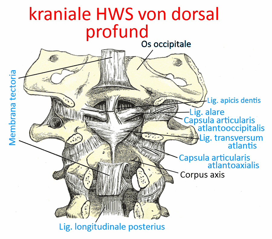
Atlantoaxial ligament
The atlantooccipital ligament consists of the anterior atlantoaxial ligament and the posterior atlantoaxial ligament. These are often also referred to as the anterior atlantoaxial membrane and the posterior atlantoaxial membrane, see there.
Images:
Linkmap: Atlas, Axis from lateral
Linkmap: Cervical spine from ventral
Linkmap: cranial cervical spine from dorsal, superficial
Lig. atlantoaxiale anterius
Synonym for Membrana atlantoaxialis anterior, see there.
Posterior atlantoaxial ligament
Synonym of Membrana atlantoaxialis posterior, see there.
Lig. atlantooccipitale
The atlantooccipital ligament consists of the anterior atlantooccipital ligament and the posterior atlantooccipital ligament. These are often also referred to as the anterior atlantooccipital membrane and the posterior atlantooccipital membrane, see there.
Images:
Linkmap: Atlas, Axis from lateral
Linkmap: Cervical spine from ventral
Linkmap: cranial cervical spine from dorsal, superficial
Lig. atlantooccipitale anterius
Synonymous with Membrana atlantooccipitalis anterior, see there.
Lateral atlantooccipital ligament
The lateral atlantooccipital ligament runs from the lateral edge of the foramen magnum to the posterior arch of the atlas.
Images:
Linkmap: cranial cervical spine from dorsal, superficial
Lig. atlantooccipitale posterius
Synonym of Membrana atlantooccipitalis posterior, see there.
Lig. capitis costae intraarticulare
The capitis costae intraarticular ligament runs from the capitis costae crest through the capsule of the capitis costae articulation to the neighbouring intervertebral disc, thus dividing the costavertebral articulation into two chambers. This ligament is missing in the 1st, 11th and 12th ribs.
Images: (still without)
Lig. capitis costae radiatum
Ligament extending radially from the ventral bone surface of the rib head(caput costae) to the two thoracic vertebrae involved in the costoververtebral joint and the intervertebral disc (discus intervertebralis) between them to secure the costovertebral joint.
Image: linkmap/BWS_von_lateral.png
Linkmap: Costovertebral joint, transverse section
Linkmap: BWS, lateral
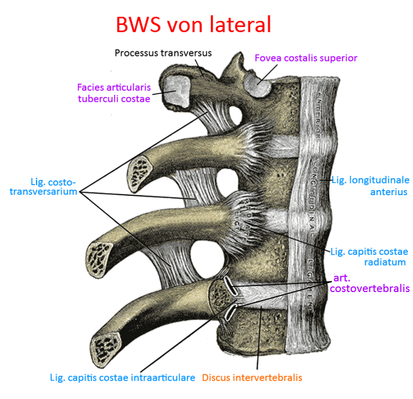
Lig. costotransversarium
Ligament from the rib to the transverse process. There are two parts:
- lateral: from the transverse process to the outside of the rib of the same segment.
- superius: from the processus transversus to the edge of the bone („crista colli costae“) at the upper edge of the rib below.
Pictures: linkmap/BWS_von_lateral.png
Lateral costotransversal ligament
The lig. costotransversarium laterale is a short ligament that runs from the transverse process to the lateral side of the rib of the same segment.
Images:
Linkmap: Costovertebral joint
Linkmap: Costovertebral joint, schematic
Linkmap: Costovertebral joint with spinal canal and ligaments
Linkmap: Costovertebral joint, transverse section
Linkmap: Torso, back, partial view, profound
Linkmap: Torso, thoracic spine, cervical spine, dorsolateral view
Lig. costotransversarium superius (tuberculum costale)
The costal tuberosity ligament runs from the tip of the transverse process of a vertebra to the corresponding costal tuberosity.
Images:
Linkmap: Costovertebral joint
Linkmap: Costovertebral joint from the ventrolateral side
Linkmap: Costovertebral joint, schematic
Linkmap: Costovertebral joint with spinal canal and ligaments
Linkmap: Dorsal view of the thoracic spine and cervical spine
Lig. cruciatum atlantis
The lig. cruciatum atlantis consists of a horizontal part, the lig. transversum atlantis (transverse ligament) and the longitudinal fasciculi longitudinales.
Lig. flavum (interosseous ligament)
Ligament formerly known as the interarcual ligament, which runs through the ventral side of the vertebral arches from the axis to the sacrum. The thickness of these ligaments increases caudally. They have a supporting effect when the spine is lifted into flexion and limit flexion; they lie between the laminae arcuum vertebrarum of neighbouring vertebral arches.
Images: linkmap/LWS_from_lateral_section.gif
Linkmap: Lumbar spine, ligaments
Linkmap: Lumbar spine from lateral
Linkmap: thoracic spine, ventral, profound
Linkmap: cranial cervical spine from dorsal, superficial
Interspinous ligament (interspinosum)
between the spinous processes of neighbouring vertebrae over the entire distance between their base and tip; limit flexion; the strength of the ligaments increases from cervical to caudal. At the tip of the spinous processes they lie against the supraspinous ligament, at the base against the flava ligament.
Images: linkmap/LWS_from_lateral_section.gif
Linkmap: Lumbar spine, ligaments
Linkmap: Trunk, lateral, very deep
Linkmap: Lumbar spine from lateral
Intertransverse ligament
Ligaments between the transverse processes; limit rotation and lateral flexion; they may be absent in the cervical spine; in some cases they are replaced by the intertransverse muscles.
Images: linkmap/Atlas_Axis_from_lateral.png
Linkmap: Lumbar spine, ligaments
Linkmap: Atlas, axis from lateral
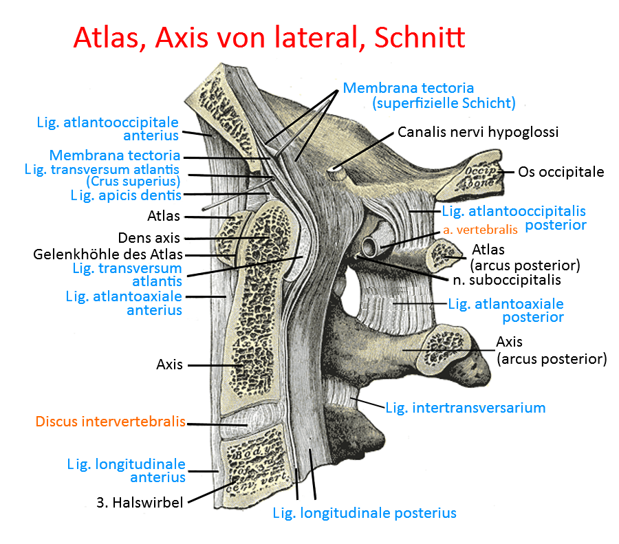
Lig. longitudinale anterius (anterior longitudinal ligament)
Anterior longitudinal ligament over the vertebral bodies and intervertebral discs ventrally from the occipital bone (base) to caudal to S1; limited extension; the fibre tracts are made of firm collagenous connective tissue and radiate into the annuli fibrosi of the intervertebral discs. The anterior longitudinal ligament is wider than the dorsal posterior longitudinal ligament. Together with the posterior longitudinal ligament, supraspinal ligament and nuchal ligament, it forms the long spinal ligaments. Haemorrhage into this ligament (Simon’s haemorrhage, triggered by tensile stress) is a typical sign of death by hanging.
Images: linkmap/BWS_von_lateral.png
Linkmap: Atlas, axis, ligaments, schematic
Linkmap: Costovertebral joint from ventrolateral view
Linkmap: Lumbar spine, ligaments
Linkmap: Trunk, posterior abdominal wall from ventral view
Linkmap: Pelvis, ligaments, ventral view
Linkmap: Lumbar spine from lateral
Linkmap: Atlas, Axis from lateral
Linkmap: Cervical spine from ventral
Linkmap: BWS from lateral
Lig. longitudinale posterius (posterior longitudinal ligament)
posterior longitudinal ligament, lies in the spinal canal on the dorsal side of the vertebral bodies. It lies against these and the intervertebral discs and forms the ventral lining of the spinal canal, while the lig. flavum lies ventrally against the vertebral arches and thus forms the dorsal lining of the spinal canal. It is only loosely connected to the vertebral bodies, but firmly connected to the intervertebral discs. It extends from the axis to the sacrum. The posterior longitudinal ligament limits flexion; together with the anterior longitudinal ligament, supraspinal ligament and nuchal ligament, it forms the long spinal ligaments.
Images: linkmap/ligamentum_longitudinale_posterius.gif
Linkmap: Costovertebral joint
Linkmap: Atlas, axis, ligaments, schematic
Linkmap: Ligaments, atlas and axis
Linkmap: Costovertebral joint with spinal canal and ligaments
Linkmap: Lumbar spine, ligaments
Linkmap: Cervical spine from cranial, profound
Linkmap: Lumbar spine from lateral
Linkmap: Atlax, Axis from lateral
Linkmap: BWS, Lig. longitudinale posterius
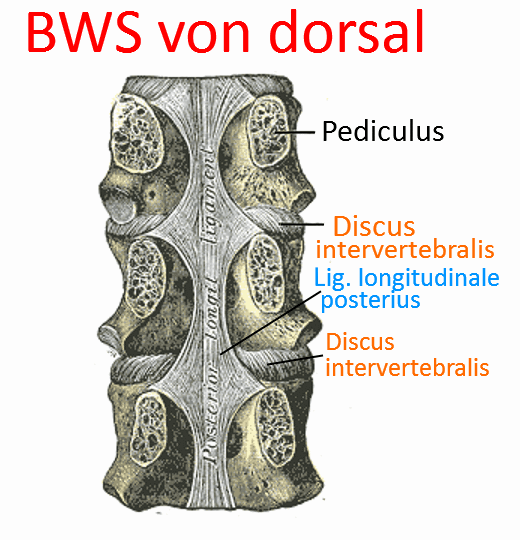
Lumbocostal ligament
The lumbocostal ligament reinforces the thoracolumbar fascia and runs with strong fibres from the 12th rib to the costal processes of L1 and L2.
Images:
Linkmap: Trunk, dorsal, profound
Linkmap: Trunk, lateral, very deep
Lig. nuchae (nuchal ligament)
Cervical ligament, limited flexion; between the external occipital protuberance over the spinous processes of the cervical spine. The lig. nuchae is the cervical continuation of the lig. supraspinale. Together with lig. longitudinale anterius, lig. supraspinale and lig. nuchae, it forms the long spinal ligaments.
Images: linkmap/LWS_von_lateral_Schnitt.gif
Linkmap: Torso, back, partial view, profound
Linkmap: Torso, thoracic spine, cervical spine from dorsolateral view
Linkmap: Torso, lateral, very profound
Linkmap: Dorsal view of thoracic spine and cervical spine
Supraspinous ligament
limited flexion; over the tips of the spinous processes from C7 to the sacrum. Fibrocartilage is formed where it touches the spinous processes. It increases in thickness caudally and is fused with neighbouring fasciae. Together with the anterior longitudinal ligament, posterior longitudinal ligament and nuchal ligament, it forms the long spinal ligaments. This ligament is the origin of the greater rhomboid. In the area of the cervical spine it merges into the nuchae ligament.
Images: linkmap/LWS_from_lateral_section.gif
Linkmap: Atlas, axis, ligaments, diagram
Linkmap: Lumbar spine, ligaments
Linkmap: Trunk, lateral, very deep
Linkmap: Pelvis, ligaments, dorsal
Linkmap: Lumbar spine from lateral
Lig. transversum atlantis (transverse ligament of the first cervical vertebra)
In addition to the longitudinal fasciculi, the transverse atlantis ligament is part of the cruciform atlantis ligament. It connects the two lateral parts of the atlas and can withstand 350 N and can be overstretched by up to 8 mm. In the centre it connects to the longitudinal fasciculi, which run from the ventral edge of the foramen magnum to the dorsal edge of the vertebral body of the axis.
Images: linkmap/cranial_spine_from_dorsal_profund.gif
Linkmap: Atlas, Axis, Ligaments, Schema
Linkmap: Atlantooccipital joint
Linkmap: Cervical spine from cranial, profound
Linkmap: Axis, dorsocranial
Linkmap: Atlas, Axis, lateral
Linkmap: Atlas, cranial
Membrana atlantoaxialis anterior (Lig. atlantoaxiale)
the atlantoaxial membrane is the equivalent of the atlantooccipital membrane in the first cervical segment and runs from the anterior arch (arcus anterior) of the atlas to the anterior edge of the body of the axis.
Images: linkmap/cranial_spine_from_dorsal_superficial.png
Linkmap: Atlas, Axis from lateral
Linkmap: Cervical spine from ventral
Linkmap: Atlas, axis, ligaments, diagram
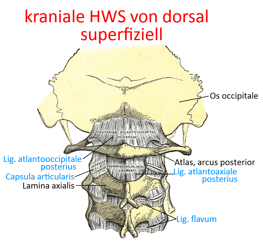
Membrana atlantoaxialis posterior (Lig. atlantoaxiale)
the atlantoaxial membrane is the equivalent of the atlantooccipital membrane in the first cervical segment and runs from the posterior arch (arcus posterior) of the atlas to the upper edge of the laminae of the axis.
Images: linkmap/cranial_spine_from_dorsal_superficial.png
Linkmap: Atlas, Axis, Ligaments, Schema
Membrana atlantooccipitalis anterior (Lig. atlantooccipitale)
The anterior part of the atlantooccipital membrane, which originates ventrally in front of the foramen magnum and extends to the anterior vertebral arch (where the vertebral body is in other vertebrae ).
Image: linkmap/cranial_spine_from_dorsal_superficial.png
Linkmap: Atlas, axis from lateral
Linkmap: Cervical spine from ventral
Membrana atlantooccipitalis posterior (Lig. atlantooccipitale posterior)
The posterior part of the atlantooccipital membrane, which originates behind the foramen magnum and extends to the posterior arch of the atlas.
Image: linkmap/cranial_spine_from_dorsal_superficial.png
Linkmap: Atlas, axis, ligaments, schematic
Membrana tectoria
Ligament in the spinal canal in the region of the upper cervicals and continuation of the posterior longitudinal ligament. It originates on the dorsal side of the body of the second axis and runs to the basal part of the os occipitale. Some fibres connect to the joint capsule of the first upper cervical joint, others on the dorsal side merge into the hard meninges (dura mater) of the brain and still others connect to the transversum atlantis ligament. The membrana tectoria brakes rotational and flexion movements in the area of the upper cervicals and has a holding function.
Images: linkmap/cranial_spine_from_dorsal_profund.gif
Linkmap: Atlas, axis, ligaments, diagram
Linkmap: Cervical spine from cranial, profund
Linkmap: Atlas, Axis, lateral
Costovertebral joint
see under „Vertebral joint“