yoga book / effects
The effects of the postures on the muscles of the movement system are described below. Wherever possible, asanas that achieve this effect are indicated. Click on the map and then on the body region that interests you, or scroll through the effects below. Green numbers in the map stand for stretching, red for strengthening. A ventral and a dorsal view of the human body should make it possible to quickly locate all the important effects.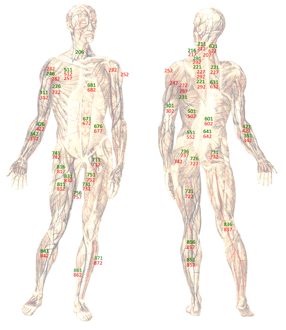
Contents on/off
- 181 Stretching the neck / cervical spine for flexion
- 182 Strengthening the neck / cervical spine for flexion:
- 186 Stretching the neck / cervical spine for reclination
- 187 Strengthening of the neck / cervical spine for reclination:
- 191 Stretching the neck for rotation:
- 192 Strengthening the neck for rotation:
- 196 Stretching the neck for lateral flexion:
- 197 Strengthening the neck for lateral flexion:
- 201 External rotation of the shoulder blade: stretching
- 202 External rotation of shoulder blade: strength
- 206 Internal rotation of shoulder blade: stretch
- 207 Internal rotation of shoulder blade: strength
- 211 Shoulder blade depression: stretching
- 212 Depression shoulder blade: strength
- 216 Elevation shoulder blade: stretch
- 217 Elevation shoulder blade: strength
- 221 Shoulder blade – stretching for protraction/retractors:
- 222 Shoulder blade: strengthening for protraction:
- 226 Shoulder blade: stretching for retraction/protractors:
- 227 Shoulder blade: strengthening for retraction/the retractors:
- 231 Shoulder: stretching for frontal abduction:
- 232 Shoulder: strengthening for frontal abduction:
- 241 Shoulder joint: stretching for frontal adduction:
- 242 Shoulder: strengthening for frontal adduction:
- 246 Shoulder joint: stretching for retroversion:
- 247 Shoulder: strengthening for retroversion/retroversion:
- 251 Shoulder: stretching for lateral abduction
- 252 Shoulder joint: strengthening for lateral abduction
- 256 Shoulder joint: stretching for lateral adduction
- 257 Shoulder: strengthening for lateral adduction/lateral adductors
- 261 Shoulder joint transverse adduction: stretching
- 262 Shoulder joint transverse adduction: force
- 266 Shoulder joint transverse abduction: extension
- 267 Shoulder joint transverse abduction: force
- 271 Shoulder joint: stretching for exorotation
- 272 Shoulder joint: strengthening for exorotation
- 276 Shoulder joint: stretching for final rotation
- 277 Shoulder joint: strengthening for endorotation
- 291 Stretching the trapezius:
- 292 Strengthening the trapezius:
- 301 Stretching the triceps:
- 302 Strengthening the triceps:
- 306 Stretching the biarticular triceps:
- 307 Strengthening of the biarticular triceps
- 311 Stretching the biceps and other arm flexors:
- 312 Strengthening the biceps and other arm flexors:
- 316 Stretching the biceps
- 317 Strengthening the biceps
- 321 Stretching the supinators/to pronate the forearm:
- 322 Strengthening the supinators of the forearm:
- 331 Stretching the pronators/supination of the forearm:
- 332 Strengthening for pronation/pronators of the forearm:
- 341 Stretching the dorsiflexors of the forearm (stretching the extensors):
- 342 Strengthening for dorsiflexion (strengthening the extensors):
- 351 Stretching the palmar flexors:
- 352 Strengthening the palmar flexors:
- 411 Stretching for dorsiflexion in the wrist:
- 412 Strengthening for dorsiflexion of the wrist:
- 416 Stretching for palmar flexion in the wrist:
- 417 Strengthening for palmar flexion in the wrist:
- 421 Stretching the finger flexors (finger flexors):
- 422 Strengthening the finger flexors (finger flexors):
- 423 Cramp/tonus of the finger flexors (finger flexors)
- 426 Stretching the finger extensors (finger extensors):
- 427 Strengthening the finger extensors:
- 428 Tone the finger extensors (finger extensors):
- 501 Stretching the latissimus dorsi:
- 502 Strengthening of the latissimus dorsi:
- 511 Stretching the pectoralis major:
- 512 Strengthening of the pectoralis major:
- 516 Dehnung des Pectoralis minor
- 517 Kräftigung des Pectoralis minor
- 521 Dehnung des Teres major
- 522 Strengthening the teres major
- 526 Stretching the teres major
- 527 Strengthening of the teres major
- 531 Stretching the infraspinatus
- 532 Strengthening of the infraspinatus
- 536 Stretching the supraspinatus
- 537 Strengthening of the supraspinatus
- 541 Stretching the subscapularis
- 542 Strengthening of the subscapularis
- 546 Stretching the rhomboids
- 547 Strengthening the rhomboids
- 551 Stretching the quadratus lumborum:
- 552 Strengthening of the quadratus lumborum:
- 556 Stretching the levator scapulae
- 557 Strengthening of the levator scapulae
- 601 Stretching of the erector spinae:
- 602 Strengthening of the erector spinae:
- 621 Stretching the (muscles in the area of) cervical spine:
- 622 Strengthening the (muscles in the area of) the cervical spine:
- 631 Stretching the (muscles in the area of) the thoracic spine:
- 632 Strengthening the (muscles in the area of) the thoracic spine:
- 641 Stretching the (muscles in the area of) lumbar spine:
- 642 Strengthening the (muscles in the area of) lumbar spine:
- 651 Flexion WS: stretching
- 652 Flexion WS: strengthening
- 656 WS extension: strengthening
- 657 Extension spine: strengthening
- 661 Spine: stretching for rotation
- 662 Spine: strengthening for rotation
- 666: Spine: stretching for lateral flexion
- 667 Spine: strengthening for lateral flexion
- 671 Abdominal muscles: Stretching of the rectus abdominis:
- 672 abdominal muscles: Strengthening the rectus abdominis:
- 676 Stretching the oblique abdominal muscles obliqui abdomini:
- 677 Strengthening the oblique abdominal muscles obliqui abdomini:
- 681 Stretching the intercostal muscles:
- 682 Strengthening the intercostal muscles:
- 711 Stretching the hip flexors:
- 712 Strengthening the hip flexors:
- 721 Stretching the ischiocrural group:
- 722 Strengthening of the ischiocrural group:
- 726 Stretching the short/monoarticular hip extensors:
- 727 Strengthening the short/monoarticular hip extensors:
- 731 Stretching the endorotators of the hip joint:
- 732 Strengthening the end rotators of the hip joint:
- 736 Stretching the exorotators of the hip joint:
- 737 Strengthening the exorotators of the hip joint:
- 741 Stretching the abductors:
- 742 Strengthening the abductors:
- 751 Stretching the adductors:
- 752 Strengthening the adductors:
- 756 Stretching the gracilis:
- 757 Strengthening of the gracilis:
- 811 Stretching the quadriceps:
- 812 Strengthening of the quadriceps:
- 813 Strengthening of the vastus medialis
- 816 Stretching the rectus femoris:
- 817 Strengthening of the rectus femoris
- 821 Stretching the ischiocrural group as exorotators of the knee joint
- 822 Strengthening the ischiocrural group as exorotators of the knee joint
- 826 Stretching the inner ischiocrural group as endorotators of the knee joint:
- 827 Strengthening the inner ischiocrural group as endorotators of the knee joint
- 831 Stretching of the sartorius:
- 832 Strengthening of the sartorius:
- 836 Stretching the popliteus:
- 837 Strengthening the popliteus:
- 841 Stretching the dorsiflexors:
- 842 Strengthening the foot lifters (dorsiflexors):
- 851 Stretching the foot extensors (plantar flexors / calf muscles):
- 852 Strengthening of the foot extensors (plantar flexors / calf muscles):
- 856 Stretching the gastrocnemius
- 857 Strengthening the gastrocnemius
- 861 Stretching the supinators of the ankle joint
- 862 Strengthening the supinators of the ankle:
- 871 Stretching the pronators of the ankle:
- 872 Strengthening the pronators of the ankle:
- 971 Stretching the toe flexors:
- 972 Strengthening the toe flexors:
- 981 Stretching the toe extensors (toe extensors):
- 982 Strengthening the toe extensors (toe extensors):
Contents
- 1 181 Stretching the neck / cervical spine for flexion
- 2 182 Strengthening the neck / cervical spine for flexion:
- 3 186 Stretching the neck / cervical spine for reclination
- 4 187 Strengthening the neck / cervical spine for reclination:
- 5 191 Stretching the neck for rotation:
- 6 192 Strengthening the neck for rotation:
- 7 196 Stretching the neck towards lateral flexion:
- 8 197 Strengthening the neck for lateral flexion:
- 9 201 External rotation of the shoulder blade: stretching
- 10 202 External rotation of the shoulder blade: strength
- 11 206 Internal rotation of the shoulder blade: stretching
- 12 207 Internal rotation of the shoulder blade: strength
- 13 211 Depression scapula: stretching
- 14 212 Depression of the shoulder blade: strength
- 15 216 Elevation of the shoulder blade: stretching
- 16 217 Elevation of the shoulder blade: strength
- 17 221 Scapula – stretching for protraction/the retractors:
- 18 222 Shoulder blade: Strengthening for protraction:
- 19 226 Shoulder blade: stretch for retraction/the protractors:
- 20 227 Shoulder blade: Strengthening for retraction/the retractors:
- 21 231 Shoulder: stretch for frontal abduction:
- 22 232 Shoulder: Strengthening for frontal abduction:
- 23 241 Shoulder joint: stretch for frontal adduction:
- 24 242 Shoulder: Strengthening for frontal adduction:
- 25 246 Shoulder joint: stretching for retroversion:
- 26 247 Shoulder: Strengthening for retroversion/theretrovertors:
- 27 251 Shoulder: stretching for lateral abduction
- 28 252 Shoulder joint: strengthening for lateral abduction
- 29 256 Shoulder joint: stretch for lateral adduction
- 30 257 Shoulder: strengthening for lateral adduction/the lateral adductors
- 31 261 Shoulder joint transverse adduction: stretching
- 32 262 Shoulder joint transverse adduction: strength
- 33 266 Shoulder joint transverse abduction: stretching
- 34 267 Shoulder joint transverse abduction: strength
- 35 271 Shoulder joint: stretching for exorotation
- 36 272 Shoulder joint: strengthening for exorotation
- 37 276 Shoulder joint: stretching for endorotation
- 38 277 Shoulder joint: strengthening for endorotation
- 39 291 Stretching of the trapezius:
- 40 292 Strengthening the trapezius:
- 41 301 Stretching the triceps:
- 42 302 Strengthening the triceps:
- 43 306 Stretching the biarticular triceps:
- 44 307 Strengthening the biarticular triceps
- 45 311 Stretching the biceps and other arm flexors:
- 46 312 Strengthening the biceps and other arm flexors:
- 47 316 Stretching the biceps
- 48 317 Strengthening the biceps
- 49 321 Stretching the supinators/pronating the forearm:
- 50 322 Strengthening the supinators of the forearm:
- 51 331 Stretching the pronators/supination of the forearm:
- 52 332 Strengthening the pronation/pronators of the forearm:
- 53 341 Stretching the dorsiflexors of the forearm (stretching the extensors):
- 54 342 Strengthening for dorsiflexion (strengthening the extensors):
- 55 351 Stretching of the palmar flexors:
- 56 352 Strengthening the palmar flexors:
- 57 411 Stretching for dorsiflexion in the wrist:
- 58 412 Strengthening for dorsiflexion of the wrist:
- 59 416 Stretching for palmar flexion in the wrist:
- 60 417 Strengthening for palmar flexion in the wrist:
- 61 421 Stretching the finger flexors (finger flexors):
- 62 422 Strengthening the finger flexors (finger flexors):
- 63 423 Cramp/tonus of the finger flexors (finger flexors)
- 64 426 Stretching the finger extensors (finger extensors):
- 65 427 Strengthening the finger extensors:
- 66 428 Tonus of the finger extensors (finger extensors):
- 67 501 Stretching the latissimus dorsi:
- 68 502 Strengthening the latissimus dorsi:
- 69 511 Stretching of the pectoralis major:
- 70 512 Strengthening the pectoralis major:
- 71 516 Stretching the pectoralis minor
- 72 517 Strengthening the pectoralis minor
- 73 521 Stretching the teres major
- 74 522 Strengthening the teres major
- 75 526 Stretching of the teres minor
- 76 527 Strengthening the teres minor
- 77 531 Stretching of the infraspinatus
- 78 532 Strengthening the infraspinatus
- 79 536 Stretching the supraspinatus
- 80 537 Strengthening the supraspinatus
- 81 541 Stretching the subscapularis muscle
- 82 542 Strengthening the subscapularis muscle
- 83 546 Stretching the rhomboid muscles
- 84 547 Strengthening the rhomboid muscles
- 85 551 Stretching the quadratus lumborum:
- 86 552 strengthening of the quadratus lumborum:
- 87 556 Stretching the levator scapulae
- 88 557 Strengthening the levator scapulae
- 89 601 Stretching the erector spinae:
- 90 602 Strengthening the erector spinae:
- 91 621 Stretching the (muscles in the) cervical spine:
- 92 622 Strengthening the (muscles in the) cervical spine:
- 93 631 Stretching the (muscles in the area of) the thoracic spine:
- 94 632 Strengthening the (muscles in the area of) the thoracic spine:
- 95 641 Stretching the (muscles in the area of) the lumbar spine:
- 96 642 Strengthening the (muscles in the area of) the lumbar spine:
- 97 651 Flexion WS: stretching
- 98 652 Flexion WS: Strengthening
- 99 656 Extension WS: strengthening
- 100 657 WS extension: strengthening
- 101 661 Spine: stretching for rotation
- 102 662 Spine: strengthening for rotation
- 103 666: Spine: stretching for lateral flexion
- 104 667 Spine: strengthening for lateral flexion
- 105 671 Abdominal muscles: Stretching the rectus abdominis:
- 106 672 Abdominal muscles: Strengthening the rectus abdominis:
- 107 676 Stretching the oblique abdominal muscles obliqui abdomini:
- 108 677 Strengthening the oblique abdominal muscles obliqui abdomini:
- 109 681 Stretching the intercostal muscles:
- 110 682 Strengthening of the intercostal muscles:
- 111 711 Stretching the hip flexors:
- 112 712 Strengthening the hip flexors:
- 113 721 Stretching the ischiocrural group:
- 114 722 Strengthening the ischiocrural group:
- 115 726 Stretching the short/monoarticular hip extensors:
- 116 727 Strengthening the short/monoarticular hip extensors:
- 117 731 Stretching the end rotators of the hip joint:
- 118 732 Kräftigung der Endorotatoren des Hüftgelenks:
- 119 736 Stretching the exorotators of the hip joint:
- 120 737 Strengthening the exorotators of the hip joint:
- 121 741 Stretching the abductors:
- 122 742 Strengthening the abductors:
- 123 751 Stretching the adductors:
- 124 752 Strengthening the adductors:
- 125 756 Stretching of the gracilis:
- 126 757 Strengthening the gracilis:
- 127 811 Stretching the quadriceps:
- 128 812 Strengthening the quadriceps:
- 129 813 Strengthening the vastus medialis
- 130 816 Stretching of the rectus femoris:
- 131 817 Strengthening the rectus femoris
- 132 821 Stretching the ischiocrural group as exorotators of the knee joint
- 133 822 Strengthening the ischiocrural group as exorotators of the knee joint
- 134 826 Stretching of the inner ischiocrural group group as endorotators of the knee joint:
- 135 827 Strengthening the inner ischiocrural group Group as end rotators of the knee joint
- 136 831 Elongation of the sartorius:
- 137 832 Strengthening the sartorius:
- 138 836 Stretching the popliteus:
- 139 837 Strengthening the popliteus:
- 140 841 Stretching the foot lifts (dorsiflexors):
- 141 842 Strengthening the dorsiflexors:
- 142 851 Stretching the foot extensors (plantar flexors / calf muscles):
- 143 852 Strengthening the foot extensors (plantar flexors / calf muscles):
- 144 856 Stretching the gastrocnemius
- 145 857 Strengthening the gastrocnemius muscle
- 146 861 Dehnung der Supinatoren des Fußgelenks
- 147 862 Strengthening the supinators of the ankle:
- 148 866 Stretching of the tibialis posterior
- 149 867 Strengthening the tibialis posterior
- 150 871 Stretching the pronators of the ankle:
- 151 872 Strengthening the pronators of the ankle:
- 152 971 Stretching the toe flexors:
- 153 972 Strengthening the toe flexors:
- 154 981 Stretching the extensors of the toes (toe extensors):
- 155 982 Strengthening the toe extensors (toe extensors):
181 Stretching the neck / cervical spine for flexion
The cervical spine is usually hardly capable of significant flexion, but its physiological lordosis can be reversed. The muscles covering the cervical spine can also be stretched by significantly stretching the thoracic spine, which can significantly contribute to subjectively reducing the tone of the muscles covering the cervical spine. Postures such as karnapidasana, sarvangasana, halasana and setu bandha sarvangasana are suitable for this. Here, the cervical spine lies flat on the floor at best, i.e. in an upright position.
As the cervical spine is a „potential weak point“ in the construction of the human body, a more defensive approach should be taken here with strengthening and stretching, all the more so if it is suspected of being pre-damaged. Specialist clarification should then be sought. This applies even more to stretching than to strengthening. If there is a suspected disc prolapse or disc protrusion in the cervical spine, these stretches are contraindicated.
By design, karnapidasana and halasana as well as shoulder stand are very suitable for this type of stretch. However, in sarvangasana, the extremely large lever arm and the exceptionally large partial body weight mean that even small changes in the angle of the upper body to the supporting floor surface can cause massive changes in the force arm that the cervical spine represents with the head. The structures, especially the muscles, can quickly become overloaded. This applies to the right-angled shoulder stand in a similar way. Although the vertical lever arm is much smaller, there is also a large horizontal lever that must be supported entirely by the cervical spine with the head. the two sarvangasana postures are definitely strengthening and, depending on the flexibility, also more or less clearly stretching.
Karnapidasana is the greater stretch compared to halasana and sarvangasana because the back curves more overall and the pelvis moves beyond the horizontal position of the shoulder. karnapidasana requires even more flexibility than chakrasana, albeit only for the brief moment of transition, as the entire spine including the cervical spine must be bent even further to achieve a sufficiently good gravity plumb line.
When stretching the cervical spine with the head and cervical spine on the floor, it is not uncommon for uncomfortably intense pressure to be applied to the C7, which may require the use of a softness mediator such as a blanket.
Asanas:
See also:
- 182: Strengthening the cervical spine muscles in the direction of flexion
- Movement physiology: flexion of the cervical spine
All postures with the effectiveness indicator 181.
See also the diagnostic notes on strengthening and stretching.
182 Strengthening the neck / cervical spine for flexion:
Strengthening the cervical spine should be approached a little more defensively, especially if the cervical spine is suspected of being pre-damaged. Specialist clarification should then be sought. Strengthening and stretching should be less intensive and more long-term, especially if the geometry of the cervical spine deviates significantly from anatomically zero, i.e. it is clearly flexed or reclined. Strengthening the flexors of the cervical spine, also known as the ventral neck muscles, is primarily achieved in postures in which the face points approximately towards the ceiling and the head is held in a more or less horizontal position against the effect of gravity. This can be done in bridge or in purvottanasana, for example. Alternating between loading and unloading, i.e. lifting and lowering the head according to gravity, also achieves good results. Another way of strengthening is to press your forehead against the floor while lying on your stomach or stand up and press your forehead against the wall. To avoid having to bend the cervical spine significantly due to the disturbing nose, it is advisable to place an object on the floor that is a few centimetres thick, still comfortable and only moderately compressible, such as a shoulder support plate, against which the head is pressed.
When strengthening the ventral cervical musculature, care must be taken to ensure that it is not performed as a head forward posture, i.e. as a translation of the head to ventral in relation to the thoracic spine, as is often seen in people who work on screens with too small a display.
Asanas:
- purvottanasana (when the head is held horizontally)
- urdhva dhanurasana (bridge) (when the head is raised)
- ustrasana (when the head is more or less actively raised)
See also:
- 181: Stretching the cervical spine muscles in the direction of flexion
- Movement physiology: flexion of the cervical spine
All postures with the effect index 182.
See also the diagnostic notes on strengthening and stretching.
186 Stretching the neck / cervical spine for reclination
Stretching in the direction of reclination of the cervical spine is often neglected, which can lead to imbalances in the cervical spine. It may be necessary to start exercising carefully and in small doses, rather than over a longer period of time. A rather rare cervical spondylolisthesis (cervical spondylolisthesis) or cervical spinal canal stenosis generally prohibit any reclination.
If there is no contraindication, purvottanasana in all variations is ideal, especially for the beginning, as the head sinks into reclination from pure gravity and no antagonistic muscles that could possibly develop a tendency to cramp need to be used in addition to the stretched muscles. To a lesser extent, this also applies to urdhva dhanurasana (bridge), especially if the mobility of the shoulder joints is not yet particularly pronounced but the pose can be held for a short time.
Matsyasana is one of the well-known stretches in the direction of reclination, but due to the very short sarcomere length, it can significantly and uncomfortably increase the tonus of the agonistic muscles, possibly even causing nuchal headaches, which is why this exercise is not recommended. In addition to the sarcomere length, this effect is due to the fact that the reclination does not occur in accordance with gravity, but as a result of the work of these same muscles and against gravity.
The head up dog pose and bhujangasana can be performed with reclination of the head. However, care must be taken in both postures to ensure that this does not lead to tension in the performing neck muscles. On the one hand, the tone of these muscles is already increased in some people due to their posture, for example when working at a computer screen. On the other hand, less flexible hip flexors mean a flatter angle of the thoracic spine to the floor and therefore a greater reclination of the cervical spine required to exceed the vertical with the head. The point from which the head sinks further into reclination due to gravity is then significantly shifted towards a shorter sarcomere length, which can quickly lead to excessive strain on the muscles performing the movement and an unpleasant increase in tone.
Stretching the ventral neck muscles to promote the reclination ability of the cervical spine can lead to unexpectedly intense stretching, for example in the platysma or sternocleidomastoid.
Asanas:
- purvottanasana
- urdhva dhanurasana (bridge) (only slightly effective with good flexibility)
See also:
- 187: Strengthening the cervical spine muscles in the direction of reclination
- Movement physiology: reclination
All postures with the effectiveness index 186.
See also the diagnostic notes on strengthening and stretching.
187 Strengthening the neck / cervical spine for reclination:
The strength for reclination essentially comes from the autochthonous neck muscles. Strengthening particularly short sarcomere lengths should be avoided if the muscles become subjectively uncomfortably tight. This can be the case with matsyasana, as already described in key figure 186. Similarly, strengthening under large sarcomere lengths such as in the right-angled shoulder stand is only indicated with caution and only for advanced practitioners, see the derivation in key figure 181. Autochthonous back muscles in the cervical spine and trapezius have the character of „holding“ muscles rather than fast-moving muscles due to their predominant fibre type 1. Due to their course entirely dorsal to the rotation centres of the cervical spine segments, they also have a reclining function.
Holding the head means work for the cervical spine muscles, strength endurance deficiencies and hypertonus can be detected in various postures, as these can often also be used for strengthening. However, this requires careful handling of the cervical spine and its musculature. These postures include, in particular, sarvangasana and right-angled sarvangasana. In these poses, it must be noted that the supported body mass and the lever arm are very large, so that overloading of the cervical spine and its muscles can easily occur. In sarvangasana, it is the immense vertical lever arm, so that even the smallest changes in the angle of the upper body to the floor can represent a massive increase in the load on the cervical spine due to its much shorter lever arm. In the case of right-angled sarvangasana, it is the horizontal lever arm of the legs that is many times greater than the supporting cervical spine. Here, too, the supported partial body weight is very large. Even the smallest changes in the angle of the upper body to the floor or a slight change in the extension of the upper body have a massive effect on the force lever.
Pressing your head against the floor and head against the wall are also good for strengthening. The sarcomere length is quite neutral, which may also reduce excessive tonus. There are fewer risks here than in the first-mentioned postures, which is why they are the exercises of choice for beginners. Later on, the first exercises mentioned can be used for more advanced strengthening.
As explained in 186, the postures head up dog pose and bhujangasana are less unproblematic if they are performed with reclination of the head. This is all the more true for less mobile hip flexors. The benefits and possible side effects are therefore not in a favourable relationship here, especially as other postures achieve this with far fewer side effects and also more effectively.
Asanas:
- Press your head to the floor
- matsyasana (only if the short sarcomere length is tolerated.)
- Right-angled shoulder stand (only recommended for experienced practitioners)
- Press your head to the floor
- Pressing your head on the wall
See also:
- 186: Stretching the cervical spine muscles in the direction of reclination
- Movement physiology: reclination
All postures with the effect index 187.
See also the diagnostic notes on strengthening and stretching.
191 Stretching the neck for rotation:
The rotation of the cervical spine and thus the rotational movement of the head relative to the trunk occurs mainly from the effect of gravity or work of the autochthonous musculature. This relaxes or stretches the contralateral muscles, depending on the intensity of the rotation. If the upper body also performs a rotation, the opposite rotation of the cervical spine can cause a more intensive stretch than the same direction. This applies to many postures, such as jathara parivartanasana, in which the head can largely rotate in the opposite rotation to the rotation of the upper body in accordance with gravity. As the neck muscles are more like postural muscles and are of the corresponding fibre type (1), stretching here is not as quick as with the limb muscles, but requires more frequent and longer action. Excessive force may lead to undesirable effects. In addition, one part of the neck musculature is autochthonous, the other allochthonous. The latter group also includes levator scapulae and trapezius, one of whose tasks is to absorb the gravitational effect of the shoulder blade including the arm. Depending on various factors such as habitual lifting of the shoulder blade due to psychological moments such as tension or fear, these muscles sometimes have greatly increased tonus. They can then react strongly to rotation or lateral flexion with stretching sensations and rapid stretching is not possible.
Asanas:
- Turning pose (parivrtta sukhasana)
- trikonasana
- ardha chandrasana
- parivrtta trikonasana
- parivrtta ardha chandrasana
- parsvakonasana
- parivrtta parsvakonasana
- jathara parivartanasana (largely gravity-induced twist)
- maricyasana 1
- maricyasana 3
- savasana with rotated head (largely gravity-induced rotation)
- ardha vasisthasana
- vasisthasana
- parivrtta uttanasana
See also:
- 192: Strengthening the cervical spine muscles in rotation
- Movement physiology: Rotation of the spine
- Movement physiology: Rotation of the head
All postures with the effect code 191.
See also the diagnostic notes on strengthening and stretching.
192 Strengthening the neck for rotation:
As already described under 191, the two sides of the rotationally active autochthonous musculature are each (partially) antagonistic. In contrast to stretching, the rotatory muscles in the cervical spine rarely succeed in strengthening against gravity, as the effective lever arm is too small, After all, the human body has adapted to walking upright in such a way that the cavity of the head lies in anatomical zero approximately in the centre of the foramen magnum and therefore on the axis of rotation of the head. Without a reclination or flexion of the cervical spine, there would be no lever arm. As the rotatory muscles also serve at least one other dimension of movement, there are also some that get into very short sarcomere lengths during reclination or flexion of the cervical spine and quickly show a tendency to spasm. The rotatory muscles should therefore be strengthened through activity in other dimensions of movement.
Strengthening against the tension of the antagonists, which is not constant but becomes higher the stronger the agonists are used, is favourable. This provides another effective mechanism for strengthening. However, care must be taken to ensure that the antagonists do not become prone to cramping. If there is any sign of a cramp, intervene immediately by reducing the intensity or changing another parameter. If necessary, the posture must be interrupted. In the standing postures listed below, the parivrtta variations generally allow the more pronounced strengthening compared to the utthita variations because the upper body can rotate less far in the direction of the vertical, depending on the mobility.
Asanas:
- Turning pose (parivrtta sukhasana)
- trikonasana
- ardha chandrasana
- parivrtta trikonasana
- parivrtta ardha chandrasana
- parsvakonasana
- parivrtta parsvakonasana
- jathara parivartanasana (largely gravity-induced twist)
- maricyasana 1
- maricyasana 3
- ardha vasisthasana
- vasisthasana
- parivrtta uttanasana
- See also:
- 191: Stretching the cervical spine muscles in rotation
- Movement physiology: Rotation of the spine
- Movement physiology: Rotation of the head
All postures with the effect index 192.
See also the diagnostic notes on strengthening and stretching.
196 Stretching the neck towards lateral flexion:
Stretching the cervical spine in the direction of lateral flexion is best done according to gravity, as some of the antagonists usually work in such short sarcomere lengths during muscle-induced stretching that they begin to spasm. Postures in which the longitudinal axis of the head is inclined between horizontal and about 20/30° to the horizontal and the face looks in a roughly horizontal direction are therefore suitable, as is the case in trikonasana without turning the head. From this position, the head is then lowered according to gravity into a lateral flexion.
Another exercise is to pull a hand on the contralateral side of the head, which gently pulls the head into a maximally tolerable lateral flexion.
Asanas:
- trikonasana (with gravity-orientated lowering of the head)
- ardha chandrasana (when the head is lowered under gravity)
- vasisthasana (when lowering the head according to gravity)
- ardha vasisthasana (when the head is lowered under gravity)
- parsvakonasana (when lowering the head according to gravity)
See also:
- 197: Strengthening the cervical spine muscles in the direction of lateral flexion
- Movement physiology: lateral flexion (side bend)
All postures with the effectiveness indicator 196.
See also the diagnostic notes on strengthening and stretching.
197 Strengthening the neck for lateral flexion:
To strengthen the lateral flexor parts of the autochthonous musculature, the main postures available are those that work with the gravity of the head. This means that the possibility of strengthening is limited and focusses more on strength endurance than maximum strength. Postures in which the head has to be held more or less horizontally against the force of gravity are suitable.
Asanas:
– trikonasana
– ardha chandrasana
– vasisthasana
– ardha vasisthasana
– parsvakonasana
See also:
– 196: Stretching the cervical spine muscles towards lateral flexion
– Movement physiology: lateral flexion (side bend)
All postures with the effectiveness index 197.
See also the diagnostic notes on strengthening and stretching.
201 External rotation of the shoulder blade: stretching
The external rotation of the shoulder blade is a movement that is necessary to move the arm into wide frontal abduction or with exorotation in lateral abduction, whereby the limits of both movements are very similar or even identical at maximum exorotation. In order to exercise stretching in this direction, it therefore makes sense to exert force in the direction of an overhead position of the arm. The main external rotators of the shoulder blade are the trapezius with its three parts and, somewhat less importantly, the serratus anterior. The serratus anterior often has a noticeable tendency to spasm, particularly in the pars descens when elevated against resistance shoulder blade, which is certainly partly due to an increased basic tone due to habitual factors of everyday postures, so that postures with further exorotation of the upper arm from the class of elbow postures (elbow stand, right-angled elbow stand, dog elbow stand, shoulder opening on the chair) may be more favourable: the greater exorotation of the upper arm lateralises the shoulder blade further, shifting the sarcomere length of the trapezius to a slightly less short range.
Among the stretched muscles, the retractors of the shoulder blade that neutralise the external rotation are found in particular: Latissimus dorsi (indirectly), Levator scapulae, Rhomboideus major and Rhomboideus minor, but also pectoralis minor, pectoralis major (indirectly) are stretched. However, some of these muscles can also be stretched well by protraction of the shoulder blade at only 90° abduction instead of in the overhead position of the arm, such as the levator scapulae and the rhomboids: rhomboideus major and rhomboideus minor.
Asanas:
- garudasana
- gomukhasana
- elbow stand
- right-angled elbow stance
- Dog elbow pose
- dvi pada viparita dandasana
- eka pada viparita dandasana
- Shoulder opening on the chair
- Headstand
- Three-point headstand
See also:
- 202: Strengthening the external rotators of the shoulder blade
- 206: Stretching the internal rotators of the shoulder blade
- Movement physiology: protraction of the shoulder blade
- Movement physiology: protractors of the scapula
- Movement physiology: retraction of the scapula (countermovement)
- Movement physiology: retractors of the scapula (antagonists)
202 External rotation of the shoulder blade: strength
The external rotation of the shoulder blade usually occurs with further lateral abduction or frontal abduction of the arm. However, it can also be controlled independently. It is then somewhat reminiscent of trying to „flap your wings“. However, this is not easy to practise, as any effort to move the slightly abducted arms outwards against resistance tends to result in the supraspinatus and deltoid being used. However, if you abduct your arms a little and do not work against resistance, the movement is easier to find. To strengthen the external rotation, it is advisable to abduct the arms as a pure punctum mobile as far as possible in a lateral direction, i.e. to move the elbows overhead as far as possible in a medial direction. If the arms were used as a punctum fixum, for example in a handstand, the stretcher of the elbow joint would be used in the event of significant mobility restrictions when trying to move the elbows towards each other, the triceps, co-operate in the movement and the effort is at least partially kept away from the external rotators. This effect naturally depends on the given mobility and the distance between the hands.
In principle, the external rotation of the shoulder blade can be controlled and practised at will, but this requires a little body awareness. Exercising the movement with force against external resistance, on the other hand, is more difficult as this is only possible up to a small point without a resulting movement of the arm and with this the effort of the lateral abductors often makes differentiation more difficult.
Asanas:
See also
- 222: Strengthening the external rotators of the shoulder blade
- Movement physiology: protraction of the shoulder blade
- Movement physiology: protractors of the scapula
- Movement physiology: retraction of the scapula (counter movement)
- Movement physiology: retractors of the scapula (antagonists)
206 Internal rotation of the shoulder blade: stretching
Stretching in the direction of internal rotation, i.e. stretching the external rotators of the shoulder blade may be necessary, if parts of the trapezius have too much tone for habitual reasons and the shoulder blade can therefore never return to full internal rotation. The muscles responsible for this are the levator scapulae and the rhomboids: rhomboideus major and rhomboideus minor. However, these are also elevators of the shoulder blade. Complaints about tight trapezius muscles are common and are likely to be caused by everyday postural behaviour in which the shoulder blades are held slightly raised or the arms are held slightly raised and twisted in for various activities. Postures and exercises that depress the shoulder blades are helpful in these cases. Exerting force in the direction of lateral adduction and internal rotation is not easy for many people. Therefore, postures that perform these movements together should be practised. The most important internal rotators are the rhomboids, which are also retractors of the shoulder blade.
Asanas:
See also:
- 222: Strengthening the protractors of the shoulder blade
- Movement physiology: Protraction of the shoulder blade
- Movement physiology: protractors of the scapula
- Movement physiology: retraction of the scapula (counter movement)
- Movement physiology: retractors of the scapula (antagonists)
207 Internal rotation of the shoulder blade: strength
In principle, internal rotation of the shoulder blade and external rotation can be controlled and practised at will, but this requires a little body awareness. It is generally possible to perform internal rotation with force against external resistance, if the arms as well as the spine are punctum fixum and the inferior angulus of the scapula is pulled medially.
Of course, the pure powerful lateral adduction of the arm also contributes to this movement.
Asanas:
See also:
- 222: Strengthening the protractors of the shoulder blade
- Movement physiology: Protraction of the shoulder blade
- Movement physiology: protractors of the scapula
- Movement physiology: retraction of the scapula (countermovement)
- Movement physiology: retractors of the scapula (antagonists)
211 Depression scapula: stretching
Stretching that allows further depression of the shoulder blade than gravity causes in upright posture is far less common and usually required to a far lesser extent than stretching that allows further elevation. This is because the gravity of the arms pulls the shoulder blade in anatomically zero and many everyday postures and activities with drooping arms in this direction. Furthermore, unlike in the case of elevation, biarticular muscles such as the latissimus dorsi do not restrict movement. This stretching may be necessary if a person habitually holds the shoulder blades raised, which is not uncommon. In many cases, psychomental moments may be the decisive factor. If you want to improve your ability to depress, all you need to do is hold a dumbbell of an appropriate weight in your hand with your arm hanging down to stretch the elevators levator scapulae and trapezius pars descendens.
Asanas:
See also:
- 222: Strengthening the protractors of the shoulder blade
- Movement physiology: Protraction of the shoulder blade
- Movement physiology: protractors of the scapula
- Movement physiology: retraction of the scapula (countermovement)
- Movement physiology: retractors of the scapula (antagonists)
212 Depression of the shoulder blade: strength
In contrast to stretching in the direction of depression, strengthening in this direction is quite often necessary or useful. This strength is required, for example, when the body is moved relative to the arms, such as in dog head up, in tolasana, in classic dips or in pull-ups. Here, the indirect depressors such as the pectoralis major and the latissimus dorsi and the direct depressors of the shoulder blade such as dorsal the trapezius pars ascendens and ventral the pectoralis minor work quite well from the respective position of the arm. In principle, a very large partial body weight can be moved during self-weight exercises, namely the entire body weight minus that of the arms and shoulder blades with their muscles.
Asanas:
See also:
- 222: Strengthening the protractors of the shoulder blade
- Movement physiology: Protraction of the shoulder blade
- Movement physiology: protractors of the scapula
- Movement physiology: retraction of the scapula (countermovement)
- Movement physiology: retractors of the scapula (antagonists)
216 Elevation of the shoulder blade: stretching
The ability to elevate the shoulder blades when the arm is bent is mainly dependent on the flexibility of the immediate trunkoscauperior depressors, in particular the trapezius, pars ascendens and the pectoralis minor, which pull from the trunk to the shoulder blade and thus only move in the scapulothoracic gliding bearing, when the arm is raised high, but mainly by the indirect depressors, which pull from the trunk via the scapulothoracic gliding joint and the glenohumeral joint to the upper arm, primarily the latissimus dorsi and secondarily the pectoralis major. The scauplohumeral adductors of the upper arm, which pull directly from the shoulder blade to the upper arm, play no role, such as Teres major and Teres minor as well as Infraspinatus or Subscapularis.
In all cases, the position of the arm plays a more or less pronounced role. If the upper arm is raised by at least 90° to lateral or frontal (or mixed), the shoulder blade will have performed an external rotation, the further the abduction, the more. As a result, the muscles connected to the shoulder blade from the trunk are under different tension than without external rotation. The rhomboids, even moderate elevators, would not provide increased resistance to elevation, However, the pars ascendens of the trapezius and caudal fibres of the serratus anterior.
The influence of the position of the arm is much more pronounced in the case of the biarticular (scapulothoracic gliding joint and glenohumeral joint) latissimus dorsi, which acts primarily as a lateral adductor and frontal adductor and light retrovertor of large parts of the dorsal trunk to the upper arm and can therefore represent a very significant restriction against elevation when the arms are raised. The influence of the pectoralis major will be somewhat less inhibitory, as it itself tends to contribute to the first degree of frontal abduction. In most cases where the arm cannot be elevated shoulder blade 180° frontally abducted or (with sufficient exorotation) laterally abducted, the latissimus dorsi should set this restriction, just as if the shoulder blade can no longer be elevated in 180° abduction in one of the two directions, or only to a very limited extent. This becomes even clearer when the restriction of elevation decreases as the wide abduction is left.
This category 216 therefore primarily includes all postures with 180° frontal abduction and elevated shoulder blade, a combination, which can be found in many yoga poses, be it the dog pose head down, the urdhva hastasana, the handstand, the hyperbola, or the bridge.
Asanas:
See also:
- 222: Strengthening the protractors of the shoulder blade
- Movement physiology: Protraction of the shoulder blade
- Movement physiology: protractors of the scapula
- Movement physiology: retraction of the scapula (counter movement)
- Movement physiology: retractors of the scapula (antagonists)
217 Elevation of the shoulder blade: strength
In this section, the same movement is performed as in 371, but with force, i.e. against a greater partial body weight. In the handstand, for example, the aim is to lift a partial body weight, which corresponds to the entire body weight minus the two arms, approximately 1:1 against gravity. The limit of the movement is set soft-elastic, usually by the latissimus dorsi, less frequently by the pectoralis major. As explained above, these two biarticular muscles represent the greater restriction of movement compared to the monoarticular muscles, which move the shoulder blade from the trunk. In terms of posture execution, this means that – if the mobility in the direction of frontal abduction is at least moderately good – an even and barely increasing development of force against gravity occurs first, followed by rapidly increasing forces for further lifting of the body without a firm or hard limit being felt. On the other hand, the evasive movements caused by the three-dimensional mobility of the shoulder joint and the overlying muscles, which set a limit to the movement, are evident, i.e. in the two important biarticular indirect depressors of the shoulder blade, Pectoralis minor and Latissimus dorsi, an outward deviation of the upper arm and a reduction in exorotation.
The most important elevators are the pars descendens of the trapezius, which also performs the frontal abduction or lateral abduction of the arm required for external rotation of the shoulder blade, the two rhomboids, rhomboideus major and rhomboideus minor, the levator scapulae and cranial fibres of the serratus anterior.
In principle, a very large partial body weight can be moved during self-weight exercises, namely the entire body weight minus that of the arms and shoulder blades with their musculature.
Asanas:
- Handstand
- Right-angled handstand
- Elbowstand
- right-angled elbow pose
- urdhva dhanurasana
- urdhva hastasana Back stretch
See also:
- 222: Strengthening the protractors of the shoulder blade
- Movement physiology: Protraction of the shoulder blade
- Movement physiology: protractors of the scapula
- Movement physiology: retraction of the scapula (countermovement)
- Movement physiology: retractors of the scapula (antagonists)
221 Scapula – stretching for protraction/the retractors:
The main muscles involved are serratus anterior (direct) and pectoralis major (indirect, via the upper arm). These muscles are used to stabilise the shoulder blade in all forward-pushing movements, such as the bar. To stretch the antagonistic retractors such as the rhomboids, the garudasana is particularly suitable.
If, for example, garudasana does not work or does not work well because the upper arms do not adduct transversely, this is usually due to the retractors. This posture is then not only diagnostically helpful, but also helps to reduce the restriction. The effect of the posture is based on the fact that the arms are moved so far medially that this is not only possible through transverse adduction of the upper arms in the shoulder joint, but also requires the shoulder blades to be protracted. Due to the course of the levator scapulae, the shoulder blades must be depressed as much as possible for the best possible stretching.
Other ways to promote protraction include postures in which the weight-bearing arms hang vertically downwards with the upper body supported horizontally. The support can be a sufficiently high, narrow surface. A supporting contralateral arm can also be used, as is possible in dumbbell rowing. Constructions with a non-horizontal upper body and without support are also possible, such as upright rowing on machines. If the upper body is not supported on the sternum, this also provides good strengthening of parts of the autochthonous back muscles.
Subsequently, if the retractors are significantly shortened, all postures that lateralise the shoulder blades are suitable.
Asanas:
- garudasana
- elbow stand
- caturkonasana
- dog elbow pose
- Right-angled elbow position
- Shoulder opening at the chair
- Bar position
See also:
- 222: Strengthening the protractors of the shoulder blade
- Movement physiology: Protraction of the shoulder blade
- Movement physiology: protractors of the scapula
- Movement physiology: retraction of the scapula (countermovement)
- Movement physiology: retractors of the scapula (antagonists)
All postures with the action index 221
222 Shoulder blade: Strengthening for protraction:
The protractors serratus anterior (direct) and pectoralis major (indirect, via the upper arm) are used firstly to stabilise the position of the shoulder blade during all forward-pushing movements. Secondly, they are also used for lateralisation of the shoulder blade, for example to support a large partial body weight against a surface, such as in vasisthasana or thirdly without external resistance in 2. warrior pose. Of the three cases mentioned, the greatest strengthening is possible in the first case, followed by the second case. In the last case, no significant strengthening can usually be achieved.
The pectoralis minor also causes protraction, but the direction of pull of the muscle is mainly caudal-cranial, so that it only generates a rather low force for protraction. If you want to strengthen this muscle, the best way to do this is through powerful depression of the shoulder blade.
Asanas:
See also:
- 221: Elongation of the protractors of the shoulder blade
- Movement physiology: protraction of the shoulder blade
- Movement physiology: protractors of the scapula
- Movement physiology: retraction of the scapula (counter movement)
- Movement physiology: retractors of the scapula (antagonists)
All postures with the action index 222
226 Shoulder blade: stretch for retraction/the protractors:
The powerful protracting muscles serratus anterior (directly) and pectoralis major (indirectly, via the upper arm) in particular can limit retraction. In addition to occupational and sporting activities, incorrect posture with protracted shoulder blades often contributes to this. The stretching of the protractors usually takes place with retroverted arms, so that stretching sensations can also occur in the pars clavicularis of the deltoid.
Asanas:
- namaste on the back
- trikonasana, variation: hand on the inner leg
- trikonasana, variation: block in the hand, option: sinking backwards
- purvottanasana
- gomukhasana
See also:
- 227: Strengthening the retractors of the shoulder blade
- Movement physiology: retraction of the shoulder blade
- Movement physiology: retractors of the scapula
- Movement physiology: protraction of the scapula (countermovement)
- Movement physiology: protractors of the scapula (antagonists)
All postures with the effectiveness index 226
227 Shoulder blade: Strengthening for retraction/the retractors:
While the retractors are often used in sports, e.g. for rowing movements or tug-of-war, they are mainly used in asanas to stabilise the shoulder blade, for example to prevent the upper body from tipping sideways in jathara parivartanasana. Because of the large lever that the two legs represent, this is a good way to strengthen the body. The pulling movement on the contralateral knee also offers an opportunity to strengthen the retractors in the twisting pose.
Asanas:
See also:
- 226: Stretching the retractors of the shoulder blade
- Movement physiology: retraction of the shoulder blade
- Movement physiology: retractors of the scapula
- Movement physiology: protraction of the scapula (countermovement)
- Movement physiology: protractors of the scapula (antagonists)
All postures with the action index 227
231 Shoulder: stretch for frontal abduction:
The effect of postures to promote frontal abduction is dependent on the rotational situation of the upper arm: arms that are turned out further are more effective than arms that are turned out less, so the derivations of the elbow stand and the shoulder opening on the chair are the first choice here. This becomes clear when you look at the insertion of the main restricting muscles, both of which are endorotatorsadductors of the shoulder joint: Teres major and Latissimus dorsi.
Asanas:
– gomukhasana
– Hyperbel
– increased back extension
– dog head down
– Dog head down wide
– urdhva dhanurasana
– Handstand
– right-angled handstand
– elbow stand
– dog elbow stand
– right-angled elbow stand
– right-angled headstand
– dvi pada viparita dandasana
– eka pada viparita dandasana
– Shoulder opening on the chair
– caturkonasana
– Headstand
– 1. Warrior pose
– Lying on a roll
– parsvakonasana
– parivrtta parsvakonasana
– back stretch
– Elevated Back Stretch
– supta virasana
– upavista konasana with block
– ustrasana: Arms stretched overhead (urdhva hastasana)
– ustrasana: plank lean back
– parsvautkatasanakonasana
– uttanasana: (P) straighten back
See also:
– 232: Strengthening frontal abduction of the shoulder joint
– Movement physiology: frontal abduction in the shoulder joint
– Movement physiology: frontal adduction in the shoulder joint (countermovement)
– Movement physiology: frontal adductors in the shoulder joint (antagonists)
All postures with the effectiveness index 231
232 Shoulder: Strengthening for frontal abduction:
During frontal abduction, the monoarticular muscles deltoid (pars clavicularis) and coracobrachialis muscles together with the biarticularbiceps. When strengthening, a distinction must be made according to sarcomere length: in short sarcomere length, overhead postures such as handstand, dog head down, postures with exorotated upper arms work even better; In medium sarcomere length, there are only a few postures that work with a greater load than the gravity of the arms, including the three-point headstand. The sarcomere length and therefore particularly valuable is the intensely backward-pressing head up dog pose and the transitions between head up dog pose and head down dog pose and back, which have a unique selling point in the simultaneity of almost complete ROM, the lack of a construction-related limitation of the use of force and the alternating succession of concentric and eccentric contraction. In addition, when strengthening the frontal abductors, the angle in the biarticularbiceps plays an important role due to the biarticular angle. elbow joint and, to a lesser extent, the state of hyperextension of the forearm (pronation/supination).
Asanas:
– Dog pose head down
– Dog pose head down: Dips
– Dog pose head down: Transition to dog head up
– Headstand
– Three-point-Headstand
– Stick pose
– upavista konasana with block
– urdhva dhanurasana (bridge)
– urdhva dhanurasana (bridge): dips
– utkatasana
See also:
– 231: Stretching for frontal abduction of the shoulder joint
– Movement physiology: frontal abduction in the shoulder joint
– Movement physiology: frontal adduction in the shoulder joint (countermovement)
– Movement physiology: frontal adductors in the shoulder joint (antagonists)
All postures with the effectiveness index 232
241 Shoulder joint: stretch for frontal adduction:
Frontal adduction brings the arm from the frontally abducted position back towards anatomically zero, so apart from hypothetical cases of a pathological nature, no special stretching is required to bring the arm into this position. However, if the arm is moved further dorsally, this is a retroversion that requires specific mobility.
Asanas:
See also:
– 242: Strengthening the frontal adduction of the shoulder joint
– Movement physiology: Retroversion in the shoulder joint
– Movement physiology: Retroversion of the shoulder joint
– Movement physiology: frontal adduction in the shoulder joint
– Movement physiology: frontal abduction in the shoulder joint (countermovement)
– Movement physiology: frontal abductors in the shoulder joint (antagonists)
All postures with the effectiveness index 241
242 Shoulder: Strengthening for frontal adduction:
Frontal adduction essentially requires the same muscles that are specified for retroversion, into which it seamlessly transitions for Anatomical zero, but here the pectoralis major is added. Frontal adduction and retroversion are primarily performed by Teres major, Latissimus dorsi, the middle head of the triceps and the posterior head of the deltoid. In the case of the middle head of the triceps, the angle in the elbow joint and the rotation (endorotation/exorotation) in the shoulder joint must be taken into account, because when the elbow joint is more or less extended, the triceps in the retroversion becomes very short sarcomere lengths, which easily causes it to spasm. This is exacerbated by exorotation of the upper arm. The head up dog pose with inverted feet often shows this effect very clearly.
Asanas:
– Dog head down with the hands on a piece of carpet
– uttanasana: Table variation
– Back extension increased when the hands are pressed down forcefully
– tolasana
See also:
– 241: Stretch for frontal adduction of the shoulder joint
– Movement physiology: Retroversion in the shoulder joint
– Movement physiology: Retroversion of the shoulder joint
– Movement physiology: Frontal adduction in the shoulder joint
– Movement physiology: frontal abduction in the shoulder joint (countermovement)
– Movement physiology: frontal abductors in the shoulder joint (antagonists)
All postures with the effectiveness index 242
246 Shoulder joint: stretching for retroversion:
Retroversion is primarily hindered by the anterior part of the deltoid and, if the arm is extended rather than flexed, also by the biceps. In addition, a shortened pectoralis can also limit movement, especially if the arm is exorotated, as the pectoralis endorotates the arm. The other frontal abductor coracobrachialis hardly plays a role in practice as a restraining muscle.
Asanas:
– purvottanasana
– uttanasana with arms behind the body
– prasarita padottanasana with arms behind the back
– gomukhasana
– namaste
– karnapidasana
– halasana
– shoulder stand
– parsvottanasana
– setu bandha sarvangasana
– trikonasana
– uttanasana: Arms behind the body
See also:
– 247: Strengthening the retroversion of the shoulder joint
– Movement physiology: Retroversion in the shoulder joint
– Movement physiology: Retroversion of the shoulder joint
– Movement physiology: frontal adduction in the shoulder joint
– Movement physiology: frontal abduction in the shoulder joint (countermovement)
– Movement physiology: frontal abductors in the shoulder joint (antagonists)
All postures with the effectiveness index 246
247 Shoulder: Strengthening for retroversion/the
retrovertors:
The retroversion is the continuation of the frontal adduction and is performed primarily by Teres Major, Latissimus Dorsi, the middle head of the Triceps and the posterior head of the Deltoideus. In the case of the middle head of the triceps, the angle in the elbow joint and the rotation (endorotation/exorotation) in the shoulder joint must be taken into account, because when the elbow joint is more or less extended, the triceps in the retroversion becomes very short sarcomere lengths, which easily causes it to spasm. This is exacerbated by exorotation of the upper arm. The head up dog pose with the feet upside down often shows this effect very clearly.
Asanas:
– Dog head up with feet upside down
– jathara parivartanasana
– parivrtta trikonasana
– ardha chandrasana
– parivrtta ardha chandrasana
– parivrtta parsvakonasana
– maricyasana 3: Pressing only with the arm against the thigh
See also:
– 246: Stretching for retroversion of the shoulder joint
– Movement physiology: Retroversion in the shoulder joint
– Movement physiology: Retroversion of the shoulder joint
– Movement physiology: frontal adduction in the shoulder joint
– Movement physiology: frontal abduction in the shoulder joint (countermovement)
– Movement physiology: frontal abductors in the shoulder joint (antagonists)
All postures with the effectiveness index 247
251 Shoulder: stretching for lateral abduction
The ability of the upper arm to abduct laterally clearly depends on the rotation of the upper arm: maximum abduction is only possible with more or less complete exorotation. In the endorotated state, the joint structure does not allow lateral abduction that goes much beyond 90°. Any further movement of the upper arm relative to the trunk would then come from the external rotation of the shoulder blade. The 180° lateral abducted position with exorotation is identical to the 180° frontal abducted position. With good mobility, however, further degrees of abduction are possible in both dimensions of movement, meaning that the limit values are not identical. A precise distinction between postures that specifically promote frontal and lateral abduction ability is hardly possible according to the above facts, which is why reference is made here to the list of postures that promote frontal abduction ability under (231).
Asanas: see under 231
See also:
– 252: Strengthening the lateral abduction of the shoulder joint
– Movement physiology: lateral abduction in the shoulder joint
– Movement physiology: lateral adduction in the shoulder joint (countermovement)
All postures with the effectiveness indicator 251
252 Shoulder joint: strengthening for lateral abduction
lateral abduction is primarily performed by the supraspinatus for the first 10-15°, then by the deltoid pars acromialis and weakly by the long head of the biceps, from approx. 70° then from the spinal pars and clavicular pars of the deltoid.
Asanas:
– ardha vasisthasana
– vasisthasana (when the supporting hand is moved away from the feet) (when the supporting hand is pushed away from the feet)
– parsvakonasana (when the supporting hand is pushed away from the feet)
– 2. Warrior stance (rather weaker and in terms of endurance)
See also:
– 251: Stretching for lateral abduction of the shoulder joint
– Movement physiology: lateral abduction in the shoulder joint
– Movement physiology: lateral abductors of the shoulder joint
– Movement physiology: lateral adduction in the shoulder joint (countermovement)
– Movement physiology: lateral adductors of the shoulder joint (antagonists)
– Joint: shoulder joint
All postures with the effectiveness index 252
256 Shoulder joint: stretch for lateral adduction
For this stretch, the arm must be moved significantly and permanently medially, either actively, mainly from force of the pectoralis major, which then tends to develop a tendency to spasm in a very short sarcomere length close to active insufficiency, or passively through the construction of the posture as in garudasana. In this pose, the movement of the arm is due to both the lateral adduction of the arm and the protraction of the shoulder blade.
Asanas:
– garudasana
See also:
– 257: Strengthening the lateral adduction of the shoulder joint
– Movement physiology: lateral adduction in the shoulder joint
– Movement physiology: lateral adductors of the shoulder joint
– Movement physiology: lateral abduction in the shoulder joint (countermovement)
– Movement physiology: Lateral abductors of the shoulder joint (antagonists)
– Joint: Shoulder joint
All postures with the effectiveness index 256
257 Shoulder: strengthening for lateral adduction/the lateral adductors
The lateral adduction (also known as transverse adduction) is mainly performed by the powerful biarticular muscles Latissimus dorsi and Pectoralis major, which originate from the trunk. The shoulder blade muscles Teres major and Teres minor also play a role, also the upper arm muscles biceps with its short head, triceps with its middle head and coracobrachialis. The pars spinalis and pars clavicularis of the deltoid also adduct, while pars acromialis can only abduct. Of the muscles belonging to the so-called rotator cuff, the subscapularis adducts, while the infraspinatus abducts with its cranial parts and adducts with its caudal parts. This means that all strengthening of these muscles promotes the force that can be used for adduction.
If the upper arm is to be adducted beyond neutral zero, a distinction must be made between adduction in front of or behind the trunk. In adduction behind the trunk, the latissimus dorsi and the pars spinalis of the deltoid work well, while the pectoralis major and the pars clavicularis of the deltoid are more restrictive, similarly, the adduction in front of the chest is performed by the pectoralis major and pars clavicularis of the deltoid, while Latissimus dorsi and Pars spinalis of the deltoid tend to be more restrictive.
During lateral adduction, it must be remembered that the retractors and internal rotators of the shoulder blade are also addressed and strengthened in accordance with the use of the scapulohumeral lateral adductors, which hold the shoulder blade in position under the pull of the lateral adductors. This becomes particularly clear if you visualise the upper arm as a punctum fixum and then use these muscles. In practice, trunkohumeral lateral adductors are always used, i.e. Pectoralis major and Latissimus dorsi and scapulohumeral lateral adductors such as Teres major and Teres minor are addressed together, so that the force that the internal rotators and retractors have to exert does not correspond to the total lateral adductor force. If the lateral adduction is performed further medially beyond the vertical position of the upper arm, the above consideration applies.
Asanas:
- ardha vasisthasana (stabilising, rather weaker effect)
- Staff pose
See also:
- 256: Stretching for lateral abduction of the shoulder joint
- Movement physiology: lateral adduction in the shoulder joint
- Movement physiology: Lateral adductors in the shoulder joint
- Movement physiology: lateral adduction in the shoulder joint (countermovement)
- Movement physiology: lateral abductors in the shoulder joint (antagonists)
- Joint: shoulder joint
All postures with the effect index 257
261 Shoulder joint transverse adduction: stretching
A distinction must be made here: transverse adduction can occur as a pure movement in the shoulder joint until the upper arm is exactly in front of the body if the shoulder blade is consciously kept retracted. If you don’t pay attention to this, the shoulder blade will already be slightly retracted beforehand. From the position of the arm exactly in front of the shoulder joint at the latest, any further transverse adduction only occurs from the protraction. The protraction is of course mainly limited by trunkoscapular muscles the retractors, i.e. mainly the rhomboids, but also by the trapezius, very subordinately with depressed shoulder blade also by the levator scapulae. The latissimus dorsi could also be considered as a trunkohumeral muscle, although this is likely to limit protraction shoulder blade, especially in elevated. If it is very limited, it is also conceivable that the approximately transverse cranial part of the latissimus dorsi limits the protraction. As far as movement in the shoulder joint is concerned, it is primarily the posterior part of the deltoid muscle, pars spinalis, that is able to limit transverse adduction regardless of the degree of protraction. In rather rare cases, a restriction by caput longum of the triceps would also be conceivable.
Asanas:
- Dog elbow pose
- Right-angled elbow stance
- Elbow stand
- Shoulder opening on the chair
- garudasana
- transverse adduction_in_lateral position
See also:
- 262: Strengthening the exorotation of the shoulder joint
- Movement physiology: Exorotation in the shoulder joint
- Movement physiology: Exorotators of the shoulder joint
- Movement physiology: endorotation in the shoulder joint (countermovement)
- Movement physiology: end rotators of the shoulder joint (antagonists)
- Joint: shoulder joint
All postures with the effectiveness index 261
262 Shoulder joint transverse adduction: strength
transverse adduction can be made up of two parts, the protraction of the shoulder blades and the movement in the glenohumeral joint. The transverse adductors include first and foremost the scapulohumeral pectoralis major. Its dorsal counterpart as the strongest lateral adductor, the latissimus dorsi, does not belong to the transverse adductors due to its dorsal origin, but the scapulohumeral deltoid with its pars clavicularis does. A typical transveral adduction is the holding work in the bar.
Asanas:
- Handstand when the hands are pushed towards each other
- Dog head up when the hands are pushed towards each other
- Dog head down when the hands are pushed towards each other
- Dog head down: Transition to dog elbow stance when the hands are pushed towards each other
See also:
- 261: Stretching for transverse adduction in the shoulder joint
- Movement physiology: transverse adduction in the shoulder joint
- Movement physiology: transverse adductors of the shoulder joint
- Movement physiology: Transverse abduction in the shoulder joint (countermovement)
- Movement physiology: Transverse abductors of the shoulder joint (antagonists)
- Joint: shoulder joint
All postures with the effect index 262
266 Shoulder joint transverse abduction: stretching
Asanas:
See also:
- 262: Strengthening the exorotation of the shoulder joint
- Movement physiology: transverse abduction in the shoulder joint
- Movement physiology: Transverse abductors of the shoulder joint
- Movement physiology: transverse adduction in the shoulder joint (countermovement)
- Movement physiology: transverse adductors of the shoulder joint (Antagonists)
- Joint: Shoulder joint
All postures with the efficiency index 266
267 Shoulder joint transverse abduction: strength
Asanas:
- jathara parivartanasana
- Handstand (when the hands are pushed away from each other)
- Dog head up (when the hands are pushed away from each other)
- Dog head down (when the hands are pushed away from each other)
See also:
- 266: Stretching for exorotation of the shoulder joint
- Movement physiology: transverse abduction in the shoulder joint
- Movement physiology: transverse abductors of the shoulder joint
- Movement physiology: transverse adduction in the shoulder joint (countermovement)
- Movement physiology: Transverse adductors of the shoulder joint (Antagonists)
- Joint: Shoulder joint
All postures with the effectiveness index 267
271 Shoulder joint: stretching for exorotation
To improve the ability to exorotate in the shoulder joint, the antagonistic endorotators must be stretched. The most powerful of these is the subscapularis, followed by the equally powerful pectoralis major, the Teres major, pars clavicularis of the deltoid and, rather weakly, the biceps. The greatest restrictions on exorotation are to be expected from pectoralis major and, secondarily, teres major, albeit depending on the angles of lateral abduction and frontal abduction. The exorotation ability decreases with increasing frontal abduction and usually also with transverse abduction. As many postures have arms that are rotated out, those that rotate the arms out more powerfully are given first and foremost.
Asanas:
See also:
- 272: Strengthening the exorotation of the shoulder joint
- Movement physiology: Exorotation in the shoulder joint
- Movement physiology: Exorotators of the shoulder joint
- Movement physiology: Endorotation in the shoulder joint (countermovement)
- Movement physiology: endorotators of the shoulder joint (antagonists)
- Joint: Shoulder joint
All postures with the effectiveness indicator 271
272 Shoulder joint: strengthening for exorotation
Although many postures have exorotated arms, there are hardly any asanas that exercise the force of the exorotators during the posture; only when assuming dog elbow pose from dog head down, for example, is the arm exorotated with force , which is also performed by endorotationadductors of the shoulder joint such as the pectoralis major due to the fixed hands.
Asanas:
See also:
- 271: Stretching for exorotation of the shoulder joint
- Movement physiology: Exorotation in the shoulder joint
- Movement physiology: Exorotators of the shoulder joint
- Movement physiology: Endorotation in the shoulder joint (countermovement)
- Movement physiology: end rotators of the shoulder joint (antagonists)
- Joint: shoulder joint
All postures with the effect index 272
276 Shoulder joint: stretching for endorotation
Improving the ability to endorotate is achieved by stretching the muscles that cause exorotation. These are Teres minor, infraspinatus and pars spinalis of the deltoid. They are fewer and also less powerful in total than the endorotators, but sports with throwing movements can lead to an endorotation deficit (GIRD).
Asanas:
– maricyasana 1
– maricyasana 3
– trikonasana with hand on inner leg
– namaste on back
– gomukhasana
See also:
– 277: Strengthening the end rotations of the shoulder joint
– Movement physiology: endorotation in the shoulder joint
– Movement physiology: endorotators of the shoulder joint
– Movement physiology: Exorotation in the shoulder joint (countermovement)
– Movement physiology: exorotators of the shoulder joint (antagonists)
– joint: shoulder joint
All postures with the effectiveness index 281
277 Shoulder joint: strengthening for endorotation
Subscapularis, the equally strong Pectoralis major, the Teres major, pars clavicularis of the Deltoideus and, rather weakly, the Biceps. The greatest restrictions on exorotation are to be expected from pectoralis major and, secondarily, teres major, albeit depending on the angles
Asanas:
– namaste (when performed with force)
– namaste on the back (when performed with force)
See also:
– 276: Stretch for endorotation of the shoulder joint
– 277: Strengthening endorotation of the shoulder joint
– Movement physiology: endorotation in the shoulder joint
– Movement physiology: endorotators of the shoulder joint
– Movement physiology: Exorotation in the shoulder joint (countermovement)
– Movement physiology: exorotators of the shoulder joint (antagonists)
– Joint: Shoulder joint
All postures with the effectiveness index 282
291 Stretching of the trapezius:
Due to its course, the trapezius is less easy to stretch than many other muscles of the movement apparatus. As all 3 parts attach to the spina scapulae (scapular spine), they benefit from the lateralisation or protraction of the depressed (pars descendens) or scapular muscle for stretching. elevated (pars ascendens) shoulder blade. The subjectively greatest need for stretching is regularly in the upper (for the pars descendens) and middle (for the pars transversum) part of the trapezius. If the stretching effect is not sufficient with a maximally lateralisedshoulder blade, additional pressure must be exerted transversely to the muscle path. The pars ascendens of the trapezius is an important shoulder blade depressor, which means that all overhead postures with elevatedshoulder blade, those in which the upper body moves gravity away from fixed hands, for example, stretch it.e.g. increased back extension, hyperbola, significantly more than those in which the pars descendens of the trapezius performs this work, e.g. dog head down, handstand. See also the postures listed under stretching frontal abduction, which stretch the pars ascendens through their elevated shoulder blade.
Asanas:
– Head Side Bend
– karnapidasana on rolls
– Shoulderstand
– halasana
– karnapidasana
– 2. Warrior pose
See also:
– 292: Strengthening the trapezius
– Muscle: Trapezius
– Movement physiology: protraction of the shoulder blade
– Movement physiology: retraction of the shoulder blade
– Movement physiology: Depression of the shoulder blade
– Movement physiology: Elevation of the shoulder blade
– Movement physiology: external rotation of the shoulder blade
– Movement physiology: internal rotation of the shoulder blade
– Bone: shoulder blade
All postures with the effectiveness index 291
292 Strengthening the trapezius:
Pars descendens is strengthened by overhead postures with elevated shoulder blade, preferably against the body’s gravity, such as in handstand. For this part, see the postures listed under frontal abduction of the shoulder joint. Pars ascendens of the trapezius is an important shoulder blade depressor, so all postures that depress the shoulder blade strengthen it. Pars transversa is strengthened in jathara parivartanasana while maintaining protraction of the shoulder blades.
Asanas:
– Dog head up (Pars ascendens)
– Dog head down (Pars descendens)
– tolasana (Pars descendens)
– Handstand (Pars descendens)
– right-angled handstand (pars descendens)
– Elbow Stand (Pars transversa)
– jathara parivartanasana (Pars transversa)
See also:
– 291: Stretching of the trapezius
– Muscle: Trapezius
– Movement physiology: protraction of the shoulder blade
– Movement physiology: retraction of the shoulder blade
– Movement physiology: Depression of the shoulder blade
– Movement physiology: Elevation of the shoulder blade
– Movement physiology: External rotation of the shoulder blade
– Movement physiology: internal rotation of the shoulder blade
– Bone: shoulder blade
All postures with the effect index 292
301 Stretching the triceps:
In the triceps, a distinction must be made between the middle, biarticular part, which also performs the retroversion of the arm, and its two monoarticular neighbouring heads, which only stretch the elbow joint. All are stretched by wide flexion of the elbow joint, but this should not be done under heavy load. A wide frontal abduction in the shoulder joint is also required to stretch the centre of the head. The gmokukhasana is by far the best posture for stretching, which stretches both the elbow joints with complete flexion of the monoarticular heads (both arms) and the biarticular head (upper arm) through very wide frontal abduction.
Asanas:
– gomukhasana
See also:
– 302: Strengthening the triceps
– Movement Physiology: Elbow Joint
– Movement Physiology: Shoulder joint
– Muscle: Triceps
– Muscle: Biceps (most important antagonist)
All postures with the effectiveness index 301
302 Strengthening the triceps:
The triceps are strengthened in all three heads during extension of the elbow joint, whereby the extension should not be performed from a maximally or almost maximally flexed elbow joint. In addition, actively performed retroversion in the shoulder joint strengthens the middle head of the triceps, such as in dog head up with feet upside down. The sustained stabilisation of the three-point headstand also has a strengthening effect.
Asanas:
– Pole
– Dog head down: Transition to rod and back
– Dog head down: dips
– Handstand: dips
– urdhva dhanurasana: dips
– dog head up with feet upside down
– bhujangasana
– jathara parivartanasana
See also:
– 301: Stretching the triceps
– Movement physiology: elbow joint
– Movement physiology: Shoulder joint
– Muscle: Triceps
– Biceps (most important antagonist)
All postures with the effect score 302
306 Stretching the biarticular triceps:
In the triceps, a distinction must be made between the middle, biarticular part, which also performs the retroversion of the arm, and its two neighbouring heads, which only stretch the elbow joint. All are stretched by wide flexion of the elbow joint, but this must not be done under greater load. A frontal abduction in the shoulder is also required to stretch the centre of the head.
Asanas:
– gomukhasana
See also:
– 301: Stretching the triceps
– 307: Strengthening the biarticular triceps
– 302: Strengthening the triceps
– Movement Physiology: Elbow joint
– Movement physiology: Shoulder joint
– Muscle: Triceps
– Biceps (most important antagonist)
All postures with the effectiveness index 306
307 Strengthening the biarticular triceps
In addition to the postures mentioned under 302, the retroversion movement in the shoulder joint is particularly important here. This can generally be performed with the elbow joint flexed, for example with the forearms resting on the floor, but then only the biarticular middle head of the triceps is strengthened. For a more complete strengthening, postures with the elbow joint extended must be selected, such as dog head up with feet upside down. If the arms are turned out, the elbow joint is not mechanically blocked in the plane of the force vector (which would represent a load on the joint) during retroversion, but the monoarticular heads of the triceps must cancel out the flexion moments in the elbow joint caused by the retroversion when the forearm or hands are fixed, which makes them work in a very short sarcomere length. As monoarticular heads, however, they should not have a tendency to spasm, unlike the biarticular head. The smaller the angle of frontal abduction in the shoulder joint and, moreover, the larger the angle of retroversion in the shoulder joint, the greater the tendency to spasm. The triceps are primarily strengthened when the elbow is extended, although the elbow should not be extended with the elbow flexed to the maximum or almost to the maximum. In addition, actively performed retroversion in the shoulder joint strengthens the centre head of the triceps.
Asanas:
– Dog Head Up with Feet Inverted
– bhujangasana
– jathara parivartanasana
See also:
– 307: Strengthening the biarticular triceps
– 301: Stretching the triceps
– 306: Stretching the biarticular triceps
– Movement Physiology: Elbow joint
– Movement physiology: Shoulder joint
– Muscle: triceps
– Muscle: biceps (most important antagonist)
All postures with the effectiveness index 307
311 Stretching the biceps and other arm flexors:
Since the biceps are biarticular in both heads, requires retroversion in the shoulder joint with the elbow joint extended or an extension of the elbow joint in wide retroversion in the shoulder joint. The brachialis is already stretched when the elbow joint is fully extended, but it is fully mobile in most cases, not least because many people spend a significant amount of time with their arms more or less extended. One reason for a shortened brachialis would be, for example, frequent heavy carrying with bent arms. The arm flexor brachioradialis is best stretched when the arm is extended with pronation, i.e. when sleeping on the stomach or back with pronated arms. In the first case, the hands point towards the bed, in the second towards the ceiling.
Asanas:
– uttanasana: Arms behind the body
– prasarita padottanasana: Arms behind the back
– purvottanasana: all variations with arms outstretched
– setu bandha sarvangasana with arms outstretched
See also:
– 312: Strengthening the biceps
– Muscle: biceps
– Muscle: Triceps (most important antagonist)
– Joint: Elbow joint
– Joint: Shoulder joint
All postures with the effectiveness index 311
312 Strengthening the biceps and other arm flexors:
There are few yoga poses for strengthening the biceps, as working against the body’s gravity (without a pull-up bar or similar) requires stretching and no flexing movements of the elbow joint. This means that the only resistance that can be used is your own body, i.e. pulling on the leg or foot in forward bends, as well as using the biceps in its function as an antevertor (frontal abductor).
Asanas:
– Dog head up
– uttanasana with pull on the lower legs
– twisting pose
– Tighten biceps
– Shoulder stand
– setu bandha sarvangasana
– janu sirsasana
– ardha baddha padma pascimottanasana
– pascimottanasana
– tryangamukhaikapada pascimottanasana
– prasarita padottanasana with upraised hands
See also:
– 311: Stretching the biceps
– Muscle: biceps
– Muscle: Triceps (most important antagonist)
– Movement physiology: elbow joint
– Movement physiology: shoulder joint
All poses with the effectiveness index 312
316 Stretching the biceps
Since the biceps is biarticular in both heads, requires retroversion in the shoulder joint with the elbow joint extended or an extension of the elbow joint in wide retroversion in the shoulder joint.
Asanas:
– uttanasana: Arms behind the body
– prasarita padottanasana: Arms behind the back
– purvottanasana: all variations with arms outstretched
– setu bandha sarvangasana with arms outstretched
See also:
– 317: Strengthening the biceps
– Muscle: biceps
– Muscle: Triceps (most important antagonist)
– Movement physiology: Elbow joint
– Movement physiology: Shoulder joint
All postures with the effectiveness index 311
317 Strengthening the biceps
There are few yoga poses for strengthening the biceps, as working against the body’s gravity (without a pull-up bar or similar) requires stretching and no flexing movements of the elbow joint. This means that the only resistance that can be used is your own body, i.e. pulling on the leg or foot when bending forwards, as well as using the biceps in its function as an antevertor (frontal abductor).
Asanas:
– Dog head up
– uttanasana with pull on the lower legs
– twisting pose
– Tighten biceps
– Shoulder stand
– setu bandha sarvangasana
– janu sirsasana
– ardha baddha padma pascimottanasana
– pascimottanasana
– tryangamukhaikapada pascimottanasana
– prasarita padottanasana with upraised hands
See also:
– 316: Stretching the biceps
– Muscle: biceps
– Muscle: Triceps (most important antagonist)
– Movement physiology: elbow joint
– Movement physiology: shoulder joint
All postures with the effect index 317
321 Stretching the supinators/pronating the forearm:
There are no postures in which forceful pronation of the forearm occurs from or with the aid of external forces that would stretch the supinators. The possibilities are limited to the stretch limited by the force exerted by the pronators. Postures such as dog head down, dog head up, handstand, right-angled handstand, dog elbow stand achieve this, urdhva dhanurasana (bridge), vasiasthasana, ardha vasiasthasana and poses that include namaste on the back, such as snamaste on the back.e.g. parsvottanasana. In these poses, the elbow joint is extended and the hand joints are more or less in dorsiflexion.
This is why they have a slightly different effect to those poses in which the
elbow joint is bent, such as elbow stand, right-angled elbow stand, three-point headstand.
Asanas:
– Dog head up
– Dog head down
– Handstand
– right-angled handstand
– dog elbow stand
– elbow stand
– right-angled elbow stance
– urdhva dhanurasana (bridge)
– vasiasthasana
– ardha vasiasthasana
– Three-point headstand
– parsvottanasana
– namaste on the back
See also:
– 322: Strengthening the supinators of the forearm
– Movement physiology: Supination of the forearm
– Movement physiology: Supinators of the forearm (Agonists)
– Movement physiology: pronation of the forearm (countermovement)
– Movement physiology: pronators of the forearm (antagonists)
– joint: wrist
All postures with the effectiveness index 321
322 Strengthening the supinators of the forearm:
Reaching for the lower leg to rotate it and therefore the whole leg in the hip joint allows you to strengthen the supinators in poses such as trikonasana standing against the wall, as part of the upper limb has to work against resistance from the lower limb. The situation is similar in hip opening 4.
The supinators are strengthened in forward bends in which the hands are placed on the floor facing backwards, such as uttanasana 1 or prasarita_padottanasana with hands on top, not by rotating the leg but by keeping pressure of the inner hand on the floor.
Asanas:
– trikonasana standing against the wall
– hip opening 4
– uttanasana 1
– prasarita_padottanasana with hands on top
See also:
– 321: Stretching the supinators of the forearm
– Movement physiology: Supination of the forearm
– Movement physiology: Supinators of the forearm (Agonists)
– Movement physiology: pronation of the forearm (countermovement)
– Movement physiology: pronators of the forearm (antagonists)
– joint: wrist
All postures with the effect index 322
331 Stretching the pronators/supination of the forearm:
A certain degree of stretching of the pronators is achieved with postures such as prasarita padottanasana, uttanasana 1 or other forward bends in which the hands are placed on the floor facing backwards in a supinated position. However, these postures are less effective at stretching the pronato teres, which originates from the medial epiconlylus of the humerus, as the elbow joint is flexed in these postures. If this is to be included, the elbow joint would have to be more or less extended, for example in urdhva dhanurasana (bridge) with inverted hands. In all the postures listed, there is no stretching beyond that which is created and maintained by the hands once they are fixed at the beginning of the posture.
Asanas:
– prasarita padottanasana
– uttanasana 1
– urdhva dhanurasana (bridge) with inverted hands
See also:
– 332: Strengthening the pronators of the forearm
– Movement physiology: Wrist
– Movement physiology: Pronation of the forearm
– Movement physiology: Pronators of the forearm (agonists)
– Movement physiology: Supination of the forearm (countermovement)
– Movement physiology: Supinators of the forearm (antagonists)
– Joint: Wrist joint
All postures with the effectiveness index 331
332 Strengthening the pronation/pronators of the forearm:
Trying to keep the base joints of the index fingers pressed to the floor provides an opportunity to strengthen the pronators in many postures with hands on the floor. Other, non- final strengthening poses are not known except for the variations of the handstand and its derivatives, in which the hands are not turned forwards but to other angles.
Asanas:
– Dog Head Down
– Dog Elbow Stand
– Elbow Stand
– right-angled elbow stand
– right-angled handstand
– handstand with hands placed at different angles
See also:
– 331: Stretching the pronators of the forearm
– Movement physiology: pronation of the forearm
– Movement physiology: pronators of the forearm (agonists)
– Movement physiology: Supination of the forearm (countermovement)
– Movement physiology: supinators of the forearm (antagonists)
– Joint: wrist joint
All postures with the effectiveness index 332
341 Stretching the dorsiflexors of the forearm (stretching the extensors):
The classic way of stretching the dorsiflexors is the dorsal forearm stretch, whereby the closed fists stretch the finger extensors more than the actual (non-finger-moving) dorsiflexors. As a rule, a lot of work is required on the stretching ability of the finger extensors before the actual dorsal flexors can also be stretched in this posture. If the finger extensors are to be bypassed, the fist must be opened. This usually increases the achievable angle of dorsiflexion by more than 10-20° and enables efficient stretching of the actual dorsiflexors. No other postures for stretching the dorsiflexors are known, which is due to the fact that leaning on the back of the hand is neither particularly physiological nor particularly comfortable.
Asanas:
– dorsal forearm stretch with open fist
See also:
– 342: Strengthening the dorsiflexion of the wrist
– Movement physiology: Dorsiflexion of the hand
– Movement physiology: Dorsal flexors of the hand (agonists)
– Movement physiology: Palmar flexion of the hand (countermovement)
– Movement physiology: Palmar flexors of the hand (antagonists)
– Joint: Wrist
All postures with the effectiveness index 341
342 Strengthening for dorsiflexion (strengthening the extensors):
In contrast to the palmar flexors, there is no asana that strengthens the dorsiflexors. For example, the back of the hand would have to be pressed against the floor, another object or a part of the body. At best, moderate strengthening can be achieved by gripping the lower leg with the hand in trikonasana and pulling the lower shoulder forwards with a retroversion movement. This can be achieved much more effectively with functional weight training, for example with bicep curls in the overhand grip. This can also be used as light regenerative training for tennis elbow.
Asanas:
See also:
– 341: Stretching the dorsiflexion of the wrist
– Movement physiology: Dorsiflexion of the hand
– Movement physiology: Dorsiflexors of the hand (Agonists)
– Movement physiology: Palmarflexion of the hand (countermovement)
– Movement physiology: Palmar flexors of the hand (antagonists)
– Joint: Wrist
All postures with the effect index 342
351 Stretching of the palmar flexors:
The palmar flexors are stretched in various postures supported by the arms, such as head-up dog poseright-angled handstandhandstand. In urdhva dhanurasana, the angle of 90° dorsiflexion can also be significantly exceeded depending on shoulder mobility. Another option is namaste on the back, where the angle of 90° dorsiflexion can be exceeded if the hands are held lower than normal.
Asanas:
– head up dog pose
– right-angled handstand
– handstand
– namaste on the back
– forearm stretch palmar in upavista konasana
– forearm stretch palmar
See also:
– 352: Strengthening the palmar flexors of the wrist
– Movement physiology: Palmar flexion of the hand
– Movement physiology: palmarflexors of the hand (agonists)
– Movement physiology: dorsiflexion of the hand (countermovement)
– Movement physiology: Dorsal flexors of the hand (antagonists)
– Joint: Wrist
All postures with the effect index 351
352 Strengthening the palmar flexors:
In addition to all postures supported on the floor and on the wall with a dorsiflexed wrist at around 90°, such as head-up dog pose, right-angled handstand and handstand, in which strength of the dorsal flexors is used to achieve a stable posture on the one hand, but also a certain movement of the shoulder (in the direction of further frontal abduction) on the other, the tolasana is also suitable, in which the balance work is dependent on the .is performed by the palmar flexors. These muscles also work continuously in the three-point headstand to maintain balance.
Asanas:
- Head up dog pose
- Right-angled handstand
- handstand
- tolasana
- Three-point headstand
- Bicep curls in the underhand grip
See also:
– 351: Stretching the palmar flexors of the wrist
– 352: Strengthening the palmar flexors of the wrist
– Movement physiology: Palmar flexion of the hand
– Movement physiology: palmarflexors of the hand (agonists)
– Movement physiology: dorsiflexion of the hand (countermovement)
– Movement physiology: Dorsal flexors of the hand (antagonists)
– Joint: Wrist
All postures with the effect index 352
411 Stretching for dorsiflexion in the wrist:
Here, the wrist palmar flexors together with the finger flexors are boundary-setting. It is not uncommon for the finger flexors (usually the superficial ones) to set the boundary a little earlier than the palmar flexors, which then becomes visible in the proximal finger joints lifting off the floor.
Asanas:
– palmare forearm stretch
– palmare forearm stretch in upavista konasana
– dog head up
– vasisthasana
– ardha vasisthasana
– Handstand
– right-angled handstand
– parsakonasana
– urdhva dhanurasana
– purvottanasana with outstretched arms, both hand positions
– Three-point-headstand
See also:
– 412: Strengthening the dorsiflexors of the wrist
– Movement physiology: Dorsiflexion of the hand
– Movement physiology: Dorsal flexors of the hand (agonists)
– Movement physiology: Palmar flexion of the hand (countermovement)
– Movement physiology: Palmar flexors of the hand (antagonists)
– Joint: Wrist
All postures with the effectiveness index 411
412 Strengthening for dorsiflexion of the wrist:
Dorsiflexors are not clearly strengthened in yoga postures except to stabilise the wrist, e.g. the lower arm in trikonasana, when the upper body is rotated from force of the arm, and in arm balances such as tolasana for balance work. Fist poses also use the dorsiflexors to stabilise the wrist.
Asanas:
– trikonasana
– tolasana
See also:
– 412: Elongation of the dorsiflexors of the wrist
– Movement physiology: Dorsiflexion of the hand
– Movement physiology: Dorsiflexors of the hand (Agonists)
– Movement physiology: Palmarflexion of the hand (countermovement)
– Movement physiology: Palmar flexors of the hand (antagonists)
– Joint: Wrist
All postures with the effect index 412
416 Stretching for palmar flexion in the wrist:
The ability to palmarflexion clearly depends on the state of the fingers: in a flexed state, i.e. with a closed fist, the finger extensors place the greatest restriction on movement in the direction of palmarflexion. If the fingers are more or less extended or at least not actively flexed, the movement is only restricted by muscles that do not move the fingers, such as flexor carpi ulnaris, flexor carpi radialis, palmaris longus. Accordingly, the dorsal forearm stretch is the most important posture for stretching; if you don’t want to stretch the finger flexors, you can simply open your fist.
Asanas: – Dorsal forearm stretch
See also:
– 417: Strengthening the palmar flexors of the wrist –
All postures with the effectiveness index 416
417 Strengthening for palmar flexion in the wrist:
All postures that provide powerful support on the floor are relevant here, and the more balancing character they have, the more so. Strengthen finger flexors and, secondarily, wrist palmar flexors.
Asanas: – palmare forearm stretch strengthens easily – palmare forearm stretch in upavista konasana strengthens easily – dog head up – vasisthasana – ardha vasisthasana – handstand – right-angled handstand – urdhva dhanurasana with both hand positions – purvottanasana with outstretched arms, both hand positions
See also:
– 416: Stretching the palmar flexors of the wrist
All postures with the effectiveness index 417
421 Stretching the finger flexors (finger flexors):
Good stretching of the finger flexors requires the fingers to be stretched as fully as possible plus a wide dorsiflexion of the wrist, as performed by the functional exercises forearm stretch palmar and forearm stretch palmar in upavista konasana in isolation. Any mobility in the finger base joints that exceeds 180° would not be used to stretch the finger flexors for physiological reasons. Firstly, it is far too inconsistent from person to person, so it is impossible to predict its effectiveness and side effects, and secondly, there is a risk of further loosening the ligament structure of the hand. Therefore, if stretched finger joints (proximal and distal) and finger metacarpophalangeal joints are set, the stretching of the finger flexors can only be intensified by further dorsiflexion of the wrist joint. In doing so, 90° should be reached and, if possible, exceeded. In addition to the above postures, the following are also suitable such as handstand, handstand, right-angled handstand, three-point headstand, ardha vasisthasana, tolasana, dog head up. Depending on your shoulder mobility (the lower the more), urdhva dhanurasana (bridge) can also be very effective, as the angle of dorsiflexion must exceed 90° for less mobile shoulder joints.
Asanas: – forearm stretch palmar – forearm stretch palmar in upavista konasana – handstand – right-angled handstand – Three-point headstand – ardha vasisthasana – tolasana – Dog head up – urdhva dhanurasana (bridge)
See also:
– 422: Strengthening the finger flexors
– 423: Tonus of the finger flexors
– Movement physiology: finger flexors / finger flexors
– Movement physiology: finger extensors / finger extensors (antagonists)
All postures with the effectiveness index 421
422 Strengthening the finger flexors (finger flexors):
Finger flexors are mainly strengthened in variations with the fingertips raised. Postures in which pulling movements are performed with flexed fingers, such as the twisting pose or seated forward bends, are also suitable. Of course, the sarcomere length in which the finger flexors work is shorter in the latter, so in the case of increased tonus, other postures should be favoured, as otherwise a further increase in tonus and cramp can easily result.
Asanas: – Dog head down on fingertips – Dog head up on fingertips – Stick on fingertips – Handstand on fingertips – right-angled handstand on fingertips – vasisthasana on fingertips – ardha vasisthasana on fingertips – Turning Seat – janu sirsasana – pascimottanasana – tryangamukhaikapada pascimottanasana – Table variation of uttanasana – Table variation of uttanasana – parivrtta variation of uttanasana – parivrtta trikonasana
See also:
– 421: Stretching the finger flexors
– 423: Tonus of the finger flexors
– Movement physiology: finger flexors / finger flexors
– Movement physiology: finger extensors / finger extensors (antagonists)
All postures with the effect code 422
423 Cramp/tonus of the finger flexors (finger flexors)
The tone of the finger flexors can be increased to such an extent that they tend to cramp even with short-term strain. This occurs in particular when they work in a short sarcomere length, such as in the twisting seat or in seated forward bends in which the outer foot is pulled. Naturally, stretching these muscles reduces tone, but in some cases, postures in which the sarcomere length is moderate also have a positive effect. This includes all postures in which the fingers are stretched and the wrist is in any position (including dorsiflexion).
Asanas: – forearm stretch palmar – forearm stretch palmar in upavista konasana – handstand – right-angled handstand – three-point headstand – vasisthasana – ardha vasisthasana – tolasana – dog head up – urdhva dhanurasana (bridge)
See also:
– 421: Stretching the finger flexors
– 422: Strengthening the finger flexors
– Movement physiology: finger flexors / finger flexors
– Movement physiology: finger extensors / finger extensors (antagonists)
All postures with the effectiveness index 423
426 Stretching the finger extensors (finger extensors):
To stretch the finger extensors, the flexion of the fingers plus palmar flexion of the wrist is required as fully as possible. As there are no postures that support the body weight on the back of a closed fist, the functional exercise dorsal forearm stretch is particularly useful. If the fist is sufficiently closed, the finger flexors are flexed significantly more than the dorsiflexors of the wrist, as opening the fist in the posture and the immediately resulting increased dorsiflexion ability of the wrist quickly shows. The finger extensors are the greatest limitation when the wrist is flexed towards the palmar when the fist is closed.
Asanas: – Dorsal forearm stretch
See also:
– 427: Strengthening the finger extensors
– 428: Tonus of the finger extensors
– Movement physiology: finger extensors / finger extensors
– Movement physiology: finger flexors / finger flexors (antagonists)
All postures with the effectiveness index 426
427 Strengthening the finger extensors:
Strengthening the finger extensors is atypical for yoga poses. There are no postures that open a closed fist against resistance, nor are there any that rest on the back of the hand and use the finger flexors to stabilise the angle in the wrist. Only a trikonasana, for example, in which the hand resting on the lower leg pushes forcefully backwards to move the corresponding shoulder forwards, can contribute a little to strengthening the finger extensors, depending on the grip technique.
Asanas:
See also:
– 426: Stretching the finger extensors
– 428: Tonus of the finger extensors
– Movement physiology: finger extensors / finger extensors
– Movement physiology: finger flexors / finger flexors (antagonists)
All postures with the action index 427
428 Tonus of the finger extensors (finger extensors):
In addition to the stretching exercises listed above, postures with supporting fists with an extended wrist can also moderate excessive tone.
Asanas: – Dog on top of fists – right-angled handstand on fists – Handstand on fists
See also:
– 427: Strengthening the finger extensors
– 426: Stretching the finger extensors
– Movement physiology: finger extensors / finger extensors
– Movement physiology: finger flexors / finger flexors (antagonists)
All postures with the effectiveness index 428
501 Stretching the latissimus dorsi:
The latissimus dorsi is the most important and strongest adductor in the shoulder joint and is often restrictive in frontal abduction, which stretches the latissimus dorsi accordingly. The stretch benefits from the exorotation of the upper arm, as the latissimus dorsi belongs to the humerus muscles due to its attachment to the endorotator muscles. In addition to the classic postures with 180°+ frontal abduction, postures with an inflected spine and a grip on the outer foot of an outstretched leg can also cause a stretch.
Asanas: – gomukhasana – hyperbel – raised back stretch – dog head down wide – urdhva dhanurasana – Handstand – Right Angle Handstand – Elbowstand – Right Angle Elbowstand – dvi pada viparita dandasana – eka pada viparita dandasana – shoulder opening on the chair – janu sirsasana – parsva upavista konasana – parivrtta parsva upavista konasana – parivrtta janu sirsasana
See also:
– 427: Strengthening the latissimus dorsi
– Muscle: latissismus dorsi
– Movement physiology: lateral adduction in the shoulder joint
– Movement physiology: frontal adduction in the shoulder joint
All postures with the effectiveness index 501
502 Strengthening the latissimus dorsi:
Pronounced strengthening of the latissimus dorsi is atypical for yoga poses, but there are certainly opportunities to use strength for this muscle, such as seated or standing forward bends with traction on one or both legs. The pressure of the palms on each other in caturkonasana is also suitable for strengthening the latissimus dorsi, although not in the same way as pull-ups. The variation parsvakonasana with block in hand is more exotic and involves holding a dumbbell instead of a light block. This is also less of a pronounced strength training exercise and more of a strength endurance training exercise, especially as the muscle capacity of the latissimus dorsi is rather low in such a long sarcomere length as in parsvakonasanas.
Asanas: – trikonasana (as lateral flexor and when using the lower arm for adduction) – uttanasana with traction on the lower legs – ardha chandrasana – janu sirsasana – parsva upavista konasana – caturkonasana – parsvakonasana with block in hand
See also:
– 426: Stretching the Latissimus dorsi
– Muscle: Latissismus dorsi
– Movement physiology: lateral adduction in the shoulder joint
– Movement physiology: frontal adduction in the shoulder joint
All postures with the effectiveness index 502
511 Stretching of the pectoralis major:
As one of the most important adductors of the shoulder joint, the pectoralis major is stretched in wide frontal abduction or often even better in wide lateral abduction. As the pectoralis in the shoulder jointendorotates, an exorotated arm clearly supports the effect, while endorotation is often observed as a classic evasive movement. A wide retroversion can also stretch the pectoralis, especially if the arms are exorotated. The functional exercise in which a horizontally outstretched arm presses against a fixed object such as a wall while the body rotates sideways away from it is also well-known. A very similar effect is achieved with a variant of trikonasana, in which an object is held in the upper hand while the arm slowly descends dorsally.
Asanas: – namaste on the back – shoulder opening on the chair – trikonasana: block in the hand – trikonasana: Hand on inner leg – purvottanasana – setu bandha sarvangasana – shoulder stand – uttanasana with arms behind the body – prasarita padottanasana with arms behind the back
See also:
– 512: Strengthening the Pectoralis major
– Muscle: Pectoralis major
– Movement physiology: lateral adduction in the shoulder joint
– Movement physiology: frontal adduction in the shoulder joint
– Movement physiology: endorotation in the shoulder joint
All postures with the effectiveness index 511
512 Strengthening the pectoralis major:
The pectoralis major is strengthened during adducting movements, especially from lateral (i.e. as transverse adduction), e.g. from the retroversion as in the bar position. All movements that push ventrally are therefore classic. In principle, frontally adducting postures are also strengthening, such as dog head up with feet upside down or functional exercises such as pull-ups. The twisting pose strengthens the pectoralis major of the arm whose hand pushes forwards on the floor or presses against the wall.
Asanas: – Stick – Dog head down: Hands on carpet – Turning seat
See also:
– 511: Stretching the pectoralis major
– Muscle: Pectoralis major
– Movement physiology: lateral adduction in the shoulder joint
– Movement physiology: frontal adduction in the shoulder joint
– Movement physiology: endorotation in the shoulder joint
All postures with the effectiveness index 512
516 Stretching the pectoralis minor
Asanas: –
See also:
– 517: Strengthening the Pectoralis minor
– Muscle: Pectoralis minor
– Movement physiology: depression of the shoulder blade
– movement physiology: elevation of the shoulder blade (countermovement)
All postures with the effect index 516
517 Strengthening the pectoralis minor
Asanas: – Dog pose head up – tolasana
See also:
– 516: Stretching the pectoralis minor
– Muscle: pectoralis minor
– Movement physiology: Depression of the shoulder blade
– Movement physiology: Elevation of the shoulder blade (countermovement)
Asanas: –
See also:
–
All postures with the effect number 517
521 Stretching the teres major
The teres major is a scapulohumeral muscle that primarily adducts the upper arm laterally, thereby generating an internal rotation moment. It is therefore stretched by wide lateral abduction with simultaneous external rotation. During these movements, the scapula will be in its maximum external rotation, which begins at approximately 90° lateral abduction. Another important lateral adductor, which simultaneously internally rotates the upper arm, is the truncohumeral latissimus dorsi. The effect of a stretching posture can therefore be altered by the position of the scapula, ranging from greater stretching of the latissimus dorsi when the scapula is elevated scapula and increased stretching of the teres major when the depressed scapula. As the range of motion for lateral abduction and that for frontal abduction is usually roughly the same, movements that end in maximum frontal abduction the teres major very well with those where the scapula is not significantly elevated or, better still, is completely depressed. As there is scarcely any rotational capacity left in the upper arm within the glenohumeral joint when the arm is in the end-range position towards frontal abduction or lateral abduction, there is virtually no rotational capacity left in the upper arm within the glenohumeral joint, so the teres major usually stretches the teres minor as well.
Asanas:
- Shoulder opening on the chair
- Elbow plank
- right-angled elbow stand
- Dog elbow stand
- Two-Legged Viparita Dandasana
See also:
- 522: Strengthening the teres major
- Muscle: Teres major
- Muscle: Teres minor (adductor synergist)
- Exercise physiology: Lateral adduction in the shoulder joint
- Exercise physiology: lateral abduction of the shoulder joint (counter-movement)
- Exercise physiology: Internal rotation of the shoulder joint
- Exercise physiology: external rotation of the shoulder joint (counter-movement)
All positions with performance indicator 521
522 Strengthening the teres major
As an internal rotator and lateral adductor of the glenohumeral joint, the teres major is primarily responsible for lateral adduction of the upper arm, i.e. lateral adduction. In doing so, it is always supported by several synergists, which also generate significant lateral adductive moments, primarily the teres minor and the latissimus dorsi. As the latter is trunkohumeral, it always generates both its lateral adductor and internal rotator moment in the glenohumeral joint as well as a force that depresses the scapula, and a torque in the direction of internal rotation of the scapula. The action of the latissimus dorsi can therefore be broken down into four individual effects; nevertheless, given its overall mass and its wide range of origins, the latissimus dorsi may be the stronger of the two compared to the teres major. Strengthening postures include, for example, the caturkonasana arm posture or postures in which the arms are held close to the upper body whilst the hands are pressed firmly medially.
In contrast to the latissimus dorsi of the upper arm, which acts as a lateral flexor of the spine can be used to counteract the gravity-induced lateral flexion of the upper body (lateral curvature) that usually occurs, the Teres major cannot be used for this purpose, as it is not trunkohumeral but scapulohumeral. The
teres major, on the other hand, can be strengthened by pressing firmly with the hand against the ventral lower leg, in order to reduce the lateral curvature with this arm as well.
In addition to lateral adduction, frontal adduction is also suitable for strengthening the teres major, particularly as this muscle is located dorsally.
Please also refer to the comments in section 257 regarding the relationship between truncohumeral and scapulohumeral muscles and the scapula during lateral adduction.
Asanas:
- ardha vasisthasana, when the supporting hand is pressed firmly towards the feet
- vasisthasana, when the supporting hand is pressed firmly towards the feet
- Covers
- swivel seat
- Dumbbell rows
- Trikonasana, when the lower hand presses firmly against the front of the lower leg.
See also:
- 521: Stretching of the teres major
- Muscle: Teres major
- Muscle: Teres minor (adductor synergist)
- Exercise physiology: Lateral adduction in the shoulder joint
- Exercise physiology: lateral abduction of the shoulder joint (counter-movement)
- Exercise physiology: Internal rotation of the shoulder joint
- Exercise physiology: external rotation of the shoulder joint (counter-movement)
All positions with performance indicator 522
526 Stretching of the teres minor
Since the teres minor is a scapulohumeral internal rotator lateral adductor of the shoulder joint, the best way to stretch it would be to place the arm in a wide lateral abduction or, given the similarity of the threshold, also frontal abduction to rotate inwards or, conversely, to rotate the upper arm inwards and continue to abduct laterally. It is well known, however, that the combination of further abduction and internal rotation is known to cause a unstable shoulder joint to dislocate very easily, so this possibility should be ruled out.
Asanas:
See also:
- 527: Strengthening the teres minor
- Muscle: Teres minor
- Muscle: Teres major (adductor synergist)
- Exercise physiology: Lateral adduction in the shoulder joint
- Exercise physiology: lateral abduction of the shoulder joint (counter-movement)
- Exercise physiology: Internal rotation of the shoulder joint
- Exercise physiology: external rotation of the shoulder joint (counter-movement)
All positions with the performance indicator 526
527 Strengthening the teres minor
As an internal rotator and lateral adductor of the glenohumeral joint, the teres minor is primarily responsible for lateral adduction of the upper arm, i.e. lateral adduction. In doing so, it is always supported by several synergists, which also generate significant lateral adductive moments, primarily the teres major and the latissimus dorsi. As the latter is trunkohumeral, it always generates both its lateral adductor and internal rotator moment in the glenohumeral joint as well as a force that depresses the scapula, and a torque in the direction of internal rotation of the scapula. The action of the latissimus dorsi can thus be broken down into four individual effects; nevertheless, given its overall mass and its wide range of origins, the latissimus dorsi may be the stronger of the two compared to the teres minor. Strengthening postures include, for example, the caturkonasana arm posture or postures in which the arms are held close to the upper body whilst the hands are pressed firmly medially.
Please also note the comments in section 257 regarding the relationship between the truncohumeral and scapulohumeral muscles and the scapula during lateral adduction.
Asanas:
See also:
- 526: Strengthening the teres minor
- Muscle: Teres minor
- Muscle: Teres major (adductor synergist)
- Exercise physiology: Lateral adduction in the shoulder joint
- Exercise physiology: lateral abduction of the shoulder joint (counter-movement)
- Exercise physiology: Internal rotation of the shoulder joint
- Exercise physiology: external rotation of the shoulder joint (counter-movement)
All positions with performance indicator 527
531 Stretching of the infraspinatus
The infraspinatus is part of the rotator cuff and the strongest external rotator of the shoulder joint. This makes it the direct antagonist of the subscapularis, with which together it forms the most important part of the stabilising system for the rotations of the arm. To stretch this muscle, the arm must be rotated inwards significantly. This must not be done with the arm in an overhead position, as an unstable shoulder joint would then be exposed to a high risk of dislocation. Furthermore, the range of motion in the direction of the two rotations overhead is extremely limited anyway. Internal rotation can usefully be practised between approximately 0° and 90° lateral abduction, but also at 90° frontal abduction. One exercise that is simple to perform without any equipment is the infraspinatus stretch. Also in Maricyasana 1, Maricyasana 3 and Namaste on the back, there is a distinct internal rotation which, if insufficient, can also impair the execution of the posture. The same applies to trikonasana with the hand on the inner thigh and malasana with the hands behind the back.
Asanas:
- namaste.htm#on_your_back
- Infraspinatus stretch
- Supine infraspinatus stretch
- Gomukhasana
- Trikonasana: Hand on the inner thigh
- maricyasana 1
- maricyasana 3
See also:
- 532: Strengthening the infraspinatus
- Muscle: Infraspinatus
- Muscle: Subscapularis (Antagonist)
- Exercise physiology: External rotation of the shoulder joint
- Exercise physiology: internal rotation of the shoulder joint (counter-movement)
All positions with performance indicator 531
532 Strengthening the infraspinatus
The infraspinatus is the strongest external rotator of the upper arm in the shoulder joint, its main antagonist being the subscapularis. The latter originates from the ventral surface of the scapula, whilst the infraspinatus originates from the dorsal surface below the spina scapulae. Together, these two muscles form the most important part of the rotator system governing the rotation of the upper arm, which is why both are classified as part of the rotator cuff. The rotational effect of the infraspinatus is stronger when the arm is held close to the torso than when the arm is laterally abducted or frontally abducted, furthermore, the capacity for rotation decreases significantly with increasing abduction in either direction or a combination of both, until it is virtually zero at maximum lateral abduction or frontal abduction. In various asanas, the arms are forcefully rotated outwards, partly against the tension of the powerful internal rotators adductors of the shoulder joint such as the latissimus dorsi, pectoralis major and teres major, partly against the friction of the floor, or against both. In postures such as the Downward-Facing Dog and several others, the exorotation also occurs against the gravity-loaded and powerfully pronated hand-forearm system. Pronation of the forearm and external rotation in the shoulder joint limit each other here. If equipment such as a cable machine, resistance band or dumbbell is available, there are several other ways to strengthen the infraspinatus. In the contralateral side-lying position, with the upper arm resting against the upper body, a dumbbell can be lifted against gravity. Whilst sitting or standing, with the arm in 90° lateral abduction and the elbow joint bent at 90°, a dumbbell can be lifted against gravity. The same exercise also works with 90° frontal abduction. A dumbbell can also be lifted whilst lying on your back at 90° lateral abduction, although, depending on the external rotation capacity of the shoulder joint, not everyone can achieve a starting position close to the floor; furthermore, with the forearm vertical, the gravity-induced torque that must be worked against is already zero. If cable machines or resistance bands are available, it is easier to perform the desired movement, usually over a larger range of motion as well. One option that requires no equipment at all is a kind of negative lift-off test, in which the back of one hand is pressed against the back in the region of the upper lumbar spine. However, this requires the upper arm to remain in its original position, so that a forward movement of the elbow does not result instead of a large exorotatory torque.
Asanas:
See also:
- 531: Stretching the infraspinatus
- Muscle: Infraspinatus
- Muscle: Subscapularis (Antagonist)
- Exercise physiology: External rotation of the shoulder joint
- Exercise physiology: Internal rotation of the shoulder joint (counter-movement)
All positions with the performance indicator 532
536 Stretching the supraspinatus
The supraspinatus should be in a sufficiently stretched state in most people, as can be seen from the position and function of the muscle. If the arm hangs down next to the upper body in an unstretched position, the supraspinatus has a greater sarcomere length. It only makes sense to perform a stretching exercise in cases where there are frequent activities and postures that increase the tension of this muscle due to significant and prolonged lateral abduction, or subjectively an increased feeling of tension, usually associated with pressure pain during palpation. Depending on the tension, it is not enough to place the upper arm directly against the upper body, but a certain lateral adduction must be performed, preferably directly in front of or behind the upper body. To increase the effect, a higher tendon strength/tension can be generated in the muscle, for example by applying a lateral adduction moment in this position, as in the functional exercise Supraspinatus training. A comparable effect can be achieved while standing or sitting with a cable pulley or resistance band that pulls the arm medially.
Asanas:
Siehe auch:
- (537) strengthening the supraspinatus
- muscle: supraspinatus
- lateral adduction in the shoulder joint
- lateral abduction in the shoulder joint
All postures with the effectiveness index 536
537 Strengthening the supraspinatus
The supraspinatus is the initial lateral abductor of the shoulder joint, which is active up to 10° or 15° of lateral abduction, before the pars acromialis and later the pars clavicularis and pars spinalis of the deltoid generate the main lateral abductor moment. Once the arms have been raised a long way, the load on the supraspinatus and therefore the strengthening that takes place is no longer worth mentioning, partly because it is actively insufficient there and partly because it is supported by stronger synergists. Strengthening must therefore be performed to the first degree or even from a lateral adduction. The Supraspinatus exercise, for example, offers this, as do variations of exercises with the hands clasped behind the back when they are pulled forcefully away from each other, as in uttanasana or supraspinatus is often the first muscle of the rotator cuff, the tendon of which becomes damaged with increasing age, possibly as a result of overuse. as a result of overloading, but usually due to a lack of training stimuli. Damage is more likely to occur to the tendon of this muscle than to many others, as its executing tendon is a sliding tendon; it lies against the humeral head, which impairs its supply due to pressure. In addition, if there is insufficient space between the humeral head and acromion, it is often exposed to pressure, which is caused by muscular dysbalances (relative hypertonicity of the deltoid compared to the muscles that caudalise the humeral head) or an abnormal shape of the acromion. Synergists for the initial lateral abduction hardly have the supraspinatus: just the pars acromialis of the deltoid, as it is redirected via the humeral head and, according to recent EMG studies, the upper fibres of the subscapularis.
In addition to the dedicated and largely exclusive response of the supraspinatus, it is also possible to strengthen the supraspinatus in poses in which both hands are shoulder-width apart on the floor without that there is frontal abduction in the shoulder joint, for example in the dog head up, but not in the dog head down when the hands are pressed forcefully outwards (laterally) against the resistance of the floor. Tolasana and bhujangasana are other good examples of this option.
Asanas:
See also:
All postures with the effect code 537
541 Stretching the subscapularis muscle
The subscapularis is the strongest internal rotator of the upper arm in the shoulder joint, its main antagonist being the infraspinatus. The latter originates from the dorsal surface of the scapula below the spina scapulae, whilst the subscapularis originates from the ventral surface. Together, these two muscles form the most important part of the rotator system governing the rotation of the upper arm, which is why both are classified as part of the rotator cuff. The rotational effect of the subscapularis is strongest when the arm is barely abducted; the further it is frontally abducted or laterally abducted, the less torque the subscapularis generates. Consequently, stretching exercises with the arm in an overhead position or when the arm is widely abducted are not very effective. This works best with minimal abduction or with the arm held close to the body. A simple exercise involves lying on your back with the arm resting alongside the body, which, using a dumbbell with the elbow joint bent at 90°, rotates outwards under the influence of gravity. As the subscapularis is also a lateral adductor, positions in which the arm is rotated outwards arm, such as the elbow stand and its variants right-angle a41> is adopted, such as the elbow stand and its variants right-angled elbow stand and dog elbow stand.
Asanas:
See also:
- 542: Strengthening the subscapularis
- Muscle: Subscapularis
- Muscle: Infraspinatus (Antagonist)
- Exercise physiology: Internal rotation of the shoulder joint
- Exercise physiology: external rotation of the shoulder joint (counter-movement)
All positions with performance indicator 541
542 Strengthening the subscapularis muscle
The subscapularis is the strongest internal rotator of the upper arm in the shoulder joint, its main antagonist being the infraspinatus. The latter originates from the dorsal surface of the scapula below the spina scapulae, whilst the subscapularis originates from the ventral surface. Together, these two muscles form the most important part of the rotator system governing the rotation of the upper arm, which is why both are classified as part of the rotator cuff. The rotational effect of the subscapularis is strongest when the arm is barely abducted; the further it is frontally abducted or laterally abducted, the less torque the subscapularis generates. Among the asanas, there are none involving forceful internal rotation of the upper arm. In postures that exceed approximately 90° lateral abduction, this would also carry a risk of dislocation. Internal rotation is therefore only suitable for arms that are more or less close to the body. A good functional exercise is horizontal internal rotation using a resistance band or cable machine. The belly press test is a good way to assess existing strength, but can also be used as a strengthening exercise if no equipment is available. If the internal rotation is not practised with the arms laterally abducted at 90° and the elbow joint bent at 90°, whilst lying on your back, care must be taken to ensure that the scapulae do not begin to protrude or elevate in the process, which frequently occurs, particularly due to carelessness or insufficient strength of the internal rotators. As regards the range of motion, this exercise is very beneficial because it primarily works with greater sarcomere lengths.
Asanas:
See also:
- 541: Stretching of the subscapularis
- Muscle: Subscapularis
- Muscle: Infraspinatus (Antagonist)
- Exercise physiology: Internal rotation of the shoulder joint
- Exercise physiology: external rotation of the shoulder joint (counter-movement)
All positions with the performance indicator 542
546 Stretching the rhomboid muscles
The two rhomboid muscles rhomboideus major and rhomboideus minor are the retractors of the scapula situated between the medial margin of the scapula and the spine. Their course from the medial margin laterally to the medially and further cranially situated spinous processes makes them not only retractors, but also to antagonists of the external rotation of the scapula, which the scapula must perform to allow for a wide abduction of the arm. Furthermore, due to their course, they tend to act as elevators rather than depressors, but are not usually counted among the elevators. To stretch these muscles, protraction of the scapula is particularly important; depression is of secondary importance. Both movements can be found, for example, in garudasana. With regard to protrusion and retraction, it should be borne in mind that they are largely independent of the movement of the upper arm in the glenohumeral joint, however, very wide retraction does not permit very wide transverse adduction of the arm, as a residual movement of the upper arm towards medial originates from the protraction of the scapula and vice versa; very wide transverse abduction is only possible with a retracted scapula. The garudasana is characterised by a specific way of crossing the arms in front of the body, which ensures both movements: protraction and depression of the scapula. However, there must also be sufficient external rotation of the upper arm in the shoulder joint, otherwise one hand cannot reach the other. Another, simpler exercise for protraction without any aids involves using one arm to pull the other, which is abducted 90° forward, as far as possible into transverse adduction. In this exercise, both movements are performed to full range of motion: that of the upper arm in the shoulder joint and that of the scapula in the scapulothoracic gliding joint.
Asanas:
See also:
- 547: Strengthening the rhomboids
- Exercise physiology: Rhomboids
- Muscle: Rhomboid major
- Muscle: Rhomboideus minor
- Muscle: Anterior serratus (Antagonist)
- Exercise physiology: Retraction of the scapula
- Exercise physiology: protraction of the scapula (counter-movement)
All positions with the performance indicator 546
547 Strengthening the rhomboid muscles
The two rhomboid muscles rhomboideus major and rhomboideus minor are the retractors of the scapula situated between the medial margin of the scapula and the spine. Their course from the medial margin laterally to the medially and further cranially situated spinous processes makes them not only retractors, but also to antagonists of the external rotation of the scapula, which the scapula must perform to allow for a wide abduction of the arm. Furthermore, due to their course, they tend to act as elevators rather than depressors, but are not usually counted among the elevators. To strengthen these muscles, the scapula must be forcefully retracted or resist a protruding force, such as that involved in a strong forward pull of the arms, for example when rowing or in the jathara parivartanasana.
Asanas:
See also:
- 546: Stretching of the rhomboid muscles
- Exercise physiology: Rhomboids
- Muscle: Rhomboid major
- Muscle: Rhomboideus minor
- Muscle: Anterior serratus (Antagonist)
- Exercise physiology: Retraction of the scapula
- Exercise physiology: protraction of the scapula (counter-movement)
All positions with the performance indicator 547
551 Stretching the quadratus lumborum:
As the quadratus lumborum pulls from the pelvic crest dorsally to the medial 12th rib, it easily supports backbends. Rib, it easily supports backbends, i.e. is stretched by WS-flexion, especially if they have a rotational component such as parsva upavista konasana. Furthermore, trunk side bends in particular are a good way to stretch it, such as parivrtta parsva upavista konasana or sitting trunk side bend.
Asanas: – Sitting Torso Side Bend – parsva upavista konasana (through the pelvic position slanting towards the front leg) – janu sirsasana – parivrtta parsva upavista konasana – parivrtta janu sirsasana – karnapidasana – parsva karnapidasana – Roll up your back
Siehe auch:
• 552: Kräftigung des Quadratus lumborum
• Bewegungsphysiologie: Lateralflexion der WS
• Bewegungsphysiologie: Extension der WS
• Bewegungsphysiologie: Flexion der WS (Gegenbewegung)
Alle Haltungen mit der Wirkungskennzahl 551
552 strengthening of the quadratus lumborum:
The quadratus lumborum is involved in maintaining the extension of the back and in extending or reducing its flexion, for example by strengthening it when assuming and exiting the 3. Warrior pose backwards against the wall3. Warrior pose, cross lift, utkatasana, right-angled uttanasana, salabhasana, urdhva dhanurasana. It is also strengthened by postures that prevent the body from falling into lateral flexion due to gravity, such as trikonasana, ardha chandrasana, ardha vasisthasana, vasisthasana.
Asanas: – 1. warrior pose: transition to parsvottanasana – 1. warrior pose: transition to hip opening 1 – 1. Warrior pose: transition to hip opening 5 – halasana – 3. Warrior pose backwards against the wall – 3. Warrior pose – cross lift – utkatasana – right-angled uttanasana – salabhasana – urdhva dhanurasana – trikonasana – ardha chandrasana – ardha vasisthasana – vasisthasana
See also:
– 551: Stretching the quadratus lumborum
– Movement physiology: lateral flexion of the WS
– Movement physiology: extension of the WS
– Movement physiology: flexion of the WS (countermovement)
All postures with the effect index 552
556 Stretching the levator scapulae
As the levator scapulae pulls down from its origin at the cervical spine to the upper part of the margo medialis, maximum stretch is achieved by shoulder blade depression with simultaneous protraction. The latter pulls the shoulder blades on the dorsal thorax in an arc forwards and outwards, the former is a prerequisite for achieving elongation, as the levator scapulae extends primarily longitually, i.e. from cranial to caudal. Its main function is to raise the shoulder blades, which is why many people who keep them slightly raised in everyday life for endogenous reasons, not only suffer from tension or hypertonicity of the most important elevator, the trapscius pars descendens, but also from increased tonus in the levator scapulae. The garudasana is the exercise with the best effect for stretching the levator scapulae. In addition, external pressure can be applied to the muscle in the form of a pressure massage.
Asanas:
See also:
Movement physiology: Depression of the shoulder blade
557: Strengthening the Levator scapulae
Muskel: Levator scapulae
557 Strengthening the levator scapulae
The leaver scapulae is the most powerful shoulder blade elevator after the trapsius pars descendens, so to strengthen it, the shoulder blade must be lifted against significant resistance. This can be the gravity of a large partial body weight as in the handstand or rectangular handstand, the elbow stand and the rectangular elbow stand and, to a lesser extent, the dog head down, but it is also possible to perform them with the body upright by raising the shoulder blades while holding the weights with the hands. The levator scapulae works in the aforementioned inverted postures due to the elevation of the shoulder blades in the shortest sarcomere length, which is not recommended in the presence of hypertonicity. Instead, the dynamic variants described as shrugging are suitable.
Asanas: –
Handstand
Rectangular handstand
Elbow stand
Rectangular elbow position
Dog position head down
See also:
Movement physiology: Depression of the shoulder blade
556: Stretching of the levator scapulae
Muskel: Levator scapulae
601 Stretching the erector spinae:
The erector spinae or back extensor is a colloquial term for the autochthonous back muscles, the developmentally oldest and „actual“ back muscles in the sense that they control the spinal column. All other back muscles are muscles that have migrated from the extremities. The functions of the autochthonous back muscles are extension of the spine, rotation of the spine, lateral flexion of the spine. This means that the term stretching of the autochthonous back muscles must refer to one or more of their functions or movement dimensions. Stretching can take place when a movement dimension is utilised as far as possible or the requirement results from a wide movement in more than one movement dimension. With regard to rotation and lateral flexion of the spine, the contralateral muscles are (partial) antagonists. The extension of the spine has no antagonists in the autochthonous back muscles. Stretching can therefore result, for example, from a maximum flexion of the spine as in karnapidasana or from a combination of two dimensions of movement as in . dimensions of movement such as further flexion plus rotation or further flexion plus lateral flexion. With wide lateral flexion, as in sitting trunk side bend, the oblique abdominal muscles often set a limit rather than the autochthonous back muscles. They also tend to limit rotation.
Asanas: – karnapidasana (relating to flexion) – malasana forward bend (relating to flexion) – parsva upavista konasana – Sitting Torso Side Bend (Related to lateral flexion) – Turning seat (Regarding rotation)
See also:
– 602: Strengthening the autochthonous back muscles
– Movement physiology: autochthonous back muscles (also: erector spinae, back extensor)
– Movement physiology: extension of the WS
– Movement physiology: rotation of the WS
– Movement physiology: lateral flexion of the WS
– Movement physiology: flexion of the WS (countermovement)
Alle Haltungen mit der Wirkungskennzahl 601
602 Strengthening the erector spinae:
In line with the explanations under 601, we must differentiate between the strengthening of the mainly extensor, mainly lateral flexor and mainly rotator muscles, although their functions mostly overlap. The extensor component is strengthened through active backbends and holding the upper body or lifting the upper body with the back pointing upwards against gravity, such as in the right-angled uttanasana or the 3rd warrior pose backwards against the wall.
Asanas: – Cross Lift – 3rd Warrior Pose – 3rd Warrior Pose. Warrior pose backwards against the wall – uttanasana: table variation – right-angled uttanasana – salabhasana – handstand: dvi pada variation – trikonasana – ardha chandrasana – shoulder stand right-angled – headstand right-angled (moderately strengthening) – headstand at right angles – utkatasana – halasana – sarvangasana – urdhva_dhanurasana
See also:
– Movement physiology: autochthonous back muscles
– 601: Stretching the autochthonous back muscles
– Exercise physiology: autochthonous back muscles (also: erector spinae, back extensor)
– Exercise physiology: Extension of the WS
– Movement physiology: Rotation of the WS
– Movement physiology: lateral flexion of the WS
– Movement physiology: flexion of the WS (countermovement)
All postures with the effect index 602
621 Stretching the (muscles in the) cervical spine:
The stretching of the neck and of the cervical spine muscles in the various dimensions of movement has already been described under
181: Stretching for flexion (flexion),
186: Stretch to reclination (extension),
191: Stretch to rotation,
196: Stretch to lateral flexion have been discussed.
Asanas: – karnapidasana – Shoulder stand – halasana – setu bandha sarvangasana
See also:
– 622: Strengthening the autochthonous muscles in the cervical spine
– Movement physiology: autochthonous back muscles (also: erector spinae, back extensor)
All postures with the effectiveness index 621
622 Strengthening the (muscles in the) cervical spine:
Strengthening the neck and cervical spine muscles of the cervical spine muscles in the various dimensions of movement has already been described under
182: Strengthening for flexion (flexion),
187: Strengthening for reclination (extension),
192: Strengthening for rotation
197: Strengthening for lateral flexion have been discussed.
Asanas: – Pressing the head to the floor – Right-angled shoulder stand
See also:
– 621: Stretching the autochthonous muscles in the cervical spine
– Movement physiology: autochthonous back muscles (also: erector spinae, back extensor)
All postures with the effectiveness index 622
631 Stretching the (muscles in the area of) the thoracic spine:
When stretching the muscles in the area of the thoracic spine, the three dimensions of movement in which the spine can move must again be taken into account: 1. flexion / extension, 2. rotation and 3. lateral flexion. Most aspects have already been discussed in 601. Especially the (slanted and straight) play a major role here.
Asanas: – karnapidasana
See also:
– 632: Strengthening the autochthonous muscles in the thoracic spine
– Movement physiology: autochthonous back muscles (also: erector spinae, back extensor)
All postures with the effectiveness index 631
632 Strengthening the (muscles in the area of) the thoracic spine:
When strengthening the muscles in the area of the thoracic spine, the three dimensions of movement in which the spine can be moved must again be taken into account: 1. flexion / extension, 2. rotation and 3. lateral flexion. Most aspects have already been discussed in 602. Especially the (slanted and straight) play a major role here.
Asanas: – Rectangular uttanasana – 3. Warrior pose – Front raise – halasana – Cross raise
See also:
– 631: Stretching the autochthonous muscles in the thoracic spine
– Movement physiology: autochthonous back muscles (also: erector spinae, back extensor)
All postures with the effectiveness index 632
641 Stretching the (muscles in the area of) the lumbar spine:
When stretching the muscles in the lumbar spine region, the three dimensions of movement in which the spine can be moved must again be taken into account: 1. flexion / extension, 2. rotation and 3. lateral flexion. Most aspects have already been discussed in 601. In the lumbar spine, some muscles come into play that play no role in the thoracic and cervical spine, such as the quadratus lumburum and the psoas major. The abdominal muscles relevant to the thoracic spine (oblique and straight) also play a role here.
Asanas: – Back Roll – janu sirsasana – pascimottanasana – tryangamukhaikapada pascimottanasana – parsva uttanasana – parsva upavista konasana – parivrtta janu sirsasana – parivrtta parsva upavista konasana – karnapidasana
See also:
– 642: Strengthening the autochthonous muscles in the lumbar spine
– Movement physiology: autochthonous back muscles (also: erector spinae, back extensor)
All postures with the effectiveness index 641
642 Strengthening the (muscles in the area of) the lumbar spine:
When strengthening the muscles in the lumbar spine area, the three dimensions of movement in which the spine can be moved must again be taken into account: 1. flexion / extension, 2. rotation and 3. lateral flexion. Most aspects have already been discussed in 601. Some muscles that do not play a role in the thoracic and cervical spine, such as the quadratus lumburum and the psoas major, come into play in the lumbar spine. The abdominal muscles (oblique and straight) relevant to the thoracic spine also play a role here. The autochthonous muscles of the lumbar spine are well strengthened when they have to hold a roughly horizontal and stretched upper body with stationary legs, such as in the 3rd warrior position, but also when the upper body is stationary and roughly horizontal legs have to be held, such as in the right-angled headstand. A third option is to generate bending moments at both ends of the spine from mobility restrictions in the cervical spine/upper thoracic spine and the back of the legs, which must be stretched against as in shoulder stand and halasana.
Asanas: – 3. warrior pose – 3. Warrior pose backwards against the wall – right-angled uttanasana – cross lift – salabhasana – right-angled shoulder stand – right-angled headstand – – dvi pada variation of handstand
See also:
– 641: Stretching the autochthonous muscles in the lumbar spine
– Movement physiology: autochthonous back muscles (also: erector spinae, back extensor)
All postures with the effectiveness index 642
651 Flexion WS: stretching
652 Flexion WS: Strengthening
656 Extension WS: strengthening
657 WS extension: strengthening
661 Spine: stretching for rotation
The ability to rotate is primarily restricted by the oblique abdominal muscles and the autochthonous back muscles, and to a lesser extent by other muscles such as the quadratus lumborum. These are stretched in twisting postures in which other parts of the body or gravitytorques are generated in the vertebral segments. The force for this can come from the rotatory muscles themselves, the upper or lower extremities, both by actively using them and by using their gravity, such as in jathara parivartanasana.
Asanas: – twisting pose – maricyasana 1 – maricyasana 3 – jathra parivartanasana – parivrtta uttanasana – parivrtta trikonasana – parivrtta ardha chandrasana – twisted back stretch – parivrtta janu sirsasana
See also:
– 662: Strengthening the rotatory part of the autochthonous musculature
– Movement physiology: rotation of the WS
– Movement physiology: autochthonous back muscles (also: erector spinae, back extensors)
– Movement physiology: oblique abdominal muscles (agonists and antagonists)
All postures with the effect code 661
662 Spine: strengthening for rotation
The rotatory muscles are strengthened in any posture that actively encourages rotation of the upper body and not just from the force of the extremities. Strengthening also takes place in jathra parivartanasana, but in a very short or very long sarcomere length, and also purely through the use of gravity of the legs. In many other classic seated and standing twisting postures, the rotation of the upper body is supported by the upper limb. If you are primarily looking to strengthen the rotator muscles, the use of strength in the arms should be limited.
Asanas: – jathra parivartanasana – trikonasana – parsvakonasana
See also:
– 661: Stretching the rotatory part of the autochthonous musculature – Movement physiology: Rotation of the WS
– Movement physiology: autochthonous back muscles, also: erector spinae, back extensors (agonists)
– Movement physiology: oblique abdominal muscles (agonists and antagonists)
All postures with the effect index 662
666: Spine: stretching for lateral flexion
The lateral flexor muscles of the spine are stretched in every posture that actively (from force of the contralateral lateral flexor muscles) or passively (gravitationally or through external influence such as a supporter) brings the spine into a pronounced lateral flexion. There are very few asanas for this. In addition to parighasana, which has not yet been described, the sitting side bend does this very simply and effectively. This stretches the contralateral lateral flexor muscles in relation to the side bend, so if the upper body bends to the right, it stretches the muscles on the left side or the lateral flexion to the left. The stretched muscles are primarily the inclined abdominal muscles and the autochthonous back muscles, then – depending on the constitution, in order of probability – muscles such as quadratus lumborum, latissimus dorsi and psoas major.
Asanas: – Sitting trunk side bend
See also:
– 667: Strengthening the lateral flexor portion of the autochondral muscles and other lateral flexors
– Movement physiology: lateral flexion of the spine
– Movement physiology: autochthonous back muscles, also: erector spinae, back extensors
667 Spine: strengthening for lateral flexion
The lateral flexor muscles are strengthened in any posture that actively laterally flexes the spine or prevents gravity-induced lateral flexion, which would be the case in trikonasana, ardha chandrasana, ardha vasisthasana and vasisthasana. In all these postures there is a gravity-induced lateral flexion tendency, which must be balanced with the contralateral lateral flexors. This musculature is in the middle sarcomere length, as the position of the upper body is close to anatomically zero, so a tendency to spasm in this musculature is not to be expected unless pathological factors predispose to it.
Asanas: – trikonasana – ardha chandrasana – ardha vasisthasana – vasisthasana
See also:
– 666: Stretching of the lateral flexor portion of the autochthonous musculature
– Movement physiology: lateral flexion of the WS
– Movement physiology: autochthonous back muscles, also: erector spinae, back extensor
671 Abdominal muscles: Stretching the rectus abdominis:
The rectus abdominis inflects the lumbar spine and lower thoracic spine through its connection of the pubic bone and thoracic bone. It is therefore stretched when these two areas move away from each other, as is the case in backbends. Tensing and shortening the rectus abdominis can make inspiration more difficult.
Asanas: – Dog head up – ustrasana – urdhva dhanurasana – setu bandha sarvangasana – bhujangasana
See also:
– 672: Strengthening rectus abdominis
– Muscle: rectus abdominis
– Movement physiology: flexion of the WS
– Movement physiology: extension of the WS (countermovement)
All postures with the effect index 671
672 Abdominal muscles: Strengthening the rectus abdominis:
in accordance with the function of the rectus abdominis explained in 671, it is strengthened when it shortens the distance between the pubic bone and thoracic bone, i.e. in actively performed flexions of the lumbar spine and lower thoracic spine or during holding work to prevent extension of the lumbar spine. This tendency can be induced by gravity acting on the upper body, such as in navasana, or by gravity acting on the pelvis and legs, such as in pasva sarvangasana and tolasana. This applies not only to medium sarcomere lengths, but also to holding the upper body in long sarcomere lengths, such as in the unsupported ustrasana.
Asanas: – navasana – ustrasana: arms stretched overhead – ustrasana: plank-like lean back – parsva sarvangasana – tolasana with legs outstretched
See also:
– 671: Stretching rectus abdominis
– Muscle: rectus abdominis
– Movement physiology: flexion of the WS
– Movement physiology: extension of the WS (countermovement)
All postures with the effect index 672
676 Stretching the oblique abdominal muscles obliqui abdomini:
This list contains the exercises listed under 661. In addition, there are those listed under 671 and all postures with lateral flexion of the spine.
Asanas: – Torso Side Bend Sitting – parivrtta janu sirsasana – parivrtta parsva upavista konasana
See also:
– 661: Stretching for rotation of the WS
– 671: Stretching of the rectus abdominis
– 677: Strengthening of the obliqui abdomini
– Movement physiology: Rotation of the WS
– Movement physiology: Flexion of the WS
– Movement physiology: Lateral flexion of the trunk
All postures with the effectiveness index 676
677 Strengthening the oblique abdominal muscles obliqui abdomini:
This list contains the postures listed under 662. There are also postures with lifting or limiting the lateral flexion of the spine.
Asanas: – jathra parivartanasana – trikonasana – ardha chandrasana
See also:
– 676: Stretching the obliqui abdomini
– Movement physiology: Rotation of the WS
– Movement physiology: flexion of the WS
– Movement physiology: lateral flexion of the trunk
All postures with the effect index 677
681 Stretching the intercostal muscles:
The (expiratory) intercostal muscles are stretched during backbends of the thoracic spine, regardless of whether they are actively performed or not, whether they are actively stretched during the spinal backbend, such as urdhva dhanurasana, or passively, such as in lying on a roll or in elevated back extension. By stretching the expiratory muscles (not just the intercostal muscles), deep breathing in particular can be facilitated and increased and a greater inspiratory reserve volume and thus a greater vital capacity can be achieved. .
Asanas: – Lying on a roll – urdhva dhanurasana – dvi pada variation of handstand – ustrasana – setu bandha sarvangasana
See also:
– 682: Strengthening the intercostal muscles
– Movement physiology: Inspiration / Inhalation
– Movement physiology: Exspiration / Exhalation
All postures with the effect index 681
682 Strengthening of the intercostal muscles:
A slight strengthening of the (inpiratory) intercostal muscles is achieved when breathing in is significantly more difficult. This is particularly the case in postures with a compressed chest, which can be achieved either by pressure on the chest or by a widely inflected thoracic spine; a typical example is karnapidasana. The rectus abdominis or the inclined abdominal muscles, which can only be lifted against greater resistance, can also be used to strengthen the inspiratory muscles in postures with tense rectus abdominis or inclined abdominal muscles. The expiratory muscles are strengthened when you exhale against resistance. This is taught in a mild form as a lip brake for asthma. Other options include blowing up balloons or other exhalations against resistance. All postures in which a wide frontal abduction of the pectoralis major pulls the sternum towards the cranial are also suitable. cranial pulls
Asanas: – karnapidasana
See also:
– 681: Stretching the intercostal muscles
– Movement physiology: Inspiration / Inhalation
– Movement physiology: Exspiration / Exhalation
All postures with the effectiveness index 682
711 Stretching the hip flexors:
All postures that stretch the iliopsoas are listed here. Those that mainly stretch the rectus femoris are listed here.
Asanas: – Dog head up – setu bandha sarvangasana – urdhva dhanurasana – Hip opening 1 – Hip opening 2 – Hip opening 3 – 1. Warrior pose – Quadriceps stretch 2 – dvi pada viparita dandasana – eka pada viparita dandasana – hanumanasana – kapotasana
See also:
– 712: Strengthening the hip flexors
– Muscle: Iliopsoas
– Muscle: Rectus femoris
All postures with the effect code 711
712 Strengthening the hip flexors:
The hip flexors are usually strengthened in the asanas by lifting one or both legs against gravity, for example in navasana and tolasana with outstretched legs. However, gravity is not the only resistance here, but with increasing flexion in the hip joints, the ischiocrural group in particular provides increasing resistance to further flexion. There are also transitions that train the quick strength of the hip flexors and therefore also increase muscle performance, Examples include jump from ustrasana into dog head down and jump from supta virasana into dog head down.
Asanas: – navasana – urdhva dhanurasana: eka pada variation – setu bandha sarvangasana: eka pada variation – eka pada prasarita tadasana – tolasana with legs extended – Jump from ustrasana into dog head down – Jump from supta virasana into dog head down
See also:
– 711: Stretching the hip flexors
– Muscle: Iliopsoas
– Muscle: Rectus femoris
All postures with the effect index 712
721 Stretching the ischiocrural group:
To stretch the ischiocrural group, the hip joint must be flexed and the knee joint must be stretched as far as possible. This stretches all biarticular parts: biceps femoris (caput longum), semimembranosus, semitendinosus. The only monoarticular muscle in the ischiocrural group is the biceps femoris caput breve; this is already – and only – stretched or kept mobile by a complete extension of the knee joint. The trade-off between flexion of the knee joint and flexion of the hip joint shows that the latter is more favourable than the former due to the larger hip joint, that the latter is the more significant due to the larger lever arm, i.e. the distance of the muscle or its tendon from the centre of rotation in the joint. The rotation of the lower leg also has a minor theoretical influence, as the ischiocrural group contains the rotators of the lower leg: semimembranosus and semitendinosusendorotate the lower leg, both parts of the biceps femorisexorotate it, However, this only plays a role when the knee joint is flexed, as no rotation of the lower leg is possible when the knee joint is extended. The rotation of the thigh in the hip joint is also a rather minor influencing variable: exorotation slightly facilitates the flexion of the hip joint. The abduction in the hip joint also has a slight relieving effect. A greater stretching effect on the biceps as the only muscle of the outer ischiocrural group than on its counterpart, the inner ischiocrural group, is achieved in the two postures Gluteus stretch at the edge of the mat and 3. Hip opening. In both cases, the flexion in the knee joint of the leg in question must be kept significantly lower, In the case of gluteus stretching at the edge of the mat, 90° flexion of the knee joint should not be exceeded. If there is a lack of intensity with good flexibility, significantly larger angles in the knee joint are also possible and sensible, possibly only 20° of flexion. For the 3rd hip opening, the flexion in the knee joint should be a maximum of 60°, or significantly less if you have good flexibility.
Asanas: – uttanasana – prasarita padottanasana – parsvottanasana – parivrtta trikonasana – parivrtta ardha chandrasana – trikonasana – ardha chandrasana – 3. Warrior pose – dog head down – hip opening 4 – hip opening 5 – janu sirsasana – pascimottanasana – ardha baddha padma pascimottanasana – tryangamukhaikapada pascimottanasana – back stretch – handstand: eka pada variation – Kofpstand: eka pada variation – Elbow stand: eka pada variation – halasana – hanumanasana – hasta padangusthasana – supta padangusthasana – dandasana – upavista konasana: Forward bend – parsva upavista konasana – parivrtta janu sirsasana – parivrtta parsva upavista konasana – right-angled handstand – right-angled elbow stand – supta konasana – krouncasana – supta krouncasana – cross lift – John’s sequence – surya namaskar – vasisthasana
See also:
– 722: Strengthening the ischiocrural group
– Joint: Hip joint
– Joint: Knee joint
– Movement physiology: Ischiocrural group
– Movement physiology: extension of the hip joint
– Movement physiology: flexion of the knee joint
All postures with the effectiveness indicator 721
722 Strengthening the ischiocrural group:
To strengthen the ischiocrural group, the hip joint must be extended with a certain force or stabilised against flexionmoments. Forceful bends of the knee joint can also strengthen the ischiocrural group, although the latter are rarely found in asanas. The former are found in postures such as right-angled uttanasana, when assuming the 3rd warrior pose backwards against the wall and, to a lesser extent, in the 3rd warrior pose itself. Very powerful extensions in the hip joint include the purvottanasana in many variations, whereby the variations with bent legs allow the ischiocrural group to work at a much shorter sarcomere length and therefore tend to tense these muscles. The variants with an extended knee joint do not have this disadvantage, and the lever arm on which the hip jointextends is significantly larger, around twice as long, which means that very good strengthening is possible here. As a synergist of the gluteus maximus, the ischiocrural group is involved in all hip extensions in backbends, starting with the head-up dog pose up to urdhva dhanurasana, whereby the proportion of the work performed by the ischiocrural group should become smaller the less the hip joint is flexed. Even in postures such as utkatasana, the ischiocrural group is involved in statics, but it works even more when leaving the posture. Even in ser sarvangasana, it is involved in maintaining the angle in the hip joint at 0°, which is not possible at all in the case of shortened hip flexors, which is why a significant strengthening of the ischiocrural group is possible here. Situations like this lead to the conclusion that it is not a disadvantage if the beginner in the discipline of asanas has a certain degree of mobility restrictions. Salabhasana is, after sarvangasana, another example of possibly intensive work of the ischiocrural group in medium sarcomere length. However, one exercise that does not originate from yoga – unless you want to understand it as a dynamic variation of the right-angled uttanasana – is likely to occupy a prominent position: the cross lift. Because it utilises a larger part of the ROM and also alternates between concentric and eccentric contraction and is also scalable in terms of load, it is hard to beat in terms of effectiveness and efficiency. Performed correctly, it is even suitable for people with intervertebral discs and those with other back conditions such as facet syndrome, spinal canal stenosis or slipped vertebrae and therefore occupies a special position.
Asanas: – uttanasana right-angled – 3. warrior pose – 3. Warrior pose backwards against the wall – cross lift – sarvangasana – salabhasana – purvottanasana
See also:
– 721: Elongation of the ischiocrural group
– Joint: Knee joint
– Joint: Hip joint
– Movement physiology: Ischiocrural group
– Movement physiology: extension of the hip joint
– Movement physiology: flexion of the knee joint
All postures with the effect index 722
726 Stretching the short/monoarticular hip extensors:
By far the most important short extensor of the hip joint is the gluteus maximus. It is not purely monoarticular because part of it originates at the sacrum and thus covers the ISG. The piriformis, on the other hand, is purely monoarticular, but only flexes in the hip jointextending from 60°, before which it supports flexion. In principle, these extensors are stretched with further flexion in the hip joint, whereby in the case of the piriformis an additional exorotation has a reinforcing effect, as its function reversal at 60° also includes the change from the exorotation that precedes it to the final orotation. In the case of the gluteus maximus, it is more complex, as its area of origin is very extensive and a function is three-dimensional. The part that originates on the sacrum and the coccyx is stretched much better by an exorotation, while the part that attaches to the pelvic crest is likely to be less affected. In addition, an adduction in the hip joint also has a strengthening effect when stretching the part originating at the sacrum, as shown by postures such as parivrtta trikonasana and parivrtta ardha chandrassana. Postures for stretching that focus purely on the extensor function of the gluteus maximus must work with the knee joint bent far enough, so that the ischiocrural group does not set a restriction significantly earlier (in terms of flexion in the hip joint).
Asanas: – malasana – half lotus forward bend – hip opening at the edge of the mat – parsvakonasana – maricyasana 1 – maricyasana 3 – parivrtta trikonasana – parivrtta ardha chandrasana
See also:
– 727: Strengthening the monoarticular hip extensors
– Joint: Hip joint
– Movement physiology: extension in the hip joint
– Movement physiology: flexion in the hip joint
All postures with the effectiveness index 726
727 Strengthening the short/monoarticular hip extensors:
The function of the short hip extensors is explained in 726. Accordingly, they strengthen all postures that take place in the hip joint from a flexion in the direction of less flexion, mostly performed against gravity, as well as all postures in which the pelvis is held in position with the upper body or lower limb against the force of gravity. This also includes almost all backbends in which these muscles are used to try to increase extension in the hip joints – or, depending on mobility, to achieve this in the first place. This also includes poses with a more or less extended hip joint such as purvottanasana and salabhasana. A certain amount of strengthening is also possible in postures with a heavy load on the wide flexedhips such as utkatasana, especially if it is difficult to further reduce the flexion, as is required when straightening the pelvis in utkatasana. 3rd warrior pose, parivrtta trikonasana and parivrtta ardha chandrasana also offer a slight strengthening effect.
Asanas: – parsvakonasana – urdhva dhanurasana – setu bandha sarvangasana – utkatasana – purvottanasana (in all variations) – parivrtta_trikonasana – 3. Warrior pose – parivrtta trikonasana – parivrtta ardha chandrasana
See also:
– 726: Strengthening the monoarticular hip extensors
– Movement physiology: extension in the hip joint
– Movement physiology: flexion in the hip joint
All postures with the effect index 727
731 Stretching the end rotators of the hip joint:
The endorotators of the hip joint are stretched in exorotation. Depending on which movement they also perform, their counter-movement must also be performed. The endorotators include gluteus medius and gluteus minimus, each with their anterior fibres, tensor fasciae latae, adductor magnus (with the part that attaches to the tuberculum adductorium: In flexed, abducted thigh). This means that they are less numerous than the exorotators and also less powerful in total, especially as the exorotators include the powerful psoas major, iliacus and gluteus maximus muscles. The gluteus medius must be divided into ventral and dorsal fibres, the ventral part reacts better to adduction in the hip joint without significant flexion, the other (dorsal) part responds better to extreme flexion without abduction as in the hip opening at the edge of the mat. The gluteus minimus, which lies deeper, behaves in a similar way. The adductor magnus must also be divided according to origin into ventral (os pubis) and dorsal (os ischium) origin. Accordingly, its fibres react to changing flexion angles, but are mainly stretched by abduction. Since abduction is quite limited without exorotation, it plays a major role in stretching. The last of these four, the tensor fasciae latae, is mainly stretched by extension without abduction, as is the case in urdhva dhanurasana, setu bandha sarvangasana or ustrasana.
Asanas: – Hip opening at the edge of the mat – urdhva dhanurasana – ustrasana
See also:
– 732: Strengthening the endorotators of the hip joint
– Movement physiology: Endorotation in the hip joint
– Movement physiology: Exorotation in the hip joint
– Movement physiology: Adduction in the hip joint
– Movement physiology: Abduction in the hip joint
– Movement physiology: flexion in the hip joint
– Movement physiology: extension in the hip joint
Alle Haltungen mit der Wirkungskennzahl 731
732 Kräftigung der Endorotatoren des Hüftgelenks:
The active endorotation of the hip joint is hardly practised in any asana beyond the zero position with regard to the rotation. Only very few poses such as garudasana have rotated thighs and even in this pose the position is assumed once and then no further changes are made or force used to change it. The endorotatory muscles can therefore be better strengthened by counteracting exorotation moments. As explained in 731, the exorotators are more numerous and stronger overall. Since in postures with extension of the hip joint both the exorotatory gluteus maximus is used to increase the extension angle and reduce the hyperlordosis of the lumbar spine, which generates exorotatorymoments, as well as the hip flexoriliopsoas, which is also exorotatory and puts the thigh into rotation, these postures offer good opportunities to strengthen the endotators of the hip joint. However, the rotatory component of evasion and correction is mixed with the abductor or adductor component. Pure strengthening of the endorotators is therefore not achieved; a significant proportion of the correction is performed by the adductors. The baddha konassana with weight on the thighs is in a similar mixed situation; here too, the adductor component is greater than the endorotator component. The endorotators are slightly strengthened in one-legged standing postures in which the pelvis must be kept stable against evasive forces when one raised leg is fixed. This type is a variation of hasta padangusthasana: hasta padangusthasana with wall. Finally, you can interpret two-legged standing poses such as tadasana, uttanasana and prasarita padottanasana in such a way that the legs are maximally twisted in with force.
Asanas: – ustrasana – urdhva dhanurasana – setu bandha sarvangasana
See also:
– 731: Strengthening the endorotators of the hip joint
– Movement physiology: Endorotation in the hip joint
– Movement physiology: Exorotation in the hip joint
– Movement physiology: Abduction in the hip joint
– Movement physiology: Adduction in the hip joint
– Movement physiology: extension in the hip joint
– joint: hip joint –
All postures with the effect code 732
736 Stretching the exorotators of the hip joint:
Exorotators are not specifically stretched by endorotation in the known postures. This is not least due to the fact that endorotation is only possible to a very limited extent compared to exorotation. On the other hand, their mobility is also maintained by other postures that bring the muscles in question into large sarcomere lengths in the direction of another dimension of movement. The gluteus maximus, for example, can move in the direction of flexion in the hip joint with or without exorotation when the range of motion is utilised, but in all cases with a sufficiently flexed knee joint, can be stretched well, far more than with pure endorotation.
Asanas:
See also:
– 737: Strengthening the exorotators of the hip joint – –
All postures with the effect code 736
737 Strengthening the exorotators of the hip joint:
Exorotation in the hip joint is performed in most postures together with abduction, often in flexion. This strengthens all the relevant muscles. Most of these muscles serve more than one movement dimension anyway, in the case of the gluteus maximus there are even three. These postures include some of the important standing postures such as 2nd warrior pose, parsvakonasana, caturkonasana. This situation must not be confused with further exorotation, which is assumed passively and not forced further, as in baddha konasanaLotus and similar poses. If you try to force exorotation in these poses, you will often find that this is not very powerful and that tension can build up in the muscles used. The exorotatingobturatorius internus as abductor with inflexedhip joint still benefits most in terms of stretching from postures such as parivrtta trikonasana and parivrtta ardha chandrasana, when the hip of the back or raised leg is the back or raised leg is dropped.
Asanas: – 2. Warrior pose – parsvakonasana – counter – caturkonasana – parivrtta trikonasana – parivrtta ardha chandrasana
See also:
– 736: Stretching the exorotators of the hip joint
– joint: hip joint
– Movement physiology: Exorotation in the hip joint
– Movement physiology: abduction in the hip joint
– Movement physiology: flexion in the hip joint
All postures with the effect index 737
741 Stretching the abductors:
The abductors of the hip joint include all glutei (maximus, medius, minimus), the piriformis, the tensor fasciae latae, if the hip joint is inflected, also the obturatorius internus and, because of its lateral origin to the acetabulum, also the sartorius. Although the latter argument also applies to a lesser extent to the rectus femoris, its abducting effect is too weak to classify it as an abductor. To achieve a stretching of the abductors, one would typically try to adduct in the hip joint, however, the adduction capability in the hip joint is much less possible than abduction, at least if the latter is performed with additional exorotation. Furthermore, an adduction from anatomically zero is spatially restricted by the other leg. This is where trikonasana comes in, which avoids the latter problem by exorotatingabducting the other leg. This allows the pelvis to be tilted very far to the side in relation to the leg whose abductors are to be stretched, resulting in a wide adduction and stretching some of the abductors, in particular the smaller gluteae, but also the tensor fasciae latae, as the hip joint is not significantly inflected. In the case of the gluteus maximus, stretching is achieved in many cases via the wide flexion in the hip joint rather than via adduction. The obturatorius internus still benefits the most from postures such as parivrtta trikonasana and parivrtta ardha chandrasana when the hip of the back or raised leg is dropped.
Asanas: – parivrtta trikonasana – parivrtta ardha chandrasana
See also:
– 742: Strengthening the abductors of the hip joint
– joint: Hip joint
– Movement physiology: Exorotation in the hip joint
– Movement physiology: abduction in the hip joint
– Movement physiology: flexion in the hip joint
All postures with the effect index 741
742 Strengthening the abductors:
As with stretching, it should be noted that pure abduction without exorotation cannot be performed very far in contrast to one with exorotation, and a tendency to cramp often occurs quickly. Therefore, most of the postures that can be used for strengthening will be pure stabilisations of the thigh position in the hip joint, such as vasisthasana, ardha vasisthasana or ardha chandrasana. In parivrtta ardha chandrasana, the strengthening is less pronounced, because unlike in ardha chandrasana, the leg cannot be lifted as strongly as desired against gravity and internal body resistance, but only the position of the partial body weight must be maintained, depending on the maxim that is followed so that the pelvis is horizontal. One of the most intense strengthening exercises is parsvakonasana, in which the bent leg is pressed against the arm of the hand placed on the outer foot. Therefore, in reality there is no limit, theoretically it would lie in the friction of the hand on the surface or the sensitivity of the arm to pressure. As discussed in many cases and elsewhere, the abduction takes place according to the geometry of the hip joint and due to the foot resting on the ground in combination with the exorotation, However, this is not a disadvantage and should not be a disadvantage compared to a technically feasible pure abduction without exorotation. Other postures for strengthening include parsvakonasana, which is related to 2nd warrior pose, counter and caturkonasana. What they have in common is the strengthening in wide exorotation and wide abduction largely exclusively against internal body resistance. In the postures discussed, the strengthening takes place at significantly different sarcomere lengths. While vasisthasana, ardha vasisthasana and ardha_chandrasana form one group, in which you largely strengthen without exorotation and without flexion in the hip joint with only slight abduction, the above-mentioned postures around parsvakonasana represent the other group, in which in a very short sarcomere length with a wide exorotation, around 90° flexion and maximum abduction. This should describe the two most important design principles.
Asanas: – parsvakonasana – 2. Warrior pose – caturkonasana – counter – vasisthasana – ardha vasisthasana – ardha chandrasana – parivrtta ardha chandrasana
See also:
– 741: Stretching the abductors of the hip joint
– joint: hip joint
– Movement physiology: Exorotation in the hip joint
– Movement physiology: abduction in the hip joint
– Movement physiology: flexion in the hip joint
All postures with the effect index 742
751 Stretching the adductors:
With the adductors, a distinction must again be made between postures with and without exorotation of the thigh in the hip joint. The postures are also divided into those with and without flexion in the hip joint. Baddha konasana as well as padmasana and their respective derivatives are typical representatives of postures with exorotation, whereby different flexion angles are offered with the variants: supta padmasana and adho mukha supta padmasana have less flexion (ideally none) than padmasana, which in turn is padmasana forward bend. The same applies to supta baddha konasana, adho mukha supta baddha konasana, baddha konasana and baddha konasana forward bend. In padmasana, the exorotation must exceed 90° due to the design so that the knees are lower than the ankles. Baddha konasana against the wall with feet on a block (or several blocks) also goes beyond 90° and points the way towards the root tuber pose, which should represent the maximum exorotation requirement. Trikonasana, ardha chandrasana, parsvakonasana, 2. Warrior pose, counter and caturkonasana are standing postures that stretch the adductors in exorotation. Among these, those with an extended knee joint must be distinguished from those without, because the former usually primarily stretch the biarticulargracilis and the latter almost invariably monoarticularadductors. exorotation-free abductions such as prasarita padottanasana, prasarita savasana and upavista konasana with their derivatives are of a completely different nature, which are also primarily concerned with the gracilis due to the stretched knee joint, whereby the first two differ from the upavista konasana and its variants in that the hip joint is stretched, which allows for a better stretching effect on the pectineus, which can also be stretched in the exorotated postures listed above with the hip joint extended.
Asanas: – parsvakonasana – 2. Warrior pose – caturkonasana – Counter – upavista konasana and variations – padmasana and variations – baddha konasana and variations
See also:
– 752: Strengthening the adductors of the hip joint
– 756: Stretching the biarticular gracilis
– Joint: hip joint
– Movement physiology: Adductors of the hip joint
– Movement physiology: Exorotation in the hip joint
– Movement physiology: Flexion in the hip joint
All postures with the effectiveness index 751
752 Strengthening the adductors:
Strengthening of the adductors takes place among the standing postures, especially in the 2. warrior position to stabilise the pelvic position, albeit with a very large sarcomere length. In many postures with extension in the hip joint, i.e. backbends of various kinds such as purvottanasana, they are used intensively to limit the lateral deviation of the thighs. This typically tends to occur as a result of several factors: the moments of the gluteus maximus, which in addition to extensor also acts exorotatory and abductor, further as a result of the pull of the iliopsoas on the trochanter minor, which also generates an exorotatorymoment and finally the exorotatorymoment, generated by the lateral of the acetabulum in the rectus femoris in the hip joint. The influence of the knee-extendingrectus femoris naturally depends on the angle in the knee joint. In the case of the exorotatorymoments mentioned above, it must be borne in mind that they are partly converted into abductormoments in the aforementioned postures due to the fixed foot(s). Backbends with the knees fixed, be it with a belt around the thighs close to the knees, as often practised in urdhva dhanurasana or setu bandha sarvangasana, or by constructing the posture, as in ustrasana, do not strengthen the adductors, except in the sense that they are involved to a small extent in holding the partial body weight as minor hip flexor muscles, as in ustrasana. Postures without fixed feet, in which abductor forces in the hip joint must be absorbed, are of a completely different nature. A classic example of this is the shoulder stance, in which the powerful work of the gluteus maximus, which is necessary with limited mobility of the hip flexors, leads to exorotation and abduction. These evasive movements must then be compensated for with force from the adductor muscles, whereby the adductor magnus plays a special role, as it is the only endorotatoryadductor that can act and absorb the exorotationmoments due to its partial origin on the sitz bone. Experience has shown that this strengthening effect on the adductors is significantly less pronounced in the structurally similar headstand than in the shoulderstand, which is due to the fact that the pelvis (depending on the mobility of the hip flexors) can usually be held upright much more easily and therefore requires much less work from the gluteus maximus. This results in significantly lower abductor and exorotatormoments that need to be compensated for. While mainly symmetrical postures have been discussed so far, there are other possible uses for the adductors, although it is not possible to speak of a lasting strengthening effect in all cases. These are postures in which one leg moves laterally. These include the one-legged (eka pada) variations of many postures, such as urdhva dhanurasana or setu bandha sarvangasana as well as one-legged handstand lift with the attempt to prevent the swinging leg from spinning out.
Asanas: – urdhva dhanurasana, also the eka pada variation – setu bandha sarvangasana, also the eka pada variation – Shoulder stand and variations – 2. Warrior stance
See also:
– 751: Stretching the adductors of the hip joint
– Joint: hip joint
– Movement physiology: Adductors of the hip joint
– Movement physiology: Exorotation in the hip joint
– Movement physiology: Flexion in the hip joint
All postures with the effect index 752
756 Stretching of the gracilis:
Stretching the biarticulargracilis requires an (at least approximately) stretched knee joint, as any degree of flexion in the knee joint relieves the muscle. In principle, the degree of flexion in the hip joint also has an influence due to its ventral origin in the acetabulum, however, almost any degree of flexion can be offset by an increase in abduction, Only in the upavista konasana forward bend or the lateral counterpart parsva upavista konasana, for example, is the ischiocrural group usually subjectively felt to be predominantly stretched, which, however, speaks more in favour of the intensity of the stretching of the ischiocrural group than against the stretching of the adductors. In addition to upavista konasana, trikonasana and ardha chandrasana are the most important poses for stretching the gracilis. As this muscle is generally less robust than most other adductor muscles, it should be stretched with a little more care. In upavista konasana, it can sometimes be seen that with very large mobility restrictions, i.e. very low abduction ability in the hip joints, only short, monoarticularadductors are limit-setting before a significant stretching effect can be exerted on the gracilis. This is then the main site of action of the posture for a longer period of time, before later – other – short adductors require further exercise. Incidentally, stretching the gracilis often brings back discomfort and restricted mobility that can be traced back to old injuries. Sports, ballet, falls and slipping with an outstretched leg or even rape scenarios can be the cause. In these cases, a defensive, sustainable approach and strengthening is required, especially in longer sarcomere lengths.
Asanas: – trikonasana – ardha chandrasana – upavista konasana – parsva upavista konasana – samakonasana – 4. Hip opening – vasisthasana
See also:
– 757: Strengthening the gracilis
– Muscle: Gracilis
– Joint: Hip joint
– Movement physiology: Adduction of the hip joint
– Movement physiology: adductors of the hip joint
– Movement physiology: flexion in the hip joint
All postures with the effectiveness index 756
757 Strengthening the gracilis:
The gracilis is strengthened together with the other adductors in all the postures listed under 752. The flexion of the knee joint in the respective posture also determines the sarcomere length in which this takes place. For the strengthening asanas, please refer to the list under 752.
Asanas:
See also:
– 756: Stretching the gracilis
– 752: Strengthening the adductors
– Joint: Hip joint
– Movement physiology: Adduction of the hip joint
– Movement physiology: adductors of the hip joint
– Movement physiology: flexion in the hip joint
All postures with the effect score 757
All postures with the effectiveness score 762
811 Stretching the quadriceps:
When stretching the quadriceps, a distinction must be made between its three monoarticular parts and the one biarticular part rectus femoris. All postures that bend the knee joint as far or as much as possible stretch the single-joint knee extensors. This is achieved above all by virassana and quadriceps stretch 1 on the wall. supta virasana also has a stretching effect, but no large partial body weight 1:1 bending in the knee joint as in virasana, but only a very small partial body weightkneeling effect due to the backward leaning and supported upper body, the rest of the action stretches the biarticularrectus femoris, however, not primarily by utilising the partial body weight but more by using the hip extensors to reduce flexion in the hip joints. Asymmetrical poses such as ardha supta krouncasana and even more supta krouncasana are also excellent for stretching the rectus femoris. There are also V-variations of other postures with one or two supta-virasana or supta-krouncasana-like legs, e.g. handstand, elbow stand or headstand. As only the pressure that can be built up against the wall can have a stretching effect here, their stretching effect is naturally rather limited. The most useful of these variations are the supta-krouncasana-type variations in which an extended leg sinks gravity to the floor and helps to extend the contralateral hip joint. With all these variants, an attempt must be made to counteract the endorotation and especially the abduction of the hip joint, as this has a significantly diminishing effect. This is all the more difficult as the friction on the wall is likely to be very limited and must come almost entirely from the relevant muscles of the hip joint.
Asanas: – Quadriceps stretch 1 on the wall – Quadriceps stretch 2 on the wall – virasana – supta virasana – krouncasana – supta krouncasana – ardha supta krouncasana – headstand: krouncasana-variant – headstand: supta virasana-variant – handstand: krouncasana-variant – elbow stand: krouncasana-variant – baddha padasana
See also:
– 812: Strengthening the quadriceps
– Muscle: Quadriceps
– Muscle: Rectus femoris
– Movement physiology: extension (extension) of the knee
– Movement physiology: flexion (flexion) of the knee (countermovement)
– Movement physiology: ischiocrural group, knee flexors (antagonists)
All postures with the effectiveness index 811
812 Strengthening the quadriceps:
The majority of postures that strengthen the quadriceps are those in which a greater partial body weight must be held against gravity with one or both knee joints flexed. These include some of the major standing postures such as 1. warrior pose (leg in front), 2. Warrior pose, counter, caturkonasana, parsvakonasana and utkatasana. There are also poses in which the full extension of the knee joint must be maintained against resistance, which is usually generated by the hip flexors, which pull the hip joints into flexion and therefore the knee joint into flexion when the foot is fixed. This includes postures such as 1. warrior stance (back leg), 1. hip opening, 2. Hip opening, 3rd hip opening, head up dog pose, and also bhujangasana. The second group differs in that it strengthens the quadriceps to a great sarcomere length, which is probably not found with comparable intensity in any other sport or discipline outside of yoga, while in the first group with a right-angled bent knee joint there is a very favourable sarcomere length for the development of strength. Strengthening the quadriceps in ustrasana is of a different kind again, as the decisive factor here is not so much the partial body weight to be maintained, but the ability to do so, to work the quadriceps against the structures of the room (floor and wall) as powerfully as you like, which means that ustrasana should stand out from all other poses. Here, too, a favourable sarcomere length is present when the knee joint is bent at right angles. For postures that primarily strengthen the rectus femoris, see 817.
Asanas: – 1. Warrior pose – 2. Warrior pose – Counter – caturkonasana – parsvakonasana – utkatasana – 1. Hip opening – 2. hip opening – 3. Hip opening – Dog pose head up – bhujangasana – eka pada urdhva dhanurasana – ustrasana – navasana – dvi pada viparita dandasana – eka pada viparita dandasana – setu bandha sarvangasana
See also:
– 811: Stretching the quadriceps
– 812: Strengthening the quadriceps
– Muscle: quadriceps
– Movement physiology: hip flexors –
All postures with the effect index 812
813 Strengthening the vastus medialis
The vastus medialis is an important muscle for the health of the knee joint, particularly the patella and the femoropatellar gliding joint. Especially in people who do not have a retinaculum patellae transversale mediale as a passive protection against lateralisation of the patella, active protection by the vastus medialis is extremely important.
Among sports scientists and trainers, training on the last 30° before extension of the knee joint is considered the best way to target this part of the quadriceps in particular. It is impossible to ignore the other parts anyway (on this side of denervation and anaesthesia), so it is a question of the proportionality with which the four parts are addressed. In fact, it is above all this proportionality that determines the muscular guidance of the patella and thus whether or not it is inclined to luxate.
There are also postures in which the full extension of the knee joint must be maintained against resistance, which is usually generated by the hip flexors, which pull the hip joints into flexion and therefore the knee joint into flexion when the foot is fixed. This includes postures such as 1. warrior stance (back leg), 1. hip opening, 2. Hip opening, 3. hip opening, dog pose head up, also bhujangasana.
Asanas: – 1. hip opening – 2. hip opening – 3. Hip opening – Dog pose head up – bhujangasana – Dog pose head down with one leg lifted backwards against the wall
See also:
816 Stretching of the rectus femoris:
As the rectus femoris is both a knee extensor and a hip flexor, a significant degree of knee flexion or a significant degree of hip extension is required for stretching. However, since the latter is possible to a much lesser extent than its counter-movement, hip flexion, and also than knee flexion, most useful stretches of the rectus femoris will be based on a clear minimum degree of knee flexion. It should be noted that the lever arm in the knee joint, i.e. the distance from the centre of rotation to the course of the muscle or the force. of the force-transmitting structure, in this case the patella, is smaller than that in the hip joint, where it is the distance between the superficial muscle and the relatively deep centre of rotation. This allows two main methods of stretching the rectus femoris to be constructed: maximising hip extension (in some cases still reducing flexion in the hip joint) with less than maximum knee flexion or with complete knee flexion. In the first case, the mobility restrictions of the hip flexors located in the pelvis, i.e. the iliopsoas, may still play a significant role, which can prevent a stretching effect on the rectus femoris. This class of postures includes urdhva dhanurasana, setu bandha sarvangasana and ustrasana. The second class includes all postures with a fully flexed knee joint such as supta virasana, ardha supta krouncasana, supta krouncasana, quadriceps stretch 1 at the wall and quadriceps stretch 2 at the wall. Depending on how stretched the rectus femoris is, it may also be possible to perform postures with an extended knee joint and hip extension such as 1. Warrior pose, hip opening 1, hip opening 2, hip opening 3 to stretch the rectus femoris.
Asanas: – ardha supta krouncasana – elbow stand: krouncasana-variant – handstand: krouncasana-variant – headstand: krouncasana-variant – headstand: supta virasana-variant – quadriceps stretch 1 – quadriceps stretch 2 – supta virasana – kapotasana
See also:
– 817: Strengthening the rectus femoris – –
All postures with the effectiveness index 816
817 Strengthening the rectus femoris
Postures that primarily strengthen the rectus femoris and less the three monoarticular parts of the quadriceps, usually live constructively from a flexing movement in the hip joint against gravity, primarily when the knee joint is extended, as the lever arm is about twice as long. This means that the rectus femoris must extend in the knee joint and flex in the hip joint, which makes it work in a very short sarcomere length. This explains its frequent tendency to spasm. Examples include tadasana: eka pada variation and tolasana with outstretched legs as well as the eka pada variations of urdhva dhanurasana and setu bandha sarvangasana . To strengthen the quadriceps in general, see 812.
Asanas: – tadasana: eka pada variation – tolasana with outstretched legs – eka pada urdhva dhanurasana – eka pada setu bandha sarvangasana – eka pada viparita dandasana – navasana
See also:
– 816: Stretching the rectus femoris
– 812: Strengthening the quadriceps
– 811: Stretching the quadriceps
– Muscle: rectus femoris
– Muscle: quadriceps
All postures with the effectiveness index 817
821 Stretching the ischiocrural group as exorotators of the knee joint
The biceps femoris is the outer part of the knee flexorischiocrural group and can rotate the lower leg when the knee joint is flexed thanks to its attachment to the calf bone. At the same time, it is also the only one that performs this function. Exorotations performed with force in the knee joint hardly ever occur in asanas. Only in postures such as 2. Warrior pose and caturkonasana, one has the impression of supporting the abduction in the hip joint by attempting an exorotation in the knee joint. However, the extent to which this can actually be reproduced in clear real forces and moments is questionable. Basically, this test causes a displacement of the bone substance in the surrounding skin when the foot is fixed to the ground if both areas, heel and forefoot, are pressed onto the ground with sufficient force. Basically, this generates nothing more than a torsional moment of the tibia-fibula structure, i.e. the lower leg bones. A direct movement of the knee in further abduction cannot be inferred from this, except in the sense that the force directed inwards (in the direction of adduction) in the knee joint at a shorter lever arm to the hip joint than the outward (in the direction of abduction). This may explain the effect perceived by many test subjects that the attempt to exorotate the lower leg supports abduction in the hip joint. In addition, the inward movement of the heel may also generate a moment that tilts the lower leg from its vertical position in such a way that the knee moves further outwards in relation to the heel. Such an inclination would also tend to support the outward movement of the knee and thus the hip abduction, but this is certainly a marginal effect and does not occur over a large sarcomere length, so that no stretching effect can be expected. As for stretching the biceps femoris, see 721.
Asanas:
See also:
– 822: Strengthening the exorotators of the knee joint
– Movement physiology: Exorotation in the knee joint
– Movement physiology: abduction in the hip joint
– Movement physiology: ischiocrural group
– Muscle: biceps femoris
All postures with the effectiveness index 821
822 Strengthening the ischiocrural group as exorotators of the knee joint
The exorotation of the lower leg and its possible effect on the hip joint are described in 721 and 821. The biceps femoris is the only exorotating muscle, so if this movement is to be performed more powerfully, this muscle must be strengthened. However, this should be done less, as discussed in 821, by attempting exorotation in the knee joint (in its flexed state) against resistance, but rather, as it is much more physiological, in its function as a knee flexor and hip extensor, such as with purvottanasana, right-angled uttanasana and cross lift. See also 722.
Asanas: – 2. Warrior pose – caturkonasana – parsvakonasana – purvottanasana – right-angled uttanasana – cross lift
See also:
– 821: Elongation of the exorotators of the knee joint
– Movement physiology: Exorotation in the knee joint
– Movement physiology: Ischiocrural group
– Movement physiology: abduction in the hip joint
– Muscle: biceps femoris
All postures with the effectiveness index 822
826 Stretching of the inner ischiocrural group group as endorotators of the knee joint:
By analogy with exorotation of the lower leg in the knee joint, discussed in 821, the medial portions of the ischiocrural group rotate, i.e. semimembranosussemitendinosus the lower leg in the knee joint in. This movement is not specifically practised either, but the muscles responsible for it are subjected to extensive stretching and strengthening. For stretching, see 721.
Asanas:
See also:
– 827: Strengthening the end rotators of the knee joint – –
All postures with the effect code 826
827 Strengthening the inner ischiocrural group Group as end rotators of the knee joint
The analogy to strengthening the exorotators of the knee joint under 822 also applies here. The relevant muscles mentioned in 826 are essentially strengthened with the same postures listed under 822.
Asanas:
See also:
– 826: Stretching the end rotators of the knee joint – –
All postures with the effectiveness index 827
831 Elongation of the sartorius:
The sartorius has three functions: exorotation and flexion of the hip joint as well as flexion of the knee joint. This makes it the only muscle that performs all the functions required to sit cross-legged, which gave it its name. Incidentally, with its origin at the SIAS and insertion at the pes anserinus of the tibia, it is considered the longest muscle in the human body with an average length of around 50 cm. To stretch this muscle, it is necessary to perform opposite movements in at least two of its movements and to minimise its function in the third movement. It must be noted that the distances of the muscle or its executing tendon from the respective centre of rotation of the joint in the knee joint and hip joint are different and therefore also the lever arms and the angular dimensions that must be achieved for its stretching.
The most favourable case should result from the hip joint being extended and at best not extended, while the knee joint is extended as far as possible. This situation is achieved, for example, in a urdhva dhanurasana, where the distance between the hands and feet also determines the bending angle in the knee joint: the smaller the distance, the greater the remaining angle. On the other hand, extension in the hip joint is more important for the stretching effect than extension in the knee joint. The effect can be increased slightly in the bridge by tying the knees together, which corresponds to a slight endorotationadduction. Due to its far lateral origin, the adductor aspect of stretching also benefits.
All other sufficiently intense stretching postures should obey the same principle. The ustrasana is already slightly less intense than the urdhva dhanurasana due to the already more bent knee joint (90°). With the knee joint extended, the hip joint must be extended as far as possible in order to achieve a useful stretching effect. Despite the exorotation in the hip joint, the 1st warrior position should at least have a useful mobility-preserving effect. The 1st hip opening, 2nd hip opening and 3rd hip opening surpass the 1st hip opening. Hip opening therefore due to a lack of exorotation in the hip joint.
Asanas: – urdhva dhanurasana – 1. Warrior pose – 1. Hip opening – 2. hip opening – 3. hip opening
See also:
– 832: Strengthening the Sartorius – –
All postures with the effectiveness index 831
832 Strengthening the sartorius:
Strengthening the sartorius should not be undertaken by attempting to move a weight attached to the foot into a cross-legged position, as such an attempt will cause significant valgus stress in the knee joint. Therefore, to strengthen it, reference must be made to its two functions in the overlying joints knee joint and hip joint. Strengthening exercises must therefore powerfully bend the knee joint or powerfully bend the hip joint, ideally both at the same time. Powerful exorotation in the hip joint would also be possible. In the latter, however, the force of the gluteus maximus is an order of magnitude higher, meaning that the sartorius only contributes as a less significant synergist. In the former, the sartorius certainly plays an equally subordinate role in terms of its strength compared to muscles such as the rectus femoris or the iliopsoas. If physiological execution is important, exercises could also be constructed in which, for example one leg is bent in relation to a standing leg and the corresponding heel is pulled towards the buttocks so that flexion of the knee joint and flexion of the hip joint are realised at the same time. However, you should be careful with these types of exercises because they affect the balance of forces in the knee joint: they increase the tonus of the caput breve of the biceps femoris unilaterally, while during execution, the three other muscles of the ischiocrural group and thus all endorotators of the knee joint increase rather than decrease their sarcomere length due to the flexion in the hip joint, i.e. stretching rather than moving in a contracted direction; after all, the lever arm in the hip joint is larger than that in the knee joint, This means that when bending the hip joint with the lower leg hanging vertically, the sarcomere length of the biarticular parts of the ischiocrural group must be greater. To summarise, it can be said that hip flexions are most suitable for developing the force of the sartorius, regardless of the position of the knee joint. These work against the gravity of the leg on the one hand, and against the movement restrictions of the ischiocrural group on the other, regardless of the position of the lifted leg or legs in space. Strengthening against gravity naturally works best the closer the legs are to the horizontal. This is why tolasana with legs straight is certainly one of the best possible postures for strengthening. navasana has a less pronounced gravity effect, the better the flexibility, but is still sufficiently effective. In all these postures, however, a tendency to spasm in the hip flexor synergist rectus femoris may make it necessary to keep the knee joint slightly flexed.
Asanas: – navasana – tolasana – tolasana with outstretched legs
See also:
– 831: Stretching the sartorius – –
All postures with the effect code 832
836 Stretching the popliteus:
Asanas:
See also:
– 837: Strengthening the popliteus – –
All postures with the effect code 836
837 Strengthening the popliteus:
Asanas:
See also:
– 836: Stretching the popliteus – –
All postures with the effectiveness index 837
841 Stretching the foot lifts (dorsiflexors):
The most important postures for stretching the dorsiflexors are those in which the ventral side of the lower leg and the back of the foot lie on the floor with the ankle extended, such as in virasana, supta virasana, krouncasana, supta krouncasana, ardha supta krouncasana and baddha padasana. In these poses, the lower leg not only lies on the floor according to gravity but a large partial body weight bends the knee joint, which means that the entire foot and lower leg are pressed to the ground via the increasing stretching moment in the knee joint with the flexion. The higher the basic tension and the lower the flexibility of the quadriceps, the greater the force, with the monoarticular parts playing a particularly important role here. The basic tension of the foot-moving lower leg muscles with maximally relaxed muscles is known to result in an inversion, even if the lower leg is not hanging vertically from the knee according to gravity, but is stretched vertically upwards, as is the case in headstand and slouch stand, whereby the inversion results from plantar flexion, adduction and supination in the three jump joints. Accordingly, the more adduction and supination is required in the postures described above, the more the foot will deviate into plantar flexion, as can also be observed regularly. In order to achieve good stretching of all foot-lifting muscles, these two evasive movements must therefore be counteracted. In principle, this can be achieved to some extent if the foot is repositioned – possibly more often – as required, but in most cases it is not possible to avoid the evasive movements completely, and attempts to correct the position of the foot often quickly result in a cramp in the inner sole of the foot. This is all the more true for people who use their legs a lot in everyday life, work, sport and hobbies. The only way to largely avoid deviation is in baddha padasana, where the deviation of the foot towards medial is blocked purely mechanically by the other foot. This causes the familiar pressure sensations in the feet in this posture. Another important class of postures that stretches the foot lifts are those in which one leg is at a rather shallow angle to the floor with the sole of the foot on the floor, as is the case in 5th Hip Opening, also thanks to the large distance between the feet. Similar in construction are parivrtta trikonasana and parsvottanasana, which, however, can hardly be practised with a comparably large distance between the feet, so that the angle of the lower leg to the floor is greater here and therefore the plantar flexion remains smaller. What these postures have in common is that the tendency to deviate in the direction of supination sometimes occurs significantly and must be prevented as completely as possible to achieve a good stretching effect on the supinatingfoot lifts, i.e. the inner foot must be pressed towards the floor as much as possible.
Asanas: – virasana – supta virasana – krouncasana – supta krouncasana – ardha supta krouncasana – baddha padasana – 5. Hip opening – parivrtta trikonasana – parsvottanasana
See also:
– 842: Strengthening the foot lifts – –
All postures with the effectiveness index 841
842 Strengthening the dorsiflexors:
There are hardly any asanas for the dedicated strengthening of the foot lifts. However, if you want to strengthen them, you can consciously press the back of the foot forcefully onto the floor in the postures described in 841. Any resulting reduction in plantar flexion does not affect the postures, but it does tend to lead to further flexion of the knee joint, which increases the quadriceps-stretching effect of the posture, but in some cases can also cause or increase discomfort in the knee joint. Strengthening of the type described takes place by definition in large sarcomere lengths of the foot lifts, which minimises the likelihood of an unpleasant increase in muscle tone and thus premature signs of fatigue when walking and running.
The situation would be different if the strengthening took place over a short sarcomere length, such as when stabilising the ankle in malasana in cases of low dorsiflexion ability in the ankle and low flexibility of the trunk in the direction of the foot. of the trunk in the direction of flexion or of the hip joints in the direction of wide flexion due to stiff hip extensors.
Under this condition, malasana is often only possible with significant use of the foot lifts and prolonged performance can increase the tone to such an extent that significant signs of fatigue or even a tendency to cramp in some foot lifts occur during later walking or running. In a different way, the foot lifts are required in standing one-legged balancing postures such as vrksasana, hasta padangusthasana, 3. Warrior pose and, subsequently, in postures with both legs on the floor but a very narrow physical base of support such as parivrtta trikonasana and even more so in parsvottanasana due to the inability to rest the hand on the floor, i.e. poses in which the pelvis points towards the foot at the narrower end of the physical support base.
This is where the foot lifts are required to fulfil their other functions, which they also do, namely as pronator (extensor digitorum longus) or supinators of the ankle (tibialis anterior and extensor hallucis longus). The stabilisation work in the ankle also results in a certain amount of strengthening.
Asanas: – malasana – parivrtta trikonasana – parsvottanasana – vrksasana – hasta padangusthasana – 3. Warrior pose – tadasana eka pada prasarita
See also:
– 841: Stretching the foot lifts – –
All postures with the effectiveness index 842
851 Stretching the foot extensors (plantar flexors / calf muscles):
The calf muscles of the ticeps surae, i.e. gastrocnemius and soleus are among the exceptionally strong muscles in the human movement system. This is generally due to the usually forward movement in everyday life, in which they can contribute significantly to propulsion and must do so to achieve greater speed, but also specifically to the upright gait, which significantly intensifies the levers and requires powerful muscles both for propulsion and for a stable stance. If the quadruped is already stable when only the knee joints and hip joints are kept reasonably stable, the two-legged stance is already a balancing posture in comparison, which becomes quite obvious under special conditions such as in a storm, on slippery ground or in sports such as tug-of-war. Depending on the speed and style, walking and running do not utilise the entire ROM but only part of it, especially in slow walking, in running, which primarily draws its propulsion from the hip extensors, in slow jogging with short stride lengths. This can lead to significant shortening of these muscles due to the high number of repetitions of the movements.
The same naturally also applies to less holistic movement patterns and activities such as step aerobics. When stretching these muscles, the two large and important muscles of the ticeps surae are discussed first. As the gastrocnemius, the biarticularknee flexor and plantar flexor In addition to dorsiflexion, extension in the knee joint is also required for stretching. This does not necessarily have to be complete, but with each degree of flexion in the knee joint, the effect gradually shifts in the direction of the monoarticularsoleus. This can be easily reproduced experimentally in average test subjects. For example, in supta dandasana or supta padangusthasana, if one hand pulls the forefoot to the limit of the tolerability of the stretch sensation and the knee joint is now flexed in an attempt to keep the sensation as constant as possible, with increasing flexion of the knee joint, an increase in dorsiflexion is observed, and it is often not even possible to maintain the intensity of the stretch sensation.
This principle of shifting the stretch between the gastrocnemius and soleus with maximum dorsiflexion by bending the knee joint can be applied to many postures and situations, for example to the dog head down. The gastrocnemius is therefore stretched in dog head down, 1. Warrior pose, parivrtta trikonasana (back leg), parsvottanasana (back leg), seated forward bends with the knee extended when pulled with one or both hands on the forefoot, this is achieved in janu sirsasana, pascimottanasana, ardha baddha padma pascimottanasana, tryangamukhaikapada pascimottanasana, krouncasana, supta krouncasana, hasta padangusthasana and supta padangusthasana.
Of the other muscles involved in plantar flexion, fibularis brevis and fibularis longus are pronators and tibialis posterior and plantaris supinators of the ankle. Accordingly, they are stretched in the first case in postures with supination and dorsiflexion and in the second case in postures with pronation and dorsiflexion. The two remaining muscles Flexor digitorum longus and Flexor hallucis longus, which do not belong to either group, are stretched in postures, in which the ankleis dorsiflexed and the toes are extended, such as in the .For example, in squat stretch 1 against the wall, squat stretch 2 against the wall, setu bandha sarvangasana against the wall and supta virasana with feet up.
Asanas: – Squat 1 – Squat 2 – Dog Head Down – 1. Warrior pose – malasana – uttanasana: Ball of foot on block (with pull on forefoot) – parivrtta parsvakonasana – parivrtta trikonasana – parsvottanasana – supta dandasana (with traction on the forefoot) – janu sirsasana (with traction on the forefoot) – pascimottanasana (with traction on the forefoot) – ardha baddha padma pascimottanasana (with traction on the forefoot) – tryangamukhaikapada pascimottanasana (with traction on the forefoot) – krouncasana (with traction on the forefoot) – supta krouncasana (with traction on the forefoot) – hasta padangusthasana (with traction on the forefoot) – supta padangusthasana (with traction on the forefoot)
See also:
– 852: Strengthening the foot extensors – –
All postures with the effectiveness index 851
852 Strengthening the foot extensors (plantar flexors / calf muscles):
Asanas that strengthen the plantar flexors through heavy labour, for example by lifting the whole body weight, do not seem to exist, except perhaps through reinterpretation. The typical strengthening is done by stabilising the position of the ankle, when stability is maintained in balancing postures on one leg or in postures with a narrow physical base of support towards which the pelvis is also rotated, through small pronations and supinations movements. Probably the most intensive strengthening of this type is found in the third warrior pose and even more intensively in the third warrior pose backwards against the wall.
Of the other muscles involved in plantar flexion, fibularis brevis and fibularis longuspronators and tibialis posterior and plantaris supinators of the ankle, which are strengthened in the postures with a balancing character listed in 842 by the constant small changes in force in the direction of supination and pronation. Only Flexor digitorum longus and Flexor hallucis longus do not belong to one of the two groups. In order to strengthen them, the above postures and others that require intensive support with the forefoot must be interpreted in such a way that toes 1 – 4 are also utilised. This can also include symmetrical poses such as uttanasana with the buttocks and heels against the wall.
Asanas: – uttanasana with buttocks and heels against the wall – uttanasana eka pada prasarita – third warrior pose – third warrior pose backwards against the wall – ardha chandrasana – eka pada viparita dandasana – John’s sequence – 1. Warrior position – 3. Warrior pose – malasana – parivrtta ardha chandrasana – parivrtta parsvakonasana – parivrtta trikonasana
See also:
– 851: Stretching the foot extensors –
All postures with the effectiveness index 852
856 Stretching the gastrocnemius
The general aspects of stretching the triceps surae and the differentiation between gastrocnemius and soleus are described in 851. Only those poses that stretch the gastrocnemius, i.e. that have stretched the corresponding knee joint, are listed here.
Asanas: – Dog head down – Dog head down: eka pada prasarita – 1. Warrior pose – parivrtta parsvakonasana – parivrtta trikonasana – parsvottanasana
See also:
– 857: Strengthening the gastrocnemius – –
All postures with the effect code 856
857 Strengthening the gastrocnemius muscle
The postures for strengthening the triceps surae are described under 852. In contrast to stretching, it is almost impossible to differentiate between the effect on the gastrocnemius and on the soleus.
Asanas: – parsvottanasana – uttanasana: Ball of foot on block – ardha chandrasana – Dog head down: eka pada prasarita variation – 1. Warrior pose – 3. Warrior pose – parivrtta parsvakonasana – parivrtta trikonasana
See also:
– 856: Stretching the gastrocnemius – –
Alle Haltungen mit der Wirkungskennzahl 857
861 Dehnung der Supinatoren des Fußgelenks
The supinators of the ankle include: triceps surae with gastrocnemius and soleus, tibialis anterior, Tibialis posterior, Flexor digitorum longus and Flexor hallucis longus. Many postures have a more neutral position with regard to the tilting of the foot around its longitudinal axis, such as tadasana, uttanasana, 3. Warrior pose and garudasana or a more or less wide supination such as parsvakonasana, 2. Warrior pose, trikonasana and prasarita padottanasana. An exception is malasana, which has a clear pronation when the knees are wide open and the feet are more or less closed, thus contributing to the mobility of the supinators. Among the postures described above, with the exception of the dorsiflexortibialis anterior, there are only those that perform plantar flexion. Since in the ankle the lever arm for pronation and supination is certainly not larger than that for plantar flexion, these muscles are more likely to be stretched in practice by the postures with wide plantar flexion, as discussed in 851. The intensification of this would be plantar flexion combined with pronation, but this does not occur in asanas. The only supinating dorsiflexor, the tibialis anterior, is stretched excellently in some of the postures described in 841.
Asanas: – malasana
See also:
– 856: Strengthening the supinators of the foot – –
All postures with the effectiveness index 861
862 Strengthening the supinators of the ankle:
The supination in the ankle is generated in most postures by gravity, either by means of mobility restrictions in the hip joint in the sense of exorotation and external resistance such as a thigh or the floor such as in padmasana or baddha konasana or by placing the soles of the feet on the floor with the legs straddled as in parsvakonasana, 2. Warrior pose, trikonasana and prasarita padottanasana. The supinators are by no means a strengthening effort. Among these poses, parsvakonasana still requires the most active work to create enough supination to press the outer foot to the floor with the same force as the inner foot. The prominent position of parsvakonasana is partly due to the fact that the pelvis is usually held lower in practice than in 2. Warrior pose, so that the leg is flatter in relation to the floor and the supination is therefore greater; on the other hand, the significant free partial body weight of the 2. Warrior stance does not act vertically downwards but is supported by an arm, so that its supinating effect on the ankle of the outstretched leg is almost completely lost and the supination must actually be produced by muscular work. All further strengthening of the supinators takes place in postures with a balancing character, in which many small pronations and supinations movements ensure stability, above all 3. Warrior pose, ardha chandrasana and parsvottanasana. Apart from this, the postures mentioned in 861 also strengthen the supinators by allowing these muscles to work in their other functions or dimensions of movement, i.e. primarily in the direction of plantar flexion.
Asanas: – parsvakonasana – 3. Warrior pose – ardha chandrasana – parsvottanasana
See also:
– 851: Stretching the supinators of the foot – –
All postures with the effectiveness index 862
866 Stretching of the tibialis posterior
If the posterior tibial muscle is to be stretched, this could in principle be achieved via dorsiflexion at the ankle joint, as it is a plantar flexor. However, this is complicated by the fact that, alongside the gastrocnemius and soleus strong plantar flexors exist, which presumably absorb a large part of the potential effect on the tibialis posterior. In any case, the knee joint would need to be sufficiently flexed, ideally by at least 30°, so that of the two, only the soleus and not also the knee-flexing gastrocnemius reduces the effect on the posterior tibialis. It therefore makes sense to stretch this muscle via its second plane of movement. As a supinator, it reacts to pronation of the ankle joint. Since the lever arm is greater in this direction than in the direction of plantar flexion, the exact angle of flexion is of secondary importance. The question is rather how a sufficient degree of pronation can be achieved; after all, this movement is, on the one hand, scarcely established in human movement patterns and, on the other hand, geometrically not easy to implement in a stable and effective posture.
Asanas:
See also:
All positions with the performance indicator 866
867 Strengthening the tibialis posterior
The posterior tibial muscle supinates at the subtalar joint and plantarflexes at the ankle joint. With its course around the medial malleolus, it possesses a significantly longer lever arm than the triceps surae, which is, however, the significantly stronger muscle, particularly as a plantar flexor. In the literature, the ratio of the tibialis posterior in the directions of plantar flexion to supination is reported in the literature with values ranging from 1:1.5 to 1:4. This muscle is therefore considered the most important in maintaining the correct position of the heel by preventing calcaneus valgus. Although the triceps surae also acts physiologically against a valgus position of the calcaneus, this is, however, solely due to the fact that the midpoint of the Achilles tendon lies slightly medially to the axis of rotation of the subtalar joint. An existing pes planus can negate this effect or even reverse it, as it shifts the insertion of the Achilles tendon shifts laterally, so that the triceps surae becomes a varus-forming muscle of the calcaneus, which tends to exacerbate the flat foot with every step. This effect cannot occur with the posterior tibial muscle, as it runs tightly around the medial malleolus. In the case of an existing flat foot, the posterior tibial muscle therefore becomes the most important muscle for intervention. Otherwise, the only remaining supinators are the foot lifters tibialis anterior and the extensor hallucis longus, but these cannot be activated during plantar flexion and therefore cannot act as an antagonist to a pathologically varus-deviating triceps surae. If one now attempts to strengthen the posterior tibialis, there are two options available: through plantar flexion as in heel raises, it is, however, significantly weaker than the triceps surae, which may be the primary muscle performing this movement, meaning that the tibialis posterior does not play a sufficient role, or through supination as in supination on the slant board, for which the tibialis posterior does indeed have a longer lever arm than the triceps surae, but in absolute terms it is still quite short. To achieve this, the load would need to be significantly increased to at least one’s own body weight. However, a biomechanical trick offers another good option: supination of the foot is always accompanied by adduction of the foot, and in this case the lever arm is considerably longer. So, for example, if resistance is applied to the metatarsophalangeal joint of the big toe whilst the heel is fixed to the floor, the adduction of the foot provides a good option that also spares the triceps surae. If this were also utilised for supination, a correspondingly large moment in the direction of plantar flexion would simultaneously develop, causing the heel to lighten or even lift off the ground. Only the foot-raising supinators tibialis anterior and extensor hallucis longus can still contribute to supination, which is avoided when no dorsiflexion is performed. Here too, when innervated, the muscle can only ever generate moments in both dimensions of movement simultaneously, never in one alone. This characteristic is utilised in posterior tibialis training with resistance bands, which, according to a study, is very successful.
Asanas:
- Heel raises
- Tibialis posterior Training am Slantboard
- Posterior tibial muscle Training with a resistance band
See also:
All positions with the performance indicator 867
871 Stretching the pronators of the ankle:
The most intense direct stretch of the pronators, which include fibularis longus, fibularis brevis, Fibularis tertius, Extensor digitorum longus, offer the numerous postures with wide supination. These include parsvakonasana, 2. Warrior pose and prasarita padottanasana, but also padmasana or baddha konasana and its derivatives, depending on the mobility restrictions (the greater the intensity). There are also postures that stretch the pronators through the other dimensions of movement in which they are agonists. Since fibularis longus, fibularis brevisplantarflex, they are stretched in dorsiflexion, e.g. in dorsiflexion.For example, in malasana, quadriceps stretch 1 on the wall. The fibularis tertius and extensor digitorum longus, on the other hand, dorsiflex the ankle and are stretched in postures with the foot extended such as virasana and baddha padasana.
Asanas: – parsvakonasana – 2. Warrior pose – prasarita padottanasana (at a great distance) – padmasana – baddha konasana – malasana – Quadriceps stretch 1 on the wall – Quadriceps stretch 2 on the wall – virasana – baddha padasana
See also:
– 872: Strengthening the pronators of the foot – –
All postures with the effectiveness index 871
872 Strengthening the pronators of the ankle:
There are probably no postures in which the pronators are strengthened directly and intensively, which is probably due not least to the nature of our movement apparatus, which is designed for bipedal stance and gait and has not had to develop a greater pronation ability because it has no practical use in any everyday activities. However, the pronators contribute to the stabilisation of the ankle and sufficient pressure of the inner foot (especially the metatarsophalangeal joint area) in many postures with a balancing character, and this is a consequence of precisely this imbalance between pronation and supination ability. In this sense, they are almost omnipresent in standing postures.
Due to the unequal range of motion, the minimal muscle tension of the muscles of the lower leg moving in the ankle corresponds to an inversion and therefore also a supination. If the involuntarily innervated foot is slowly placed on the ground with the lower leg vertical, the outer edge of the foot touches the ground first. This then absorbs more and more pressure and serves as an abutment on which the foot is successively tilted further and further out of supination towards the neutral position. At a certain point, the inner edge of the foot finally touches the ground and subsequently absorbs more and more pressure. Even when using the entire body weight, however, there will still be an uneven distribution of weight between the inner and outer edges of the foot, so that an even distribution can only be achieved by using the pronators. To be precise, if the lower leg is not held vertically but at an angle in the sense of the knee being further medial than the foot, an angle of the lower leg to the floor can be found at which this effect no longer occurs, namely when the lower leg is at an angle that corresponds to the natural supination inclination. The same applies to plantar flexion, which is gradually reduced with the forefoot as an abutment.
Perhaps the most interesting exercises in the direction of pronation are poses such as 5th hip opening, parsvottanasana and malasasna. While the latter involves pronation of the foot after contortion, in many cases this will be on the edge of the given flexibility, especially in the malasasna forward bend, so that the pronators must be used to ensure that the inner foot presses firmly enough on the floor. Although the former two lack pronation, the supination inclination is significantly more pronounced in the given wide plantar flexion than it would be in a lower one, which also requires intensive use of the pronators if the inner edge of the foot is to press sufficiently heavily on the floor.
Asanas: – malasana – malasasna forward bend – 5th hip opening – parsvottanasana
See also:
– 871: Stretching the pronators of the foot – –
All postures with the effectiveness index 872
971 Stretching the toe flexors:
The flexors of the toes are stretched very well in postures that require 90° extension in the metatarsophalangeal joints such as quadriceps stretch 1 on the wall, setu bandha sarvangasana with wall or supta virasana with feet up. This applies without restriction in the case of the intrinsic toe flexors, However, metatarsalgia or events in the joint area such as hallux valgus and its advanced forms hallux limitus and hallux rigidus can make walking very painful and prohibit it. In the case of the extrinsic toe flexors, stretching is favoured when there is little or no plantar flexion or even dorsiflexion of the ankle instead.
Another excellent stretch for the intrinsic toe flexors is working with the small fascia roller.
Asanas: – Quadriceps stretch 1 on the wall – Quadriceps stretch 2 on the wall – setu bandha sarvangasana with wall – supta virasana with feet up
See also: – 972: Strengthening the toe flexors – –
All postures with the impact indicator 971
972 Strengthening the toe flexors:
Typically, standing postures avoid pressing the toes to the floor. The reasons for this are explained in the FAQ. If you still want to strengthen the toe flexors, many standing postures can be used to do this by consciously pressing the toes onto the floor with force. However, care must be taken to ensure that the toes remain as stretched as possible and not just the tips of the toes (distal phalanges) are pressed onto the floor. The postures that are best suited to strengthening the toe flexors in this way are those in which you would tend to use the toes for support anyway due to a physical support base that is shifted far towards the balls of the feet and toes. These include uttanasana with the buttocks and heels against the wall and 3rd warrior pose backwards against the wall. But even a simple uttanasana can be used for this purpose by shifting the pelvis forwards as much as possible, even more so the right-angled uttanasana.
Asanasanas: – uttanasana with buttocks and heels against the wall – 3. warrior pose backwards against the wall – uttanasana – right-angled uttanasana
See also:
– 971: Stretching the toe flexors – –
All postures with the effect index 972
981 Stretching the extensors of the toes (toe extensors):
The extensors of the toes are typically not the target of the asanas, neither stretching nor strengthening. Only the extrinsic flexors, i.e. flexor hallucis longus and flexor digitorum longus, which are located in the lower leg, are stretched in postures with the foot extended, such as virasana, which 841: Stretching the foot lifts. In everyday life, they are also only stretched in a few situations as foot lifts, but not in the actual movement of the toes. The intrinsic muscles, i.e. those located in the foot itself, can be stretched passively by grasping and flexing the toes from the back of the foot with the hand. This should be done with a dorsiflexed or neutral angle in the ankle so that the extrinsic toe extensors do not absorb any significant forces.
Asanas:
See also:
– 982: Strengthening the toe extensors – –
All postures with the effectiveness index 981
982 Strengthening the toe extensors (toe extensors):
When strengthening the toe extensors, a distinction must also be made between intrinsic and extrinsic, but both groups can be strengthened if the toes are pressed to the floor in postures with the foot stretched out and the back of the foot resting on the floor, as is sometimes the case in supta virasana.This is sometimes done in supta virasana, for example, to reduce the tendency of the intrinsic toe flexors in the medial sole of the foot, such as the flexor hallucis brevis, to spasm. See also 842: Strengthening the foot lifts
Asanas:
See also:
– 981: Stretching the toe extensors – –
All attitudes with the impact indicator 982